

 PDF
PDF ePub
ePub Citation
 Print
Print


Abstract
Purpose
To report a case of rapid progression of the epiretinal membrane following intravitreal aflibercept injection in a patient with exudative age-related macular degeneration.
Case summary
An 82-year-old female presented with a complaint of decreased visual acuity in her left eye for 1 month. The initial best-corrected visual acuity was 0.2 in the left eye. Fundus examination and optical coherence tomography revealed soft drusen with retinal hemorrhage of the macula and a transparent epiretinal membrane in the left eye. Fluorescein angiography and indocyanine green angiography showed retinal angiomatous proliferation (RAP) of the left eye, so intravitreal aflibercept injection was performed. One month after the first injection, intraretinal cystic macular edema decreased, while transparency of the epiretinal membrane decreased and reflectivity and thickness of the membrane increased. After two additional injections of aflibercept, RAP showed improvement, whereas the epiretinal membrane progressed. Visual acuity of the left eye decreased to 0.1 and vitrectomy of the membrane was performed.
Figures and Tables
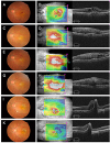 | Figure 1Fundus photographies and optical coherence tomographies during treatment. At the time of initial examination, a large number of soft drusens and retinal hemorrhages were observed in the fundus photography (A). Optical coherence tomography (OCT) revealed intraretinal cysts, pigmented epithelium detachment (PED), and epiretinal membrane (ERM), resulting in a central macular thickness (CMT) of 405 µm (B). 1 month after the first injection, the transparency of the ERM decreased in the fundus photography (C). In the OCT, the intraretinal cysts disappeared and height of PED decreased. But the reflectivity and thickness of the ERM increased, resulting in a CMT of 457 µm (D). 1 month after the second injection, the ERM became more opaque in the fundus photography (E), and CMT increased to 506 µm (F). 1 month after the third injection, the ERM remained thick in the fundus photography and OCT (G, H). Pars plana vitrectomy for ERM was done. The membrane was removed well though retinal pigment epithelial tear was observed at postoperative 10 days (I, J). 1 month after the fourth injection, fovea remained stable with scarring (K, L).
|
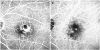 | Figure 2Fluorescein angiography and indocyanine green angiography of the patient at the initial visit. Late phase fluorescein angiography showed hyperfluorescence and pooling of dye at the fovea with a blocked fluorescence due to retinal hemorrhages (A). Indocyanine green angiography revealed hot spots (B).
|
References
1. Ambati J, Ambati BK, Yoo SH, et al. Age-related macular degeneration: etiology, pathogenesis, and therapeutic strategies. Surv Ophthalmol. 2003; 48:257–293.

2. Falavarjani KG, Nguyen QD. Adverse events and complications associated with intravitreal injection of anti-VEGF agents: a review of literature. Eye (Lond). 2013; 27:787–794.
3. Wickham L, Konstantinidis L, Wolfensberger TJ, et al. Epiretinal membranes, vitreoretinal traction, and cystoid macular edema. In : Schachat AP, editor. Ryan's Retina. 6th ed. Philadelphia: Elsevier Inc.;2017. 3:chap. 120.
4. Chen YS, Hackett SF, Schoenfeld CL, et al. Localisation of vascular endothelial growth factor and its receptors to cells of vascular and avascular epiretinal membranes. Br J Ophthalmol. 1997; 81:919–926.
5. Abouammoh MA, Belliveau MJ, Almeida DR, et al. Ranibizumab for idiopathic epiretinal membranes: a retrospective case series. Saudi J Ophthalmol. 2013; 27:79–82.
6. Marticorena J, Romano MR, Heimann H, et al. Intravitreal bevacizumab for retinal vein occlusion and early growth of epiretinal membrane: a possible secondary effect? Br J Ophthalmol. 2011; 95:391–395.
7. Kuiper EJ, Van Nieuwenhoven FA, de Smet MD, et al. The angio-fibrotic switch of VEGF and CTGF in proliferative diabetic retinopathy. PLoS One. 2008; 3:e2675.
8. Zhang Q, Qi Y, Chen L, et al. The relationship between anti-vascular endothelial growth factor and fibrosis in proliferative retinopathy: clinical and laboratory evidence. Br J Ophthalmol. 2016; 100:1443–1450.
9. Chu SJ, Zhang ZH, Wang M, Xu HF. Effect of bevacizumab on the expression of fibrosis-related inflammatory mediators in ARPE-19 cells. Int J Ophthalmol. 2017; 10:366–371.
10. Zhang JJ, Chu SJ, Sun XL, et al. Bevacizumab modulates retinal pigment epithelial-to-mesenchymal transition via regulating Notch signaling. Int J Ophthalmol. 2015; 8:245–249.
 XML Download