Abstract
Tracheal agenesis is an extremely rare and typically fatal congenital anomaly, with only scattered case reports attesting to of its successful management. This condition usually presents with severe respiratory distress and aphonia after birth. Failed attempts at intubation make neo natal resuscitation difficult. This condition appears to be under-recognized, and there is a lack of con sensus regarding the optimum approach for managing this lethal condition. We report herein a rare case of tracheal agenesis and describe our experience, following its successful treatment through surgical management.
REFERENCES
1). Payne WA. Congenital absence of the trachea. Brooklyn Med J. 1900. 14:568.
2). Lander TA., Schauer G., Bendel-Stenzel E., Sidman JD. Tracheal agenesis in newborns. Laryngoscope. 2004. 114:1633–6.

3). de Groot-van der Mooren MD., Haak MC., Lakeman P., Cohen-Overbeek TE., van der Voorn JP., Bretschneider JH, et al. Tracheal agenesis: approach towards this severe diagnosis. Case report and review of the literature. Eur J Pediatr. 2012. 171:425–31.

4). Jung YM., Kim JE., Son DW., Kim HN., Hwang HY. MDCT findings of tracheal agenesis: a case report. J Korean Soc Radiol. 2009. 60:23–6.

5). Park KM., Suh YL., Khang SK., Lee JG. Tracheal agenesis: report of an autopsy case. Korean J Pathol. 1992. 26:283–7.
6). Sung IK., Chun CS., Cho SH., Kim SY., Kim SJ., Lee WB, et al. A case of tracheal agenesis with tracheoesophageal fistula. Korean J Perinatol. 1998. 9:320–4.
7). Lee HJ., Park EA., Lee SJ., Kim MJ., Seong SH. An autopsy case of tracheal agenesis type 2. Korean J Pediatr Soc. 1996. 39:1759–62.
8). Lee JY., Kim SY., Lee KY., Moon SH. Tracheal agenesis: a case report. Korean J Anesthesiol. 1998. 34:187–91.

9). Floyd J., Campbell DC Jr., Dominy DE. Agenesis of the trachea. Am Rev Respir Dis. 1962. 86:557–60.
10). Fraser N., Stewart RJ., Grant J., Martin P. Gibbin KP, Padfield CJ. Tracheal agenesis with unique anatomy. J Pediatr Surg. 2005. 40:e7–10.
11). Altman RP., Randolph JG., Shearin RB. Tracheal agenesis: recognition and management. J Pediatr Surg. 1972. 7:112–8.

12). Hedrick MH., Ferro MM., Filly RA., Flake AW., Harrison MR., Adzick NS. Congenital high airway obstruction syndrome (CHAOS): a potential for perinatal intervention. J Pediatr Surg. 1994. 29:271–4.

13). Evans JA., Greenberg CR., Erdile L. Tracheal agenesis revisited: analysis of associated anomalies. Am J Med Genet. 1999. 82:415–22.

14). Ergun S., Tewfik T., Daniel S. Tracheal agenesis: a rare but fatal congenital anomaly. Mcgill J Med. 2011. 13:10.
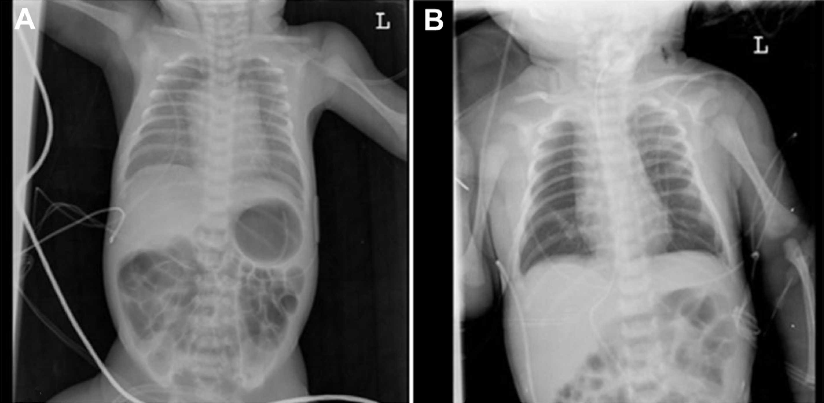
Fig. 1
Initial infantogram (A) and infantogram after tracheostomy (B). (A) Infantogram shows quite good air content in both lungs, but with fine granular appearance. (B) The lungs have increased air content after tracheostomy.

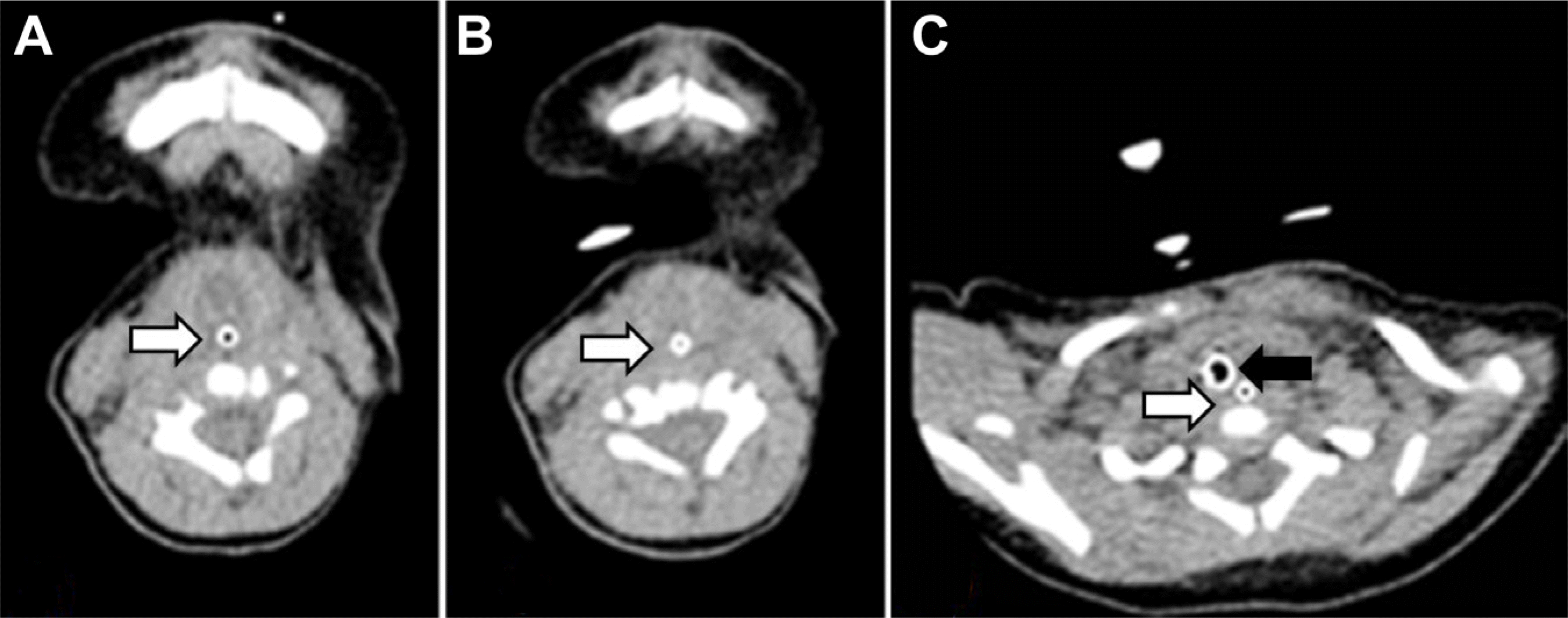
Fig. 2
Pharyngeal non-contrast computed tomography showed no visualization of trachea. (A) At level of C3 vertebral body, (B) at level of C5 vertebral body, (C) at level of C7 vertebral body, tra cheostomy tube insertion site. White arrow indicates esophagus and black arrow indicates tra cheostomy tube.

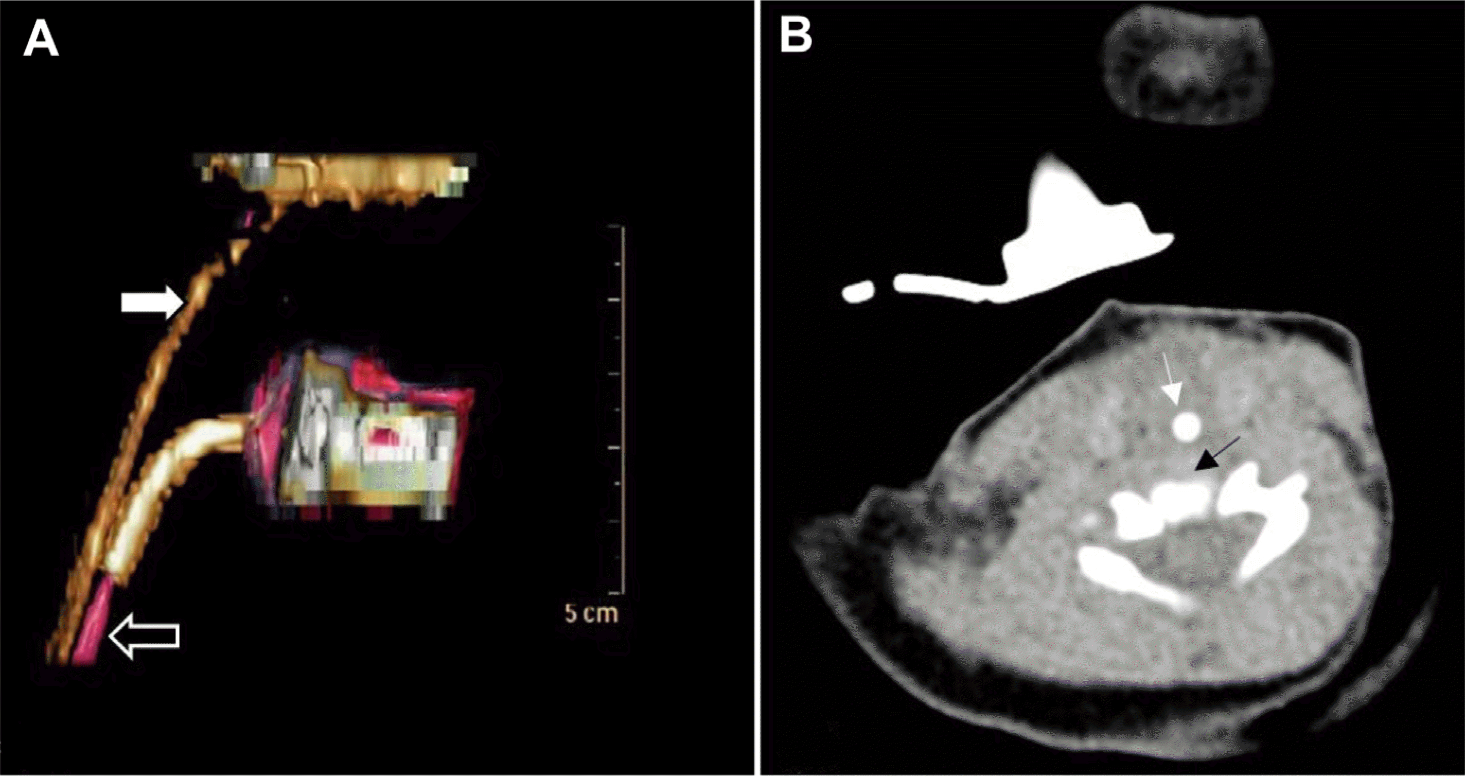
Fig. 3
(A) Pharynx 3 dimension computed tomography showed no visualization of proximal trachea: white arrow indicates esophagus and black arrow indicates distal trachea. (B) Pharyngeal non-contrast computed tomography showed no visualization of trachea at level of C5 vertebral body: thin black arrow indicates C5 vertebral body and thin white arrow indicates esophagus and no visualization proximal trachea.

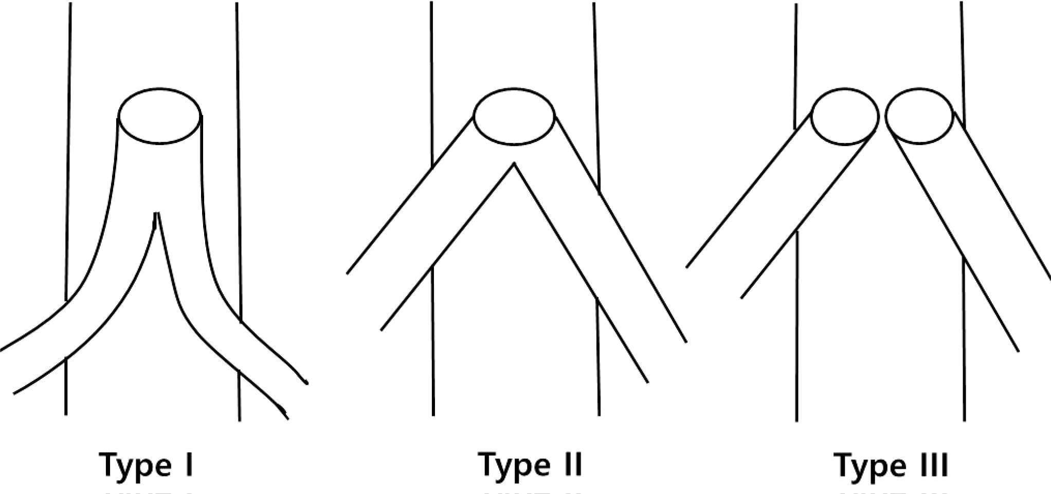
Fig. 4
Floyd's classification of tracheal agenesis by Floyd et al.16 Three types of tracheal agenesis. Type I: atresia of the proximal trachea with presence of the distal trachea and carina and two completely formed bronchi. Type II: the bronchi join in the midline and communicate with the esophagus as a common fistula. Type III: independent communication between the bronchi and esophagus.

Table 1.
Comparison: Five Cases of Tracheal Agenesis Reported in Korea


 PDF
PDF ePub
ePub Citation
 Print
Print


 XML Download