

 PDF
PDF Citation
 Print
Print


VIDEO CLIP
Video can be found with this article online at https://ejgo.org/src/sm/jgo-29-e87-s001.mp4.
1. Clinical setting
The number of robotic surgery has been rapidly increasing in gynecologic cancers [1]. In spite of the advanced application of robotic surgery, upper paraaortic lymph node dissection (UPALD) to the infrarenal level remains one of the most challenging robotic procedures in the gynecologic field. Because of the limitation of robotic arm mobility, the da Vinci S or Si robotic systems (Intuitive Surgical, Inc., Sunnyvale, CA, USA) do not allow reaching to the whole abdominal cavity without relocating the robotic column. Furthermore, the robotic system is too bulky to be relocated freely in an operation room. There have been several reports that showed techniques to perform UPALD [234]. However, these techniques are potentially limited by placement of robotic ports, patients' body habitus, experienced operating room staffs, and adequate spacing in operating suites. This surgical video introduces a novel robotic approach, Lim's lower pelvic port placement (LP3), to perform optimally and simultaneously both UPALD and pelvic procedures using da Vinci Xi system (Intuitive Surgical, Inc.).
2. Procedure
The patient presented with high-grade endometrial cancer. She underwent robotic surgical staging operation. Overall procedures were composed of 2 parts. One was upper abdominal procedures, including paraaortic lymph node dissection (PALD) to the infrarenal level and omentectomy. The other was pelvic procedures, including hysterectomy, bilateral salpingo-oophorectomy, and pelvic lymph node dissection (PLD). Of these, this video shows PALD and PLD.
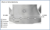
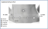
For the setup of the LP3, a line was drown between both anterior superior iliac spines. At 3 cm below this line, another line was drown and 4 robotic ports were placed on this line (Fig. 1). After appropriate LP3, the small bowel mesentery was reflected cephalad. It is important to take down all the adhesions of the small bowel from the pelvic brim and anterior abdominal wall so that the small bowel mesentery can be reflected cephalad. The mesenteric fold of small bowel was identified and exposed. It ran parallel to the vena cava and abdominal aorta. The peritoneum overlying the sacral promontory 2 cm medial to the right common iliac artery was grasped. A reverse J-shaped peritoneum incision was made initially cephalad along the mesenteric fold parallel to the vena cava to the level of duodenal recess. The duodenal recess was mobilized ventrally to expose the underlying left renal vein. The peritoneal incision was extended inferiorly about 5 cm below the sacral promontory which would facilitate the left infra inferior mesenteric artery (IMA) lateral displacement (LD). After completion of the peritoneal incision, the incised peritoneum was retracted ventrally to create a ‘tenting effect’ which would prevent the small bowel from falling into the operative field. Once this has been achieved, the PALD was carried out systematically. Right infra IMA LD was achieved. The bedside assistant grasped the right lateral edge of the incised peritoneum and retracted laterally and ventrally to expose the right infra IMA nodes. The right abdominal ureter which remained attached to the peritoneum was identified and mobilized laterally. The right ovarian vein which was superior to the ureter was also identified and mobilized laterally. The LD commenced at the proximal portion of the right common iliac vessel and the lymph node bearing tissue was retracted ventrally and laterally and incised. The dissection was performed medially to laterally and cephalad once the anterior caval wall was identified. Right supra IMA LD was carried out. The assistant grasped and retracted the superior edge of the right incised peritoneum to expose the right supra IMA while the third robotic arm grasped the left lateral incised peritoneum and retracted ventrally and laterally. The right ovarian artery was identified as it crossed the vena cava and ran parallel to the right ovarian vein. The right ovarian vein was then identified as it entered the vena cava. Left infra IMA LD was performed. The dissection began by identifying the left common iliac artery. Then left lower abdominal ureter which was attached to the lateral peritoneum was identified and mobilized laterally along with the left ovarian vein. The dissection was performed medially to laterally removal of the anterior nodes followed by paraaortic nodes. The dissection was carried out cephalad to the level of IMA. Left supra IMA LD was performed. The third robotic arm was placed again to grasp the left incised peritoneal edge to retract the bowel of the operative field while the assistant grasped the right incised peritoneal edge for optimal operative field exposure. The dissection was carried out medially to laterally skeletonizing the lymph nodes anterior to the aorta follow by the paraaortic nodes. The left ovarian vein was identified follow by the left upper abdominal ureter which was inferior to the left ovarian vein. The dissection was continued cephalad until the renal vein was identified. After PALD was completed, the boom of robotic system was rotated 180° to retarget for the PLD. Robotic ports were placed and docked again (Fig. 2). The PLD was achieved as a usual manner.
The LP3 was feasible for performing simultaneously optimal UPALD as well as procedures in pelvic cavity in gynecologic cancer patients. The advantage of LP3 technique is the robotic port placement that affords for multi-quadrant surgery, abdominal and pelvic dissection. The LP3 is facilitated by utilizing advanced technology of Xi system, including the patient clearance function, the rotating boom, and ‘port hopping’ that allows using every ports for a camera. The patient clearance function extends the range of motion of robotic arms. The rotating boom feature facilitates the pelvic dissection to be performed in an efficient manner. In addition, the LP3 affords the surgeon to give the same operative view as open technique for PALD with port hopping. Consequently, the PALD can be achieved safely and optimally. The LP3 will enable surgeons to extend the surgical indication of robotic surgical system in the gynecologic oncologic field.
 XML Download