

 PDF
PDF Citation
 Print
Print


INTRODUCTION
Behcet's disease (BD) is a rare disorder that causes blood vessel inflammation throughout the body. Although the disease leads to numerous signs and symptoms, recurrent oral aphthous ulcers, uveitis, skin rashes and lesions as well as genital ulcers are the classical signs. Central nervous system involvement in BD has remained one of the most serious complications of the disease because it is closely related to morbidity and mortality [1]. The frequency of neuro-Behcet's disease (NBD) has been reported to be approximately 3.5%–14.3%, according to the diagnostic criteria for NBD [2]. The most common initial symptom of NBD is headache, which is accompanied by other neurological symptoms, such as gait disturbance, dizziness, and dysarthria [2]. These symptoms are associated with magnetic resonance imaging (MRI) lesions, usually involving specific areas of the brain, including the thalamus, the internal capsule and the brainstem [3]. Nevertheless, several case reports and series showed that neurocognitive deficits and behavioral changes may be the first signs of NBD [45678]. Most NBD patients have good clinical outcomes after steroid therapy, but long-term functional and clinical outcomes for patients with the malignant relapse-remission pattern are not well-known [28]. We encountered an NBD patient who was referred for functional deterioration, and we report a 4-year follow-up experience of the patient's rehabilitation.
CASE REPORT
A 34-year-old male presented with abrupt onset memory disturbance, aggressive behavior, and gait disturbance in January 2014. He was a military officer with no specific past medical history. Recurrent oral and genital ulcers, eye pain, diplopia, and ptosis were observed in a comprehensive physical examination and on neurologic examination. On the fluid-attenuated inversion recovery sequence of brain MRI, high signal intensity was shown in the bilateral basal ganglia, left thalamo-mesencephalic territory, and bilateral midbrain (Fig. 1). Laboratory tests did not show inflammatory findings, and HLA-B51 and cerebrospinal fluid were negative. Upon consultation with the rheumatology department and according to International Study Group criteria, BD was diagnosed on the basis of oral ulcers recurring more than 3 times a year, recurrent genital ulcers, and eye lesions [9]. Definitive NBD was determined according to international consensus recommendation criteria on the basis of BD with neurological syndromes such as cognitive dysfunction, optic neuropathy, dysphagia, abnormal neuroimaging, and absence of a better explanation for the neurological findings [10].
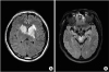 | Fig. 1The axial sections of fluid-attenuated inversion recovery imaging of the patient. Multifocal high signal intensity lesions are observed in (A) the bilateral basal ganglia and left thalamo-mesencephalic area and (B) in the bilateral midbrain.
|
Steroid therapy was started with IV methylprednisolone infusion (1,000 mg/day), slowly tapering to oral steroid (10 mg/day). The neurologic symptoms completely recovered after that first steroid therapy, but relapse and remission repeated 5 times within 2 years after the first onset (2, 18, 20, 23, 27 months postonset). Although high-dose steroid therapy (1,000 mg/day) was performed with each relapse, the cognitive impairment and gait disturbance due to ataxia/motor weakness became more severe.
Because severe dysphagia and motor weakness accompanied by cognitive deficits were not improved despite steroid therapy at the 5th recurrence, the patient was referred to the department of rehabilitation.
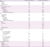
At 27 months after first onset, the time of the 5th recurrence, motor weakness, ataxia, and balance dysfunction of bilateral limb were observed. Fugl-Meyer Assessment (FMA) score was 71 in the motor function domain and motor weakness was not severe. However, the Berg Balance Scale (BBS) score was 9 and the Korean version of the Scale for the Assessment and Rating of Ataxia (K-SARA) score was 30, meaning severe ataxia such that he could not walk. The Caregiver-Administered Neuropsychiatric Inventory (CGA-NPI) was 86. Delusions, depression, anxiety, euphoria, apathy, disinhibition, aberrant motor behavior and night-time behaviors were observed. The Korean version of the Mini-Mental State Examination (K-MMSE) score was 23, demonstrating abnormalities in attention, recall, and language domains. The Korean version of the Modified Barthel Index (K-MBI) score was 27, indicating severe dependency in gait and activities of daily living (ADL). The modified Rankin Scale (mRS) score was 5, suggesting severe disability such that he was bedridden, incontinent and requiring constant nursing care and attention. At the time of referral, the patient was tube feeding. His swallowing function was evaluated by a videofluoroscopic swallowing study (VFSS). Endotracheal aspiration was found when tested with liquid. The total score of the Videofluoroscopic Dysphagia Scale (VDS) was 50, suggesting severe dysphagia, and the subject continued tube feeding [11] (Table 1).
Table 1
Summary of evaluation tools

FMA, Fugl-Meyer Assessment; BBS, Berg Balance Scale; K-SARA, Korean version of the Scale for the Assessment and Rating of Ataxia; CGA-NPI, Caregiver-Administered Neuropsychiatric Inventory; K-MMSE, Korean version of the Mini-Mental State Examination; K-MBI, Korean version of the Modified Barthel Index; mRS, modified Rankin Scale; VDS, Videofluoroscopic Dysphagia Scale.
![]()
To evaluate the domain of cognitive function in detail, the Seoul Neuropsychological Screening Battery (SNSB) was performed. The percentile scores were corrected for age, education and gender. In the case of this patient, percentile scores were applied to the 45-year-old norm. The 50th percentile score corresponds to the average of people with age and educational levels similar to those of the patient. If the percentile score is lower than 16%, it can be interpreted as abnormal [12]. SNSB showed marked abnormalities in digit span test, verbal learning test, Rey Complex Figure Test (RCFT), Controlled Oral Word Association Test (COWAT), and Korean-Color Word Stroop Test (K-CWST). These findings suggested severe impairments in the domains of attention, memory and executive function (Table 2).
Table 2
Summary of the SNSB
SNSB, Seoul Neuropsychological Screening Battery; SVLT-E, Seoul Verbal Learning Test-Elderly's version; RCFT, Rey Complex Figure Test; COWAT, Controlled Oral Word Association Test; K-CWST, Korean-Color Word Stroop Test; WR, word reading; CR, color reading.
![]()
He began comprehensive rehabilitation therapies consisting of physical, occupational, cognitive, and dysphagia therapy twice a day combined with medical therapy. Balance exercise for weight bearing and shifting and gait training were performed in physical therapy. In occupational and cognitive therapy, cognitive function training for calculation and memory, ADL training for communication and self-care, and the Comcog® attention and memory program were conducted. Oromotor facilitation, oropharyngeal muscles training, postural compensatory strategies and transcutaneous neuromuscular electrical stimulation were also carried out for dysphagia.
After discharge, the patient received rehabilitation continuously on an outpatient basis 3 times per week with the same programs. His function gradually improved during the following 23 months without relapsing. When the patient was reevaluated at 50 months after the first onset, he could walk under supervision without assistance tools. FMA was 88 in the motor function domain. BBS was 46 and K-SARA was 9. CGA-NPI was 32, meaning a decrease in behavior symptoms. K-MMSE was 26, with improvement in attention, recall, and language domains. K-MBI was 77, indicating mild dependency in gait and ADL. The mRS was 3, suggesting moderate disability requiring some help, but he was able to walk without assistance. In VFSS, at 43 months after first onset, aspiration was not observed, and VDS was 25. Therefore, tube removal was performed, and oral feeding started with a modified diet (Table 1). In SNSB, the average improvement was 10% to 15% in the domains of attention, memory, and executive function (Table 2).
DISCUSSION
In this case, cognitive impairment, motor weakness, ataxia, and dysphagia caused serious functional deterioration of ADL during 4 years of follow-up.
The current treatments for NBD are high-dose steroid injections during the acute phase, followed by tapering doses [13]. The majority of patients usually show symptomatic improvement after the initial treatment and have a good prognosis [28]. Although it is rare for patients to experience prolonged neurological impairment in spite of treatment, repetitive involvement of NBD can cause irreversible brain damage [3]. Brainstem involvement appears to give a worse prognosis because of its possible association with progressive or more resistant disease [18]. The patients with progressive courses and repeated relapses are recommended to receive concomitant cyclophosphamide and steroid therapy during the early course of the disease; nevertheless, the effects are debated [2].
In the reported patient, severe cognitive deficits in the areas of attention, memory, and frontal executive function were identified by neuropsychological screening tests, including digit span, verbal learning test, RCFT, COWAT, and K-CWST. These dysfunctions induced many behavioral problems, thereby making it difficult for the patient to return to work and live with family members. Nevertheless, language and visuospatial functions were relatively preserved, and these features were similar to those of previous studies [45678]. The mechanisms of these characteristics are unclear, but several hypotheses can be proposed based on the studies of functional neuroimaging. For example, using single photon emission computed tomography in NBD patients, Mimura et al. [6] proposed that the thalamus and related subcortical–frontal connection were responsible for amnesia and frontal/executive deficits. Another study using functional MRI reported that ventrolateral and anterior thalamic involvements directly affected attention and higher-level cognition [14]. Therefore, the main neurocognitive symptoms in this case were primarily assumed to have originated from lesions in the thalamus.
Noel et al. [15] analyzed long-term outcomes according to the mRS in NBD patients. If the patients had hemiparesis or paraparesis and brainstem localization on MRI, the authors found that these factors were associated with mRS 3 or more, that is, poor outcome, after a median follow-up of 73 months. In the present case, the patient was expected to have a poor outcome because of brain lesions, motor weakness and mRS 5. At 50 months after first onset, because of the improved motor weakness and mRS 3 in rehabilitation therapy, continuous rehabilitation had a significant role in the long-term outcome.
In this respect, rehabilitation can help to maintain and improve overall functional levels, including cognitive and motor functions for high-risk NBD patients. Previous studies regarding the effectiveness of rehabilitation in refractory NBD patients are very rare. A case report of a spinal cord NBD patient presenting with paraplegia demonstrated that early aggressive treatment and continuous rehabilitation in conjunction with medication provided good prognosis with excellent clinical outcome [16]. This patient was in a complete paraplegic state due to myelopathy of multiple thoracic spinal cord levels, but they recovered complete independence in ADL and long-distance ambulation after 1 year without relapse by means of continuous rehabilitation therapy [16]. Similarly, in our case, comprehensive rehabilitation therapies were administered starting after the 5th recurrence. Cognitive impairments, gait disturbance, and dysphagia were gradually ameliorated, and the patient regained functional independency during the follow-up period. There have been no case reports related to motor function and other capabilities in brain-involved NBD; therefore, the effect of rehabilitation in our patient was indirectly estimated by referring to the previous case report to see the change in functional level in NBD.
In summary, cognitive impairments in the domains of attention, memory, and executive function, as well as motor weakness, ataxia, and dysphagia are major problems that cause serious functional deterioration in patients with NBD. Despite adequate steroid therapy, some patients follow a chronically progressive course with repeated relapses and remissions. We would like to highlight that steadily performing rehabilitation therapy combined with medication can be helpful to improve long-term function and prognosis in refractory NBD patients.
 XML Download