

 PDF
PDF ePub
ePub Citation
 Print
Print


Introduction
Cigarette smoking is one of the primary causes of increased health-care costs and decreased life expectancy.12 Epidemiological studies have focused on the deleterious effects of smoking on human health. In particular, a number of epidemiological studies reported that cigarette smoke decreases bone mineral density (BMD), increases the risk of bone fracture, and is a risk factor for osteoporosis.345 In the United States, the prevalence of maternal smoking during pregnancy decreased slightly from 13.3% in 2000 to 12.3% in 2010. However, the smoking rate among pregnant women is still high at over 10%.6 In Korea, 3% of all women smoke during pregnancy.7 Maternal smoking during pregnancy can cause various problems such as an increased chance of premature birth, placental abruption, and placenta previa. Furthermore, it can affect the health of the fetus by increasing the risks of intrauterine fetal growth restriction and sudden infant death syndrome.8
Despite extensive research on maternal smoking during pregnancy, the exact effect of maternal smoking during pregnancy on the bones of the offspring remains unknown. One study reported low bone mineral content and BMD in the newborns of mothers who smoked.9 Another study that examined the long-term effects of maternal smoking during pregnancy showed that smoking has negative effects on child bone mass for 8 years.10 On the other hand, other studies reported that maternal smoking during pregnancy does not affect bone mineral content and BMD in the neonate.11 Therefore, further studies are needed to evaluate the effects of maternal smoking on the offspring's bones. The aim of the present study was to evaluate the effect of maternal smoking during pregnancy on the bones of the offspring.
Methods
1. Animals
CD-1 outbred mice were purchased from Charles River Laboratories (Charles River Laboratories, Wilmington, MA, USA) and maintained under conditions of 14 hours light, 10 hours dark at 23℃ with food and water available ad libitum. Experimental pregnant mice were obtained after mating. Day 1 of gestation (E1) was designated the first day after observation of the vaginal plug, which is formed by secretions from the male vesicular and coagulating glands and is a convenient and easily visible indicator that mating has occurred. Coeval pregnant mice were to check the health condition at all stages of the experiment. All animal procedures were approved by the Korea University Animal Ethics Committee (KUIACUC- 2012-121).
2. Exposure to cigarette smoke
Pregnant mice were randomly divided into three groups: a non-exposed group, which included control pregnant mice that received no intervention; a 1 cigarette exposed group, which included pregnant mice that were exposed individually to the smoke of 1 cigarette/day during delivery; and a 2 cigarette exposed group, which included pregnant mice that were exposed individually to the smoke of 2 cigarettes/day during pregnancy. Five pregnant mice were studied in each group. Pregnant mice in the 1 cigarette or 2 cigarette groups were exposed to 1 or 2 cigarettes per day for 5 days/week, respectively (code 3R4F reference cigarettes, produced for the University of Kentucky Tobacco and Health Research Institute, Lexington, KY, USA) from E1 to delivery. On each day of exposure, animals were placed individually inside a Plexiglas cabinet (40×90×100 mm). Cigarette smoke was delivered into the cabinet through air inflow at a rate of 1.7 mL/s by introducing a burning cigarette in the chamber; the combustion time of the cigarette was less than 3 minutes. For 2 cigarette group, cigarette smoke was delivered with no resting time between two cigarettes.
A ventilator inside the cabinet ensured rapid and equal distribution of smoke. Fresh air was delivered into the cabinet to remove the smoke. The control, non-exposed group was placed in an identical chamber and then exposed to inflow of fresh air for the same period.
3. Micro-computed tomography (CT) analysis
After delivery, nursing dams and offspring were kept together in individual cages for 4 weeks. From 5 dams for each group, 1–2 offspring for each dam were selected considering that each group can be composed of the same gender proportion in offspring. Finally, a total of eight offspring (4 males and 4 females) from each group were evaluated. Offspring were categorized into three groups as follows: control group, including offspring from the non-exposed group; 1 cigarette group, including offspring from the 1 cigarette exposed group; and 2 cigarette group, including offspring from the 2 cigarette exposed group. At 4 weeks, offspring from each dam were anaesthetized with isoflurane. The fourth lumbar vertebral body from each mouse was scanned by using a SkyScan1173 micro-CT apparatus (SkyScan, Kontich, Belgium) with an isotropic resolution of 18 µm. Micro-CT scanning operating parameters were 50 kVp volts of X-rays, 500 µA of source current, and 250 ms exposure. A 0.5-mm thick aluminum filter was used to minimize beam-hardening artifacts. During scanning, the lumbar spine was enclosed in a tightly fitting rigid plastic tube to prevent movement. The scans were taken in the distal transverse plane of the vertebral body for a 2.4-mm thick region. The number of slices was determined according to the size of the vertebrae and was approximately 200 slices per vertebral specimen. The 2-dimensional (2-D) grayscale CT images were reconstructed in 2,000×1,330 pixel matrices by using NRecon ver. 1.6.1.5 (SkyScan). Three-dimensional (3-D) reconstructions of Ø 0.61×0.71 mm of the trabecular bone on the central vertebral body were obtained from the same samples. Trabecular parameters including bone volume faction (bone volume/total volume, %), trabecular thickness (µm), separation (µm), and number (1/mm) were evaluated. The BMD (mg/cm3) was also measured.
4. Biochemical markers of bone turnover
Before obtaining micro-CT images, blood samples were collected from each offspring. To evaluate bone formation at the serum level, osteocalcin (OC) levels were measured by using a mouse OC enzyme-linked immunosorbent assay kit (Biomedical Technologies, Stoughton, MA, USA). To evaluate bone resorption at the serum level, C-terminal telopeptides of type I collagen (CTX) were measured by using the RatLaps™ enzyme immunoassay kit (Nordic Bioscience Diagnostics, Herlev, Denmark). The manufacturer's protocols were followed, and samples were assayed in duplicate. A standard curve was generated by using the provided protein and absolute concentrations from the kit, and the OC and CTX in the serum were extrapolated from a standard curve. Samples were measured at 450 nm for both the OC and CTX.
Results
The number of pups from a non-exposed, 1 cigarette exposed and 2 cigarette exposed group were different (12.25±1.39, 11.25±0.46, and 12.50±0.54, P=0.027, respectively). The number of demise pups were also different among three groups with observation only in 2 cigarette exposed group (0, 0, and 0.75±0.46, P<0.001, respectively).
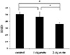
The BMD of the fourth lumbar vertebral body from each group was measured. BMD was significantly lower in the 2 cigarette group than in the control and 1 cigarette groups. However, no differences in BMD were observed between the control and 1 cigarette groups (Fig. 1).
The microarchitecture of the fourth lumbar vertebral body from each offspring was evaluated. Representative 2-D and 3- D micro-CT images of the fourth lumbar vertebral body from each group are presented in Fig. 2. The micro-CT images reveal that Perinatologythe 2 cigarette group had a smaller trabecular thickness, a decreased trabecular number, and expanded trabecular separation in the fourth lumbar vertebra compared to those in the control and 1 cigarette groups.
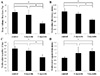
The results of microarchitecture analyses are presented in Fig. 3. Trabecular bone volume fraction, thickness, and number were significantly lower in the 2 cigarette group than in the control and 1 cigarette groups, whereas trabecular separation was higher in the 2 cigarette group than in the control group. However, no difference in trabecular separation was detected between the 1 and 2 cigarette groups. Trabecular bone volume fraction, thickness, number, and separation did not differ between the control and 1 cigarette groups.
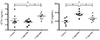
The biochemical characteristics of bone turnover were compared between the three groups. There was a significant increase of serum CTX levels in the 2 cigarette group compared with those in the control and 1-cigarette group (Fig. 4). However, no difference in serum levels was observed between the control and 1 cigarette groups (Fig. 4). Serum OC levels were higher in the 1 and 2 cigarette groups compared with those in the control group. Serum OC levels were higher in the 1 cigarette group than in 2 cigarette group (Fig. 4).
Discussion
The present study evaluated the effect of maternal smoking during pregnancy on the bones of the offspring, and the results showed that maternal smoking during pregnancy is associated with a decrease in the offspring's BMD. This result is in line with the results of previous human and animal studies, which reported that maternal smoking during pregnancy has a negative effect on the offspring's bone growth.910
BMD is the chief parameter for diagnosing osteoporosis, evaluating bone characteristics, and predicting fracture risk; BMD is known to be responsible for approximately 70–80% of the strength in bone tissue.12 Although these characteristics are related to the mechanical performance of bones, their usefulness for predicting fracture risk is limited.13 For example, 50% of individuals presenting with hip fracture have a T-score above −2.5.14 Therefore, other factors in addition to BMD need to be evaluated to determine bone health. Bone microarchitecture properties are emerging as major determinants of the risk of fracture.1516 Bone microarchitecture significantly influences bone strength, independent of the BMD.17
Several animal studies have evaluated the effects of smoking on bone microarchitecture during various stages of life and reported that smoking has a negative influence on bone microarchitecture.1218 However, little is known about the effect of maternal smoking on the offspring's bone microarchitecture. In the present study, we evaluated the effect of maternal smoking during pregnancy on bone microarchitecture in the offspring, and found that maternal smoking during pregnancy was associated with deterioration of the bone microarchitecture of the offspring. Although several studies did not report any significant differences in bone mineral content and BMD between the infants of women who smoked and those of women who did not smoke,11 maternal smoking may have a detrimental effect on the offspring's bone through its effect on the microarchitecture.
The mechanism by which maternal smoking during pregnancy affects the offspring's bones is unclear, although there are several possible explanations. First, maternal smoking during pregnancy may have a direct effect on the offspring's bones. In our study, the levels of CTX, a bone resorption marker synthesized by osteoclasts, were higher in the cigarette-smoking groups. Smoking induces bone loss by increasing bone resorption.192021 Similarly, in the present study, maternal smoking during pregnancy may have increased bone resorption in the fetus, resulting in a decrease in the BMD and damage to the microarchitecture.
Another possible mechanism is that maternal smoking during pregnancy may indirectly affect fetal bones. For example, maternal smoking during pregnancy is related to the offspring's low birth weight222324 through a decrease in protein-like maternal energy intake2324 or through changes in the placenta and its vascularization leading to changes in placental morphology.25 Birth weight is associated with peak bone mass.26 Several studies reported that low birth weight is associated with lower BMD and the bone mineral content measured in various stages of life.272829 Recently, a meta-analysis confirmed that birth weight positively influences bone health in later life.30 Therefore, factors such as smoking during pregnancy that causes poor intrauterine growth may eventually have negative effects on the growth of the skeletal system, increasing lower bone mass and the risk of hip fracture from then on to adulthood. However, more research is needed to elucidate the mechanism underlying the effect of maternal smoking on the offspring's bones.
Given that smoking reduces the activity of osteoblasts through various pathways,21 we hypothesized that maternal smoking during pregnancy may negatively affect osteoblasts, resulting in a decrease in OC levels. In addition, infants of smoker women have significantly lower OC levels in the umbilical cord than that of infants of both passive smokers and nonsmoker women.31 In the present study, the levels of OC increased in the cigarette smoking groups. Although the reason for this discrepancy is unclear, there are possible explanations. Bones are dynamic organs; homeostasis is maintained because of the balance between bone resorption by osteoclasts and bone formation by osteoblasts. Therefore, an increase in the activity of osteoclasts caused by smoking may simulate the activity of osteoblasts to compensate and maintain bone homeostasis, resulting in an increase of OC levels. Another possible reason is related to the study design including the dose of smoking. Nicotine, one of the main effective ingredients contributing to the harmful effects of smoking, affects the proliferation of osteoblast cells and the expression of markers of bone formation.32 However, nicotine at lower concentrations stimulates bone formation, whereas at higher concentrations, it inhibits bone formation.3233 Similarly, in the present study, OC levels were higher in the cigarette groups, especially in the 1 cigarette group compared with the 2 cigarette group. Therefore, the dose of smoking used in this study may have stimulated bone formation. To confirm these dose-dependent effects of maternal smoking on bone formation, we used a 3 cigarette smoke exposed model; however, most fetuses aborted and the data could not be included (data not shown).
In the present study, we selected adolescence, which is the period during which a young person develops from a child into an adult, as the period to conduct the research by using 4-week-old mice.34 Therefore, further studies are needed to determine the long-term effects of maternal smoking during pregnancy on the offspring's bones. However, after the adolescence period, different factors such as food and exercise can influence bone characteristics in adulthood. Therefore, experiments performed before adolescence may more accurately reflect the effect of maternal smoking on the bones of the offspring.
In conclusion, maternal smoking during pregnancy was shown to have a negative effect on bone health in the offspring. These results are important to emphasize the importance of quitting smoking during pregnancy.

 XML Download