

 PDF
PDF ePub
ePub Citation
 Print
Print


INTRODUCTION
Polyarteritis nodosa (PAN) is a systemic necrotizing vasculitis affecting medium-sized arteries, with occasional involvement of small arteries, resulting in secondary tissue ischemia involving the kidneys, skin, joints, muscle, peripheral nerves and gastrointestinal tract. The typical manifestations include systemic symptoms, and biopsy and/or arteriography are usually required for the definitive diagnosis [1]. Cutaneous polyarteritis nodosa (CPAN) differs from the systemic form of PAN; in that it is limited to the skin and there is a lack of visceral involvement [2]. In addition to cutaneous findings, patients with CPAN are also frequently reported to develop extra-cutaneous manifestations such as arthralgia, myalgia, and peripheral neuropathy localized to areas of skin lesions [34]. CPAN should be monitored closely over time for progression to systemic PAN, but some reports suggest that CPAN may represent a distinct subset of classic PAN [345]. Treatment with systemic glucocorticoids alone or in combination with azathioprine, methotrexate or cyclophosphamide, depending on disease severity, has been shown to be effective [36]. The use of anti-tumor necrosis factor-α therapy (anti-TNF-α therapy) (infliximab or etanercept) has been described in case reports of patients with severe symptoms and refractory to conventional treatment [78910]. Herein, we report the successful treatment with adalimumab in a patient with refractory CPAN and present similar cases reported in the literature.
CASE REPORT
A 53-year-old woman was hospitalized in a tertiary medical center in July 2015 with a 6-month history of livedo reticularis and multiple tender erythematous subcutaneous nodules on the legs and arthralgia of both ankles. Initially, she was diagnosed with varicose vein in the legs and had a ligation and stripping of varicose veins in the calf and saphenous veins. However, she suffered from persistent symptoms and was transferred to rheumatology in August 2015. There was no history of trauma, medication or chronic illness, and her family history was unremarkable. The patient had been in good health and there was no weight change. On physical examination, she looked chronically ill, with a normal body temperature, a blood pressure of 120/70 mmHg, and a pulse rate of 97 beats/min. She had a livedo reticularis and subcutaneous nodules on the legs (Figure 1A) and paresthesia to area of skin lesions. Moreover, she had tenderness of both ankles. Laboratory data were as follows: hemoglobin, 11.0 g/dL; white cell count, 7,490/µL; platelet, 204,000/µL; erythrocyte sedimentation rate (ESR), 60 mm/h; C-reactive protein (CRP), 3.88 mg/dL; blood urea nitrogen, 9 mg/dL; creatinine, 0.54 mg/dL; alanine aminotransferase, 15 IU/L; aspartate aminotransferase, 17 IU/L; and albumin, 3.3 g/dL. The test for hepatitis B virus (HBV) surface antigen and hepatitis C virus antibody were negative and that for HBV surface antibody was positive. The test for anti-nuclear antibodies, anti-double-stranded DNA antibody immunoglobulin G (IgG), anti-neutrophil cytoplasmic antibody (ANCA), rheumatoid factor, anti-cyclic citrullinated peptide antibody, cryoglobulin, lupus anticoagulant, anti-beta2 glycoprotein 1 IgM/IgG, anti-cardiolipin IgM/IgG and anti-streptolysin O were negative. Complement values were: C3, 128 mg/dL (normal range 90~180 mg/dL) and C4, 31.6 mg/dL (normal range 10~40 mg/dL). There was no hematuria and proteinuria in urinary analysis. Chest and abdomino-pelvic computed tomography showed no abnormalities. Nerve conduction velocity tests of lower extremities revealed a peripheral neuropathy pattern. Skin biopsy that included epidermis, dermis and subcutaneous fat revealed intimal thickening with neutrophils and lymphocytes infiltration (Figure 2A), and thrombotic obliteration of medium-sized vessel in the fatty layer (Figure 2B). CPAN, it is limited to the skin of legs without visceral involvement, was diagnosed. Because of severe skin lesions, paresthesia of legs and arthralgia, treatment with 45 mg/day (1 mg/kg/day) of oral prednisolone and 15 mg/week of methotrexate were initiated, producing a partial response. However, she relapsed whenever prednisolone was reduced under 25 mg/day and 4 months later, due to the persistence of lesions on the legs and significant pain, the treatment strategy was switched to 30 mg/day of oral prednisolone and 100 mg/day (2 mg/kg/day) of azathioprine. This treatment was discontinued after 5 months due to lack of efficacy and the treatment strategy was switched to 30 mg/day of oral prednisolone and 100 mg/day (2 mg/kg/day) of oral cyclophosphamide. However, cyclophosphamide was stopped after 3 months because of lack of efficacy and gastrointestinal complication (nausea and vomiting). The ESR and CRP levels were decreased by increasing the prednisolone dose to 30 mg daily, but the levels were elevated again whenever prednisolone was reduced under 25 mg/day. From September 2016 (1-year after the diagnosis), 40 mg per biweekly of adalimumab was started subcutaneously. After the fourth injection, skin lesions on legs faded (Figure 1B) and paresthesia to area of skin lesions was improved and laboratory data (ESR and CRP) returned the normal limits. Prednisolone was tapered from 30 mg/day to 10 mg/day within 4 months and was stopped after 10 months the adalimumab was begun. During 18 months after treatment with adalimumab, no adverse events have been noted and there has been no recurrence. She continues to receive adalimumab and remains in clinical and laboratory remission without prednisolone.
DISCUSSION
We describe a patient with refractory CPAN who was successfully treated with adalimumab. She had experienced refractory skin lesion, paresthesia and arthralgia despite aggressive treatment with high dose glucocorticoid, methotrexate, azathioprine and cyclophosphamide. Finally, anti-TNF-α therapy (adalimumab) sufficiently attenuated clinical symptoms and improved the laboratory data. Previous studies suggested that the symptoms of extra-cutaneous manifestations of CPAN such as peripheral neuropathy, arthralgia and myalgia were consistent with the areas of skin lesions, while, in classic PAN, such symptoms often occur in areas unrelated to the skin lesions [36]. In our patient, the symptoms of paresthesia and arthralgia were corresponding to the skin lesions. Therefore, the clinical features of this case were consistent with those of CPAN.
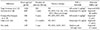
The efficacy of anti-TNF-α therapy for treating systemic vasculitis has been suggested [11]. TNF-α induces leuko-endothelial adhesion via increased expression of various adhesion molecules, such as E-selectin, Intercellular Adhesion Molecule 1 and Vascular Adhesion Molecule 1, and mediates tissue leukocyte infiltration through chemokine synthesis [12]. TNF-α induces metalloproteinase production and may also participate in endothelial cell death directly via apoptosis or indirectly via neutrophil activation [13]. In addition, TNF-α may play a role in neutrophil priming inducing membrane expression of proteinase-3 or myeloperoxidase, which are subsequently recognized by ANCA in ANCA-associated vasculitis [14]. This cytokine may thus be involved in the pathogenesis of different kind of vasculitis. In addition, binding of anti-TNF-α to membrane-associated TNF-α can have an agonistic action, initiating reverse signaling and processes such as apoptosis and cytokine suppression, which could constitute an interesting target in the treatment of vasculitis [12]. The successful use of anti-TNF-α therapy in CPAN has been described in case reports of a small number of patients. Four cases of CPAN treated with anti-TNF-α therapy have been reported (Table 1) [78910]. One patient was adult and three patients were male. All cases had received treatment with systemic glucocorticoids alone or in combination with azathioprine, methotrexate or cyclophosphamide and were refractory. Of them, two patients received infliximab and two patients received etanercept, and clinical symptoms of them were improved for follow-up durations (from 8 months to 7 years).
SUMMARY
We report a case of CPAN, refractory to treatment with high dose glucocorticoid and immunosuppressants, who was successfully treated with adalimumab. The case suggests that adalimumab could be a treatment for refractory CPAN. Further studies are required to confirm the effectiveness and safety of anti-TNF-α therapy in refractory CPAN.


 XML Download