

 PDF
PDF ePub
ePub Citation
 Print
Print


Abstract
Purpose:
To determine the reliability of edema grading of the psoas and paraspinal muscles on axial T2-weighted magnetic resonance (MR) image (T2WI) for the detection of lumbar transverse process (TP) fractures.
Materials and Methods:
A retrospective review of lumbar spine MR images of 56 patients (mean age = 56.1 years, age range = 17–87 years, male:female = 28:28) was conducted by 2 radiologists. Based on the axial T2WI at the disc level, the paraspinal muscles were classified into 4 compartments and muscle edema (increased signal intensity on axial T2WI) grading performed for each quadrant.
Results:
A total of 486 TPs (with fracture: 24, without fracture: 462) of 56 patients were evaluated. Muscle edema grade showed moderate correlation with the presence of TP fracture (ρ = 0.466). When the total score of muscle edema was 2.50, the receiver operating characteristic curve showed a sensitivity of 72.7% and a specificity of 90.7%. A higher edema grade had a significantly higher probability of concomitant TP fracture [any sided (total): odds ratio = 1.704 (95% confidence interval = 1.410–2.060)].
Go to : 
REFERENCES
1.Patten RM., Gunberg SR., Brandenburger DK. Frequency and importance of transverse process fractures in the lumbar vertebrae at helical abdominal CT in patients with trauma. Radiology. 2000. 215:831–834.

2.Miller CD., Blyth P., Civil ID. Lumbar transverse process fractures—a sentinel marker of abdominal organ injuries. Injury. 2000. 31:773–776.
3.Flanders AE., Schaefer DM., Doan HT., Mishkin MM., Gonzalez CF., Northrup BE. Acute cervical spine trauma: correlation of MR imaging findings with degree of neurologic deficit. Radiology. 1990. 177:25–33.
4.Denis F. The three column spine and its significance in the classification of acute thoracolumbar spinal injuries. Spine (Phila Pa 1976). 1983. 8:817–831.
5.Krueger MA., Green DA., Hoyt D., Garfin SR. Overlooked spine injuries associated with lumbar transverse process fractures. Clin Orthop Relat Res. 1996. 327:191–195.
6.Tucker C. The mechanics of sports injuries: an osteopathic approach. 2nd ed. Oxford: Blackwell Scientific Publica-tions;1990.
7.Bali K., Kumar V., Krishnan V., Meena D., Rawall S. Multiple lumbar transverse process stress fractures as a cause of chronic low back ache in a young fast bowler - a case report. Sports Med Arthrosc Rehabil Ther Technol. 2011. 3:8.
8.Rogers LF. Radiology of skeletal trauma. 2nd ed.New York: Churchill Livingstone;1992. p.564.
9.May DA., Disler DG., Jones EA., Balkissoon AA., Manaster BJ. Abnormal signal intensity in skeletal muscle at MR imaging: patterns, pearls, and pitfalls. Radiographics. 2000. 20 Supple 1:S295–S315.
10.Berry GE., Adams S., Harris MB., Boles CA., McKernan MG., Collinson F, et al. Are plain radiographs of the spine necessary during evaluation after blunt trauma? Accuracy of screening torso computed tomography in thoracic/lumbar spine fracture diagnosis. J Trauma. 2005. 59:1410–1413. discussion 1413.
11.Martina Udovicˇic´, Ksenija Baždaric´, Lidija Bilic´-Zulle, Mladen Petrovecˇki. What we need to know when calculating the coefficient of correlation? Biochemia Medica. 2007. 17:10–15.
12.Researchers at the University of Alabama. Spinal cord lnju-ry statistics. Available at:. www.sci-info-pages.com/fact-sheets.html. Accessed on May 22. 2008.
13.Gamanagatti S., Rathinam D., Rangarajan K., Kumar A., Fa-rooque K., Sharma V. Imaging evaluation of traumatic thoracolumbar spine injuries: radiological review. World J Radiol. 2015. 7:253–265.
14.Vaccaro AR., Oner C., Kepler CK., Dvorak M., Schnake K., Bella-barba C, et al. AOSpine thoracolumbar spine injury classification system: fracture description, neurological status, and key modifiers. Spine (Phila Pa 1976). 2013. 38:2028–2037.
15.Looby S., Flanders A. Spine Trauma. Radiol Clin N Am. 2011. 49:129–163.
16.Kwon JA., Hwang JY., Kim MJ., Kwon HY., Kim DH. Importance of Bone marrow and soft tissue edema to improve the diagnostic accuracy of lumbosacral MRI for transverse process fractures and sacral fractures. J Korean Soc Radiol. 2018. 78:107–114.
Go to :
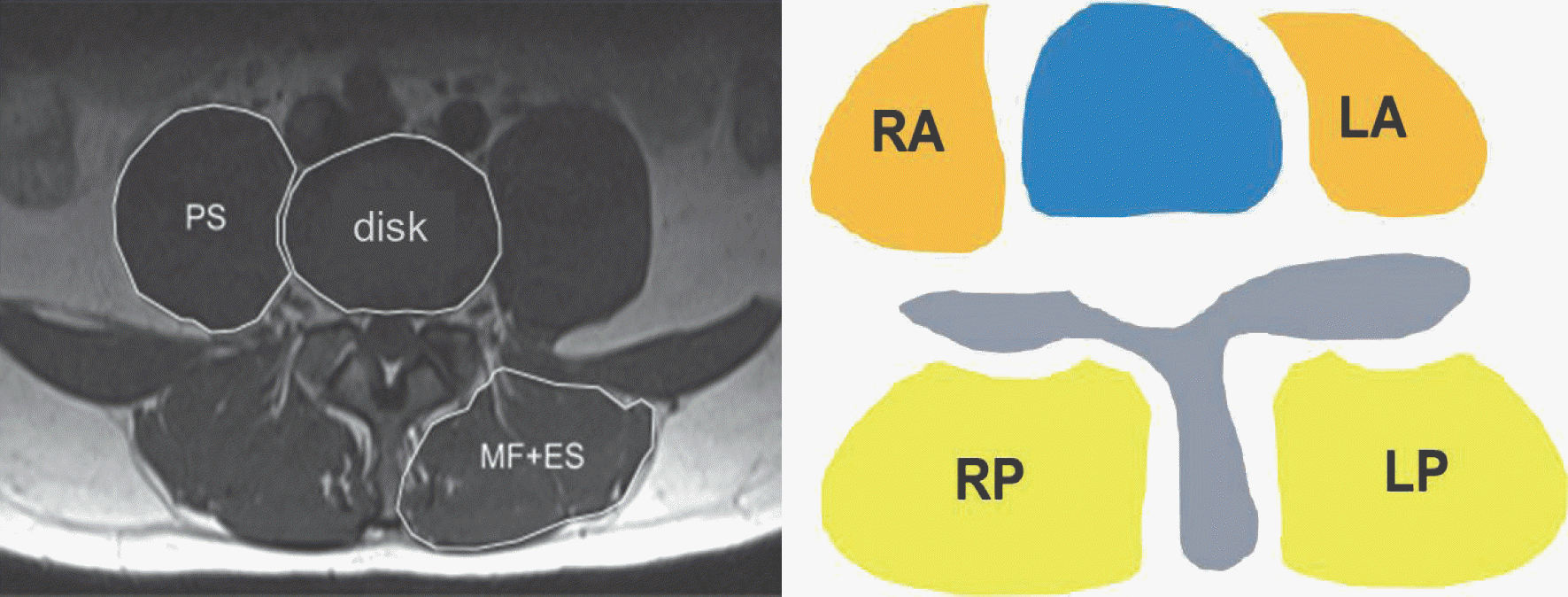 | Fig. 1Four muscular compartments around the spine. Muscles around the spine consist of 4 compartments: right psoas muscle (RA), left psoas muscle (LA), right paraspinal back muscle (RP), and left paraspinal back muscle (LP). And Paraspinal back muscle (RP or LP) consists of mulitifidus muscle (MF) and erector spinae muscle (ES). PS means the psoas muscle. |
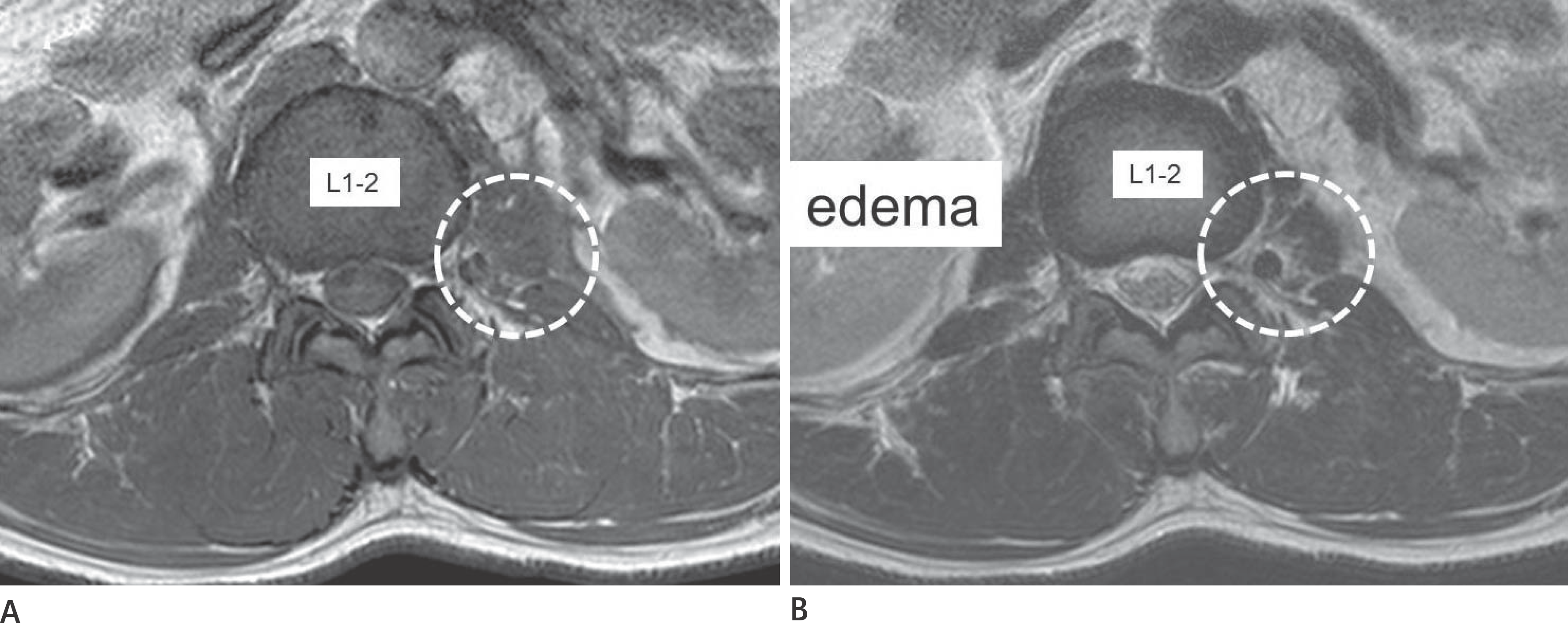 | Fig. 2Definition of muscle edema, 40-year-old man with right transverse process fracture of L3 and L4, and L3 burst fracture. A, B. Increased signal intensity is noted at left psoas muscle (L1-2 level) on T2-weighted image (B) compared with T1-weighted image (A). |
 | Fig. 3Muscle edema grading. A, B. Grade 0 and grade 1, 40-year-old man with transverse process fractures of right L3 and L4, and L3 burst fracture. (A) Grade 0, RA muscle (L2-3 level) shows no increased T2 signal intensity. (B) Grade 1, RP muscle (L1–2 level) shows increased T2 signal intensity involving less than 25 percent of the compartment. C, D. Grade 2, 23-year-old man with transverse process fractures of right L1, L2, L3, and L4. RA muscle (L2-3 level) shows increased T2 signal intensity involving more than 25 percent and less than 50 percent of the compartment (C), transverse process fracture of right L3 in the same patient (D). E, F. Grade 3, 33-year-old man with both L1 and right L2 transverse process fractures. RA muscle (L1-2 level) shows increased T2 signal intensity involving more than 50 percent of the compartment (E), transverse process fracture of right L2 in the same patient (F). RA = right psoas, RP = right paraspinal |
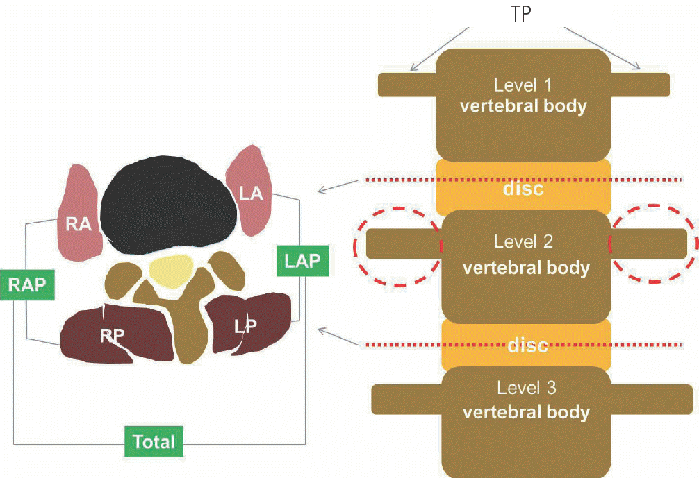 | Fig. 4Evaluation of each transverse process. For evaluation of one leveled TP in one side, two leveled (two disc levels, just cranial and caudal to a targeted TP) muscle edema grades were summed. LA = left psoas, LAP = left psoas + left paraspinal, LP = left paraspinal, RA = right psoas, RAP = right psoas + right paraspinal, RP = right paraspinal, TP = transverse process |
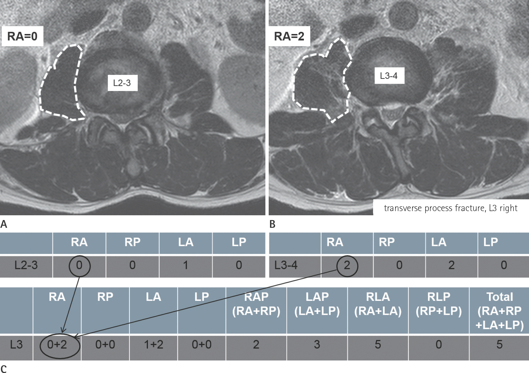 | Fig. 5Method to calculate muscle edema grading. A 40-year-old man with right transverse process fractures of L3 and L4, and L3 burst fracture. A. Muscle edema grading for each compartment at L2–3 level is RA (0), RP (0), LA (1), and LP (0). B. Muscle edema grading for each compartment at L3–4 level is RA (2), RP (0), LA (2), and LP (0). C. In the next step, the two-leveled scores for each compartment are summed. For example, L3 RA score (0 + 2) is derived from L2-3 RA score (0) plus L3-4 RA score (2). L3 RAP score (2) means RA score (2) plus RP score (0) and L3 LAP score (3) means LA score (3) plus LP score (0). L3 RLA score (5) means RA score (2) plus LA score (3) and L3 RLP score (0) means RP score (0) plus LP score (0). Finally, we can obtain total score of L3 (5) by adding RA score (2), RP score (0), LA score (3), and LP score (0) together. LA = left psoas, LAP = left psoas + left paraspinal, LP = left paraspinal, RA = right psoas, RAP = right psoas + right paraspinal, RLA = right psoas + left psoas, RLP = right paraspinal + left paraspinal, RP = right paraspinal |
Table 1.
Number of Patients with and without Transverse Process Fractures, Their Mean Age and Age Range
| Fracture (+) | Fracture (-) | p | |
|---|---|---|---|
| Age∗ | 45.2 (23–71) | 58.5 (17–87) | 0.042 |
| Gender† | 0.081 | ||
| Male | 8 (80) | 20 (43.5) | |
| Female | 2 (20) | 26 (56.5) |
Table 2.
Number and Mean Score (RA, RP, LA, LP, RAP, LAP, RLA, RLP, and total) of TP with and without Fracture
Table 3.
Spearman Correlation Coefficients between Edema Grade of Each Muscle Compartment and Transverse Process Fracture
 XML Download