Abstract
Purpose:
To evaluate the detection performance of hepatocellular carcinoma and image quality in patients with chronic liver disease with quadruple arterial MR imaging using radial volumetric imaging breath-hold examination (VIBE) with k-space weighted image contrast (KWIC).
Materials and Methods:
Forty-four patients underwent liver MR examinations with quadruple arterial imaging using radial VIBE-KWIC sequence (full-frame and four sub-frame images). Diagnostic performance was evaluated with receiver operating characteristics (ROC) for detection of hepatocellular carcinoma. The image quality and severity of artifact were scored by using the five-point scale.
Results:
The area under the ROC curve (Az) value of Hepatocelluar Carcinoma (HCC) detectability was the highest on third sub-frame images, followed by full-frame images. The Az values of third sub-frame and full-frame about the detection of HCC were statistically significantly different from the Az value of first sub-frame images. The full-frame and four sub-frame images showed acceptable image quality and low degree artifact with rating of higher than grade 3.
Conclusion:
Quadruple arterial MRI using radial VIBE-KWIC is a feasible method for detecting hepatocellular carcinoma in patients with chronic liver disease without deterioration of image quality. The third sub-frame and full-frame image are superior to other sub-frame images in detecting hepatocellular carcinoma.
Go to : 
REFERENCES
1.Hawighorst H., Schoenberg SO., Knopp MV., Essig M., Miltner P., van Kaick G. Hepatic lesions: morphologic and functional characterization with multiphase breath-hold 3D gadolinium-enhanced MR angiography—initial results. Radiology. 1999. 210:89–96.

2.Elsayes KM., Narra VR., Yin Y., Mukundan G., Lammle M., Brown JJ. Focal hepatic lesions: diagnostic value of enhancement pattern approach with contrast-enhanced 3D gradient-echo MR imaging. Radiographics. 2005. 25:1299–1320.

3.Kim BK., Kim MJ., Park BJ., Sung DJ., Cho SB. [Triple arterial phase hepatic MRI using four dimensional T1-weighted high resolutions imaging with volume excitation keyhole techniques: feasibility and initial clinical experience in focal liver lesions]. J Korean Soc Radiol. 2013. 69:223–234.

4.Hong HS., Kim HS., Kim MJ., De Becker J., Mitchell DG., Kanematsu M. Single breath-hold multiarterial dynamic MRI of the liver at 3T using a 3D fat-suppressed keyhole technique. J Magn Reson Imaging. 2008. 28:396–402.

5.Kanematsu M., Semelka RC., Matsuo M., Kondo H., Enya M., Goshima S, et al. Gadolinium-enhanced MR imaging of the liver: optimizing imaging delay for hepatic arterial and portal venous phases—a prospective randomized study in patients with chronic liver damage. Radiology. 2002. 225:407–415.

6.Goshima S., Kanematsu M., Kondo H., Yokoyama R., Miyoshi T., Nishibori H, et al. MDCT of the liver and hypervascular hepatocellular carcinomas: optimizing scan delays for bolus-tracking techniques of hepatic arterial and portal venous phases. AJR Am J Roentgenol. 2006. 187:W25–W32.

7.Kim KW., Lee JM., Jeon YS., Kang SE., Baek JH., Han JK, et al. Free-breathing dynamic contrast-enhanced MRI of the abdomen and chest using a radial gradient echo sequence with K-space weighted image contrast (KWIC). Eur Radiol. 2013. 23:1352–1360.

8.Fujinaga Y., Ohya A., Tokoro H., Yamada A., Ueda K., Ueda H, et al. Radial volumetric imaging breath-hold examination (VIBE) with k-space weighted image contrast (KWIC) for dynamic gadoxetic acid (Gd-EOB-DTPA)-enhanced MRI of the liver: advantages over Cartesian VIBE in the arterial phase. Eur Radiol. 2014. 24:1290–1299.

9.Brodsky EK., Bultman EM., Johnson KM., Horng DE., Schelman WR., Block WF, et al. High-spatial and high-temporal resolution dynamic contrast-enhanced perfusion imaging of the liver with time-resolved three-dimensional radial MRI. Magn Reson Med. 2014. 71:934–941.

10.Zech CJ., Vos B., Nordell A., Urich M., Blomqvist L., Breuer J, et al. Vascular enhancement in early dynamic liver MR imaging in an animal model: comparison of two injection regimen and two different doses Gd-EOB-DTPA (gadoxetic acid) with standard Gd-DTPA. Invest Radiol. 2009. 44:305–310.

11.Park YS., Lee CH., Yoo JL., Kim IS., Kiefer B., Woo ST, et al. Hepatic arterial phase in gadoxetic acid-enhanced liver magnetic resonance imaging: analysis of respiratory patterns and their effect on image quality. Invest Radiol. 2016. 51:127–133.
12.Hope TA., Saranathan M., Petkovska I., Hargreaves BA., Her-fkens RJ., Vasanawala SS. Improvement of gadoxetate arterial phase capture with a high spatio-temporal resolution multiphase three-dimensional SPGR-Dixon sequence. J Magn Reson Imaging. 2013. 38:938–945.

13.Beck GM., De Becker J., Jones AC., von Falkenhausen M., Wil-linek WA., Gieseke J. Contrast-enhanced timing robust acquisition order with a preparation of the longitudinal signal component (CENTRA plus) for 3D contrast-enhanced abdominal imaging. J Magn Reson Imaging. 2008. 27:1461–1467.

14.Hadizadeh DR., Gieseke J., Beck G., Geerts L., Kukuk GM., Boström A, et al. View-sharing in keyhole imaging: partially compressed central k-space acquisition in time-resolved MRA at 3.0 T. Eur J Radiol. 2011. 80:400–406.
15.Agrawal MD., Spincemaille P., Mennitt KW., Xu B., Wang Y., Dutruel SP, et al. Improved hepatic arterial phase MRI with 3-second temporal resolution. J Magn Reson Imaging. 2013. 37:1129–1136.

16.Kim BS., Lee KR., Goh MJ. New imaging strategies using a motion-resistant liver sequence in uncooperative patients. Biomed Res Int. 2014. 2014:142658.

17.Budjan J., Riffel P., Ong MM., Schoenberg SO., Attenberger UI., Hausmann D. Rapid Cartesian versus radial acquisition: comparison of two sequences for hepatobiliary phase MRI at 3 tesla in patients with impaired breath-hold capabilities. BMC Med Imaging. 2017. 17:32.

18.Yu MH., Lee JM., Yoon JH., Kiefer B., Han JK., Choi BI. Clinical application of controlled aliasing in parallel imaging results in a higher acceleration (CAIPIRINHA)-volumetric interpolated breathhold (VIBE) sequence for gadoxetic acid-enhanced liver MR imaging. J Magn Reson Imaging. 2013. 38:1020–1026.

19.Li H., Xiao Y., Wang S., Li Y., Zhong X., Situ W, et al. TWIST-VIBE five-arterial-phase technology decreases transient severe motion after bolus injection of Gd-EOB-DTPA. Clin Radiol. 2017. 72:800. .e1-800.e6.

20.Theilmann RJ., Gmitro AF., Altbach MI., Trouard TP. View-ordering in radial fast spin-echo imaging. Magn Reson Med. 2004. 51:768–774.

21.Spuentrup E., Katoh M., Buecker A., Manning WJ., Schaeffter T., Nguyen TH, et al. Free-breathing 3D steady-state free precession coronary MR angiography with radial k-space sampling: comparison with cartesian k-space sampling and cartesian gradient-echo coronary MR angiography—pilot study. Radiology. 2004. 231:581–586.

Go to : 
 | Fig. 1Schematic timing diagram for T1-weighted quadruple arterial MR imaging using radial VIBE-KWIC. KWIC = k-space weighted image contrast, T1CE = contrast-enhanced T1-weighted image, VIBE = volumetric imaging breath-hold examination |
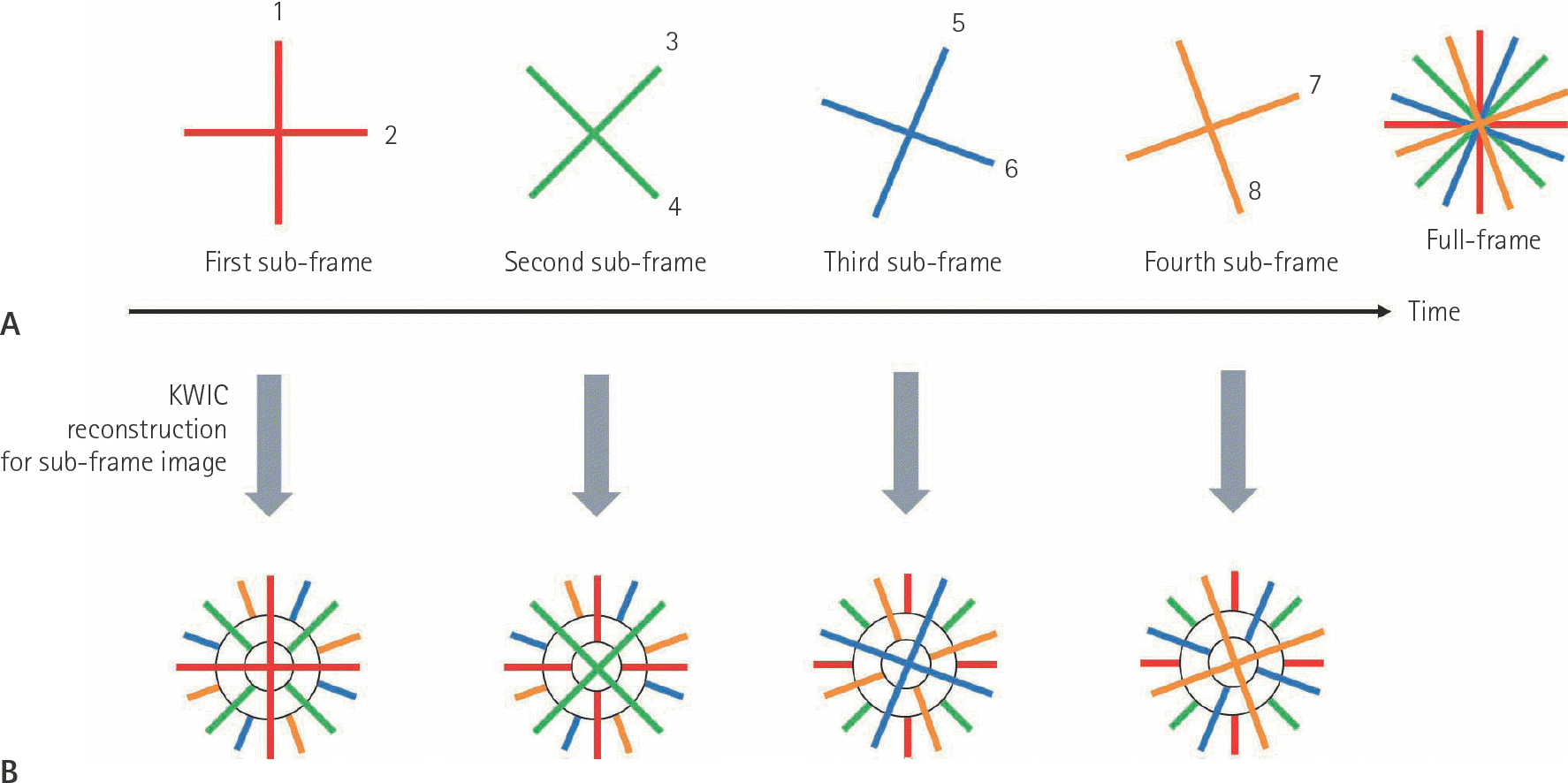
 | Fig. 2Principles of the radial acquisition with KWIC reconstruction technique. A. Schematic diagram of the four-interleaf angle-bisection reordering acquisition shows simple example composed of eight projection views. For making full-frame image, k-space data are serially obtained radially projection views which are grouped into four interleaved subsets. And data from all subsets are combined for the reconstruction of full-frame images. B. For the reconstruction of sub-frame images, KWIC divides the k-space into three regions based on the circular Nyquist radius and fills the spokes. For example, spokes obtained first among all acquired radial spokes are filled in k-space. The next spokes are filled without the center. The last obtained spokes are used to fill only the outermost part of the k-space so that it has little effect on the resolution. Therefore four sub-frame KWIC images have different k-space cores and the surrounding k-space is similar. KWIC = k-space weighted image contrast |
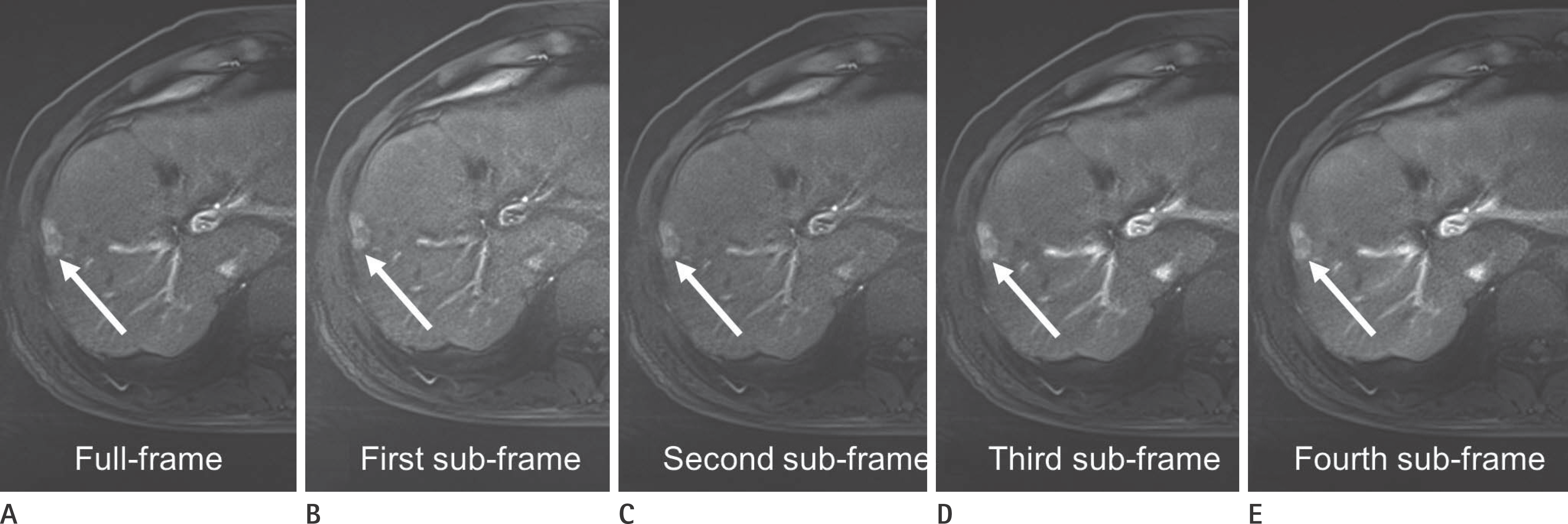
 | Fig. 3MR images obtained in a 64-year-old man with a hepatocellular carcinoma. Contrast-enhanced 3D Radial k-space weighted image contrast, volumetric imaging breath-hold examination during hepatic arterial dominant phase imaging comprised one full-frame and four sub-frame images (A-E). The full-frame and sub-frame images show a focal enhancing lesion (arrows) in right hepatic lobe. Third sub-frame image (D) shows tumor with greater conspicuity and best arterial enhancement than other arterial images (A-C, E). Much better image quality and lesser artifacts were seen on full-frame image. The arterial enhancing lesion was pathologically proved to be a hepatocellular carcinoma after right anterior sectionectomy. |
 | Fig. 4MR and CT images obtained in a 63-year-old man with a hepatocellular carcinoma. Full-frame and four sub-frame images using contrast-enhanced 3D Radial k-space weighted image contrast, volumetric imaging breath-hold examination during multiple hepatic arterial dominant phases at 5-second temporal resolution (A-E), show gradually increasing enhancing lesion (arrows) in left hepatic lobe. Third sub-frame image (D) depicts an enhancing tumor (arrow) more clearly with best arterial enhancement than other arterial images (A-C, E). Three months follow-up CT (F) after transcatheter arterial chemoembolization demonstrates accumulation of iodized oil (arrow) in a hepatocellular carcinoma in left hepatic lobe. TACE = transcatheter arterial chemoembolizatio |
Table 1.
Mean Area Under the ROC Curve for Detection of Hepatocellular Carcinoma
Table 2.
Composite Sensitivity, Specificity, PPV, and NPV for Detection of Hepatocellular Carcinoma
Table 3.
Results of Overall Image Quality of Quadruple Arterial MRI Using Radial VIBE-KWIC
Table 4.
Results of Artifact Severity of Quadruple Arterial MRI Using Radial VIBE-KWIC


 PDF
PDF ePub
ePub Citation
 Print
Print


 XML Download