Journal List > J Korean Foot Ankle Soc > v.22(3) > 1101928

Lee, Choi, Kang, and Ko: The Risk Factors Associated with Nonunion after Surgical Treatment for Distal Fibular Fractures

Abstract

Purpose

The purpose of this study was to evaluate the radiologic outcomes of distal fibular fractures and to analyze the risk factors associated with nonunion.

Materials and Methods

Between January 2009 and March 2016, 13 patients who had final nonunion with ankle fracture were included. In the control group, 370 patients who had undergone bony union and removed metal implants were included. All patients underwent the same surgical procedure and had the same treatment method, ultimately achieving satisfactory open reduction results with less than 2 mm fracture gap. Surgical treatment of fracture was considered to have the same effect on nonunion, and factors that might be associated with nonunion were evaluated. SPSS ver. 13.0 (SPSS Inc., USA) was used for all statistical analyses. Pearson's chi-square test and multi-variate regression analysis were performed to determine the factors affecting nonunion of distal fibular fracture. A p-value less than 0.05 was considered statistically significant, and relative risk was assessed.

Results

The mean age of 13 patients was 46.9 years (range, 16∼57 years); there were 8 men and 5 women. Among the 13 patients with nonunion, atrophic was the most common (12 cases). The association between the injury mechanism and the Lauge-Hansen classification and diabetes mellitus was not statistically significant. Distal fibular fractures with tibia shaft fracture (p=0.015) and Danis-Weber type C fracture (p=0.023), open fracture (p=0.011), and smoking (p=0.023) were significantly associated with nonunion.

Conclusion

In this study, the combined injury of the ipsilateral tibia shaft fracture, open fracture, and Danis-Weber type C fracture may increase the possibility of nonunion. Therefore, caution is advised to prevent nonunion.

References

1. Bhadra AK, Roberts CS, Giannoudis PV. Nonunion of fibula: a systematic review. Int Orhop. 2012; 36:1757–65.
crossref
2. Hak DJ, Fitzpatrick D, Bishop JA, Marsh JL, Tilp S, Schnettler R, et al. Delayed union and nonunions: epidemiology, clinical issues, and financial aspects. Injury. 2014; 45(Suppl 2):S3–7.
crossref
3. Kang CH, Jung JH, Ye HU, Cho CH. Associated factors of nonunion following conservative management of distal clavicle fractures. J Korean Orthop Assoc. 2015; 50:137–42.
crossref
4. Guo F. Observations of the blood supply to the fibula. Arch Orthop Trauma Surg. 1981; 98:147–51.
crossref
5. Walsh EF, DiGiovanni C. Fibular nonunion after closed rotational ankle fracture. Foot Ankle Int. 2004; 25:488–95.
crossref
6. Ko SB. Relationship of tibial nonunion with fibular nonunion in the tibio-fibular shaft fracture. J Korean Fract Soc. 2006; 19:153–6.
crossref
7. Shin DE, Cho DY, Yoon HK, Lee JS, Lee YS, Kim HJ. The effect of fibular malreduction on ankle joint after tibial interlocking IM nailing of tibial and fibular fractures. J Korean Fract Soc. 2005; 18:29–35.
crossref
8. Siliski JM, Healy W, Carr C, Baumgaertner M, Blitzer C. Nonunion of the fibula after ankle fracture. J Orthop Trauma. 1993; 7:198.
crossref
9. Ahmed M, Wimhurst JA, Walton NP. Non-union of Weber B fractures: a case series. Injury. 2007; 38:861–4.
crossref
10. Guille JT, Lipton GE, Bowen JR, Uthaman U. Delayed union following stress fracture of the distal fibula secondary to rotational malunion of lateral malleolar fracture. Am J Orthop (Belle Mead NJ). 1997; 26:442–5.
11. Song KS, Kim HG, Park BM, Moon CS, Kim BY. Treatment of lateral malleolar fracture associated with disruption of the deltoid ligament. J Korean Orthop Assoc. 2010; 45:210–5.
crossref
12. Yablon IG, Leach RE. Reconstruction of malunited fractures of the lateral malleolus. J Bone Joint Surg Am. 1989; 71:521–7.
crossref
13. Dodson NB, Ross AJ, Mendicino RW, Catanzariti AR. Factors affecting healing of ankle fractures. J Foot Ankle Surg. 2013; 52:2–5.
crossref

Figure 1.
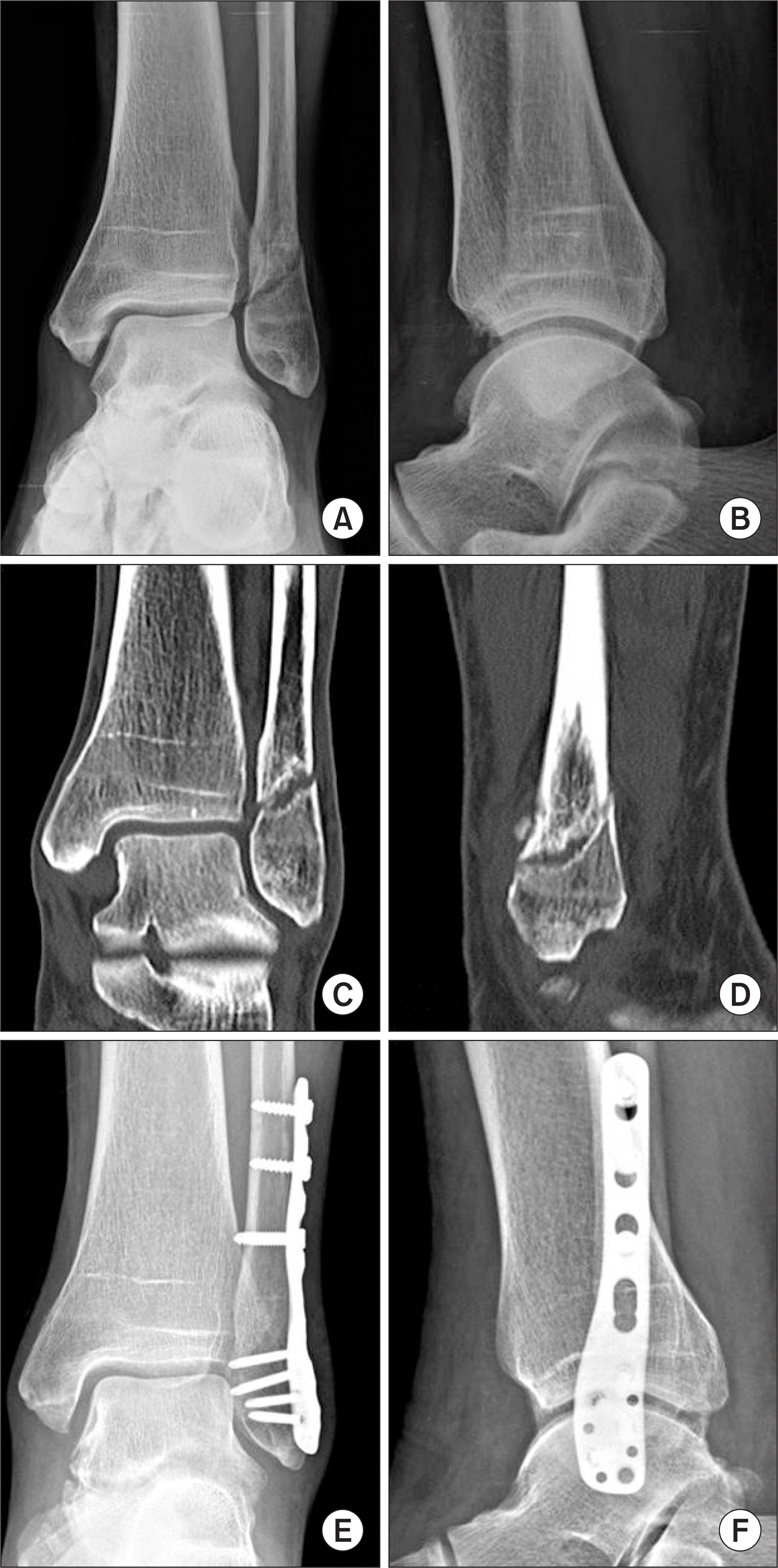
A 68-year-old male with lateral malleolar fracture (Denis-Weber type B, Lauge-Hansen supiration-external rotation type) visited our clinic after conservative treatment. (A, B) Ankle anteroposterior and lateral view taken 6 months after the injury showed lateral malleolar nonunion. (C, D) Ankle computed tomography showed sclerotic lesion of the fracture site and fracture gap. (E, F) He underwent plate and screw fixation of the fibula with autogenous iliac crest bone graft. Radiographs taken 9 months postoperatively demonstrate complete healing. He had diabetes, smoking of nonunion risk factors.
jkfas-22-95f1.tif
Figure 2.
A 40-year-old male presents for evaluation of continued ankle pain 8 months after open reduction and internal fixation of a bimalleolar fracture. (A) Initial ankle anteroposterior view at injury shows left ankle bimalleolar fracture (Denis-Weber C, Lauge-Hansen pronation-external rotation type) with syndesmosis injury. (B) Internal fixation was performed using a cannulated screw and 1/3 semitubular plate. (C, D) Radiograph and computed tomography demonstrate an atrophic nonunion. (E) The patient underwent operative revision with iliac crest bone graft.
jkfas-22-95f2.tif
Table 1.
Associated Factors to Fibular Nonunion after Treatment of Lateral Malleolar Fractures
    Nonunion (n=13) Union (n=370) p-value p-value
Injury mechanism Motor vehicle accident 7 (53.8) 89 (24.1) 0.086 0.094
Slip down 3 (23.1) 173 (46.8) 0.165 0.271
Fall down 2 (15.4) 62 (16.8) 0.594 0.341
Direct injury 1 (7.7) 46 (12.4) 0.432 0.706
Combined injury Tibia shaft fracture 8 (61.5) 64 (17.3) 0.015 0.009
Malleolar fracture 2 (15.4) 125 (33.8) 0.067 0.301
Syndesmosis injury 2 (15.4) 104 (28.1) 0.627 0.493
Deltoid ligament rupture 2 (15.4) 8 (2.2) 0.071 0.107
Denis-Weber type Type A 0 (0) 18 (4.9) 0.196 0.189
Type B 3 (23.1) 198 (53.5) 0.432 0.382
Type C 10 (76.9) 154 (41.6) 0.023§ 0.009
Lauge-Hansen type Supiration-aduction 0 (0) 15 (4.1) 0.206 0.267
Supiration-external rotation 3 (23.1) 98 (26.5) 0.108 0.362
Pronation-external rotation 1 (7.7) 85 (23.0) 0.271 0.176
Pronation-adduction 1 (7.7) 18 (4.9) 0.317 0.471
Fracture Open fracture 8 (61.5) 17 (4.6) 0.011 0.000
Comminuted fracture 5 (38.5) 58 (15.7) 0.073 0.142
Patient factor Diabetes mellitus 1 (7.7) 24 (6.5) 0.624 0.639
Smoking 7 (53.8) 54 (14.6) 0.023 0.002

Values are presented as number (%).

Denis-Weber type: Type A are caused by internal rotation and adduction that produce a transverse fracture of the lateral malleolus at or below the plafond. Type B are caused by external rotation resulting in an oblique fracture of the lateral malleolus, beginning on the anteromedial surface and extending proximally to the posterolateral aspect. Type C are divided into abduction injuries with oblique fracture of the fibula proximal to the disrupted tibiofibular ligaments (C-1) and abduction-external rotation injuries with a more proximal fracture of the fibula and more extensive disruption of the interosseous membrane (C-2).

Pearson's chi-square test.

Multi-variate regression test. Relative risk:

2.105,

§ 2.618,

4.102,

3.412.

TOOLS
Similar articles