

 PDF
PDF ePub
ePub Citation
 Print
Print


INTRODUCTION
Bronchogenic cysts are congenital abnormalities that arise from the ventral foregut. They occur most commonly within the mediastinum but rarely in the diaphragm. Up to now, there have been few reported cases of intradiaphragmatic bronchogenic cyst reported. On CT, bronchogenic cysts typically manifest as spherical masses of either water or soft-tissue attenuation (12). In this report, we report a 52-year-old man who was admitted to our hospital for evaluation of dyspnea. He had a cystic mass in left diaphragm crus, with inner calcification, and peripheral and septal enhancement on CT scan. Follow up CT scans, revealing changes in the size and density of the mass, were believed to be documentation of the healing process of an infectious bronchogenic cyst.
CASE REPORT
A 52-year-old man was admitted to our hospital emergency room due to sudden onset dyspnea in recumbent positioning. On physical examination, he complained of shortness of breath and left chest pain with breathing motion. On the laboratory test, the oxygen (O2) saturation of 92.2% (normal range 94–100%). White blood cell count (WBC, 15260/µL, normal range 3500–10000/µL), erythrocyte sedimentation rate (ESR, 26 mm/hr, normal range 0–20 mm/hr) and C-reactive protein (CRP, 10.51 mg/dL, normal range 0–0.5 mg/dL) were elevated.
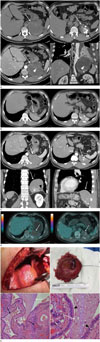
Chest radiograph revealed blunting of the left costophrenic angle and increased opacity in the left lung parenchyma (Not shown). CT scan was performed using a 128-row detector CT scanner (Somatom Definition AS, Siemens Healthcare, Erlangen, Germany). The CT scan parameters were as follows: 120 kVp tube voltages, 100 mA tube current. Nonionic contrast material (120 mL) was administered at a rate of 2.5mL/s, followed by a 30 mL saline flush. Chest CT revealed a 10 × 9.8 × 10.8 cm sized, well defined, oval shaped, heterogeneous huge cystic mass, centered in the left diaphragm crus (Fig. 1A). The mass contained multiple nodular calcifications and multiple inner septations, which inferiorly abutted the left pleura. After intravenous contrast injection, peripheral and septal enhancement was apparent in the mass. In addition, CT scan revealed left pleural effusion and passive atelectasis of adjacent lung parenchyma, which was thought to be reactive change.
At initial hospitalization, lung abscess was clinically suspected, empirical antibiotics including Piperacillin/tazobactam [Tabaxin (Penmix Ltd., Jeong-dong, Seoul, Korea), 4.5 g/vial, three times per day] and Clindamycin [Fullgram (Samjin Pharm., Seoul, Korea), 600 mg/amp, three times a day] were prescribed for 5 days. On second admission day, laboratory test values were normalized (O2 saturation: 94%, WBC: 6620/µL, ESR: 15 mm/hr, and CRP: 0.1 mg/dL).
We could receive a previous non-contrast chest CT scan of the patient performed at outside hospital about 2 years ago. This CT scan revealed a smooth, sharply margined, elliptical, homogeneous density mass with multiple calcifications in the same area (Fig. 1B). The size of the mass was significantly smaller than in the initial chest CT in our hospital. Considering the change in size, density and presence of enhancement, ultrasonography guided lung biopsy was performed under the suspicion of malignancy. However, no malignant tumor was detected on pathological analysis. The patient was discharged with a recommendation for two-week follow up chest CT.
The follow up chest CT revealed decreased size with disappearance of the inner cystic lesion (Fig. 1C). The mass exhibited a smooth margin, homogeneous density without significant enhancement. A fluorine-18 fluorodeoxyglucose (18F-FDG) PET-CT scan revealed mild marginal hypermetabolism and inner metabolic defect, which was interpreted to be likely benign nature (Fig. 1D).
However, surgical removal of the mass was performed to exclude the possibility of malignancy during his second hospitalization. The operators performed a lateral thoracostomy through the left 8th intercostal space. Grossly, the mass was located in the posterolateral side of left hemidiaphragm (Fig. 1E). The mass was soft and exhibited inner sticky material greenish in color. There was no invasion of the thoracic cavity. Final pathological diagnosis of the mass was intradiaphragmatic foregut cyst, consistent with bronchial cyst. Hematoxylin & eosin stain revealed pseudostratified ciliated respiratory epithelium which is lined mainly in normal respiratory tract (Fig. 1E).
The second follow up postoperative chest CT scan demonstrated no evidence of a remnant mass.
DISCUSSION
Bronchogenic cysts are congenital lesions that are believed to arise from an abnormally budding ventral foregut, which then develops into a blind ended fluid filled pouch (1). They occur between the 26th and 40th days of gestation. They are usually found in the mediastinum, near the tracheal carina, in 85% of patients, and 79% occur in middle mediastinum. They also may be found in the lung parenchyma, pleura, retroperitoneum, and neck (2). However, intradiaphragmatic bronchogenic cysts are exceedingly rare and only a few cases have been reported.
Symptoms are frequently nonspecific; however, when present, pain is the most common clinical symptom (13). Other presenting symptoms may include fever due to infection and symptoms ascribable to pressure on adjacent structures (4).
The pathological hallmark of bronchogenic cysts is the presence of ciliated pseudostratified columnar epithelium, cartilage, and smooth muscle within the cyst wall. Grossly, there is a variable presentation, which likely contributes to their variable radiologic appearance (1).
CT findings of bronchogenic cyst have been well described in the literature. They are usually sharply margined with soft tissue or water attenuation, with cystic characteristics. Some bronchogenic cysts may have soft tissue attenuation, and contrast enhanced CT may help in distinguishing malignancy by the lack of enhancement. Ten percent of bronchogenic cysts can have calcification (4). In our patient, the mass was a well margined, cystic lesion in the left diaphragmatic crus; these characteristics could be regarded as typical findings of bronchogenic cyst retrogradely. On the other hand, the mass showed peripheral and septal enhancement, which should be regarded as a possibility of malignancy with cystic change. After 18F-FDG PET/CT evaluation, and changes in size and attenuation in follow up chest CT, peripheral and septal enhancement of the mass were thought to be inflammatory changes rather than malignant enhancement. Therefore, we considered benign lesions in the differential diagnosis including hemangioma, rhabdomyoma, teratoma and hematoma. After confirming gross pathology, increased density of the mass on chest CT was believed to be due to the inner sticky materials.
The evaluation of bronchogenic cysts depends on the size of the cyst and patient symptoms. Small, asymptomatic cysts can be followed conservatively. However, enlargement of bronchogenic cysts over a span of years is typical, and rapid enlargement associated with pain indicates hemorrhage or infection. Because of their tendency to grow, bronchogenic cysts are traditionally treated using complete surgical resection (5). On occasion, bronchogenic cysts may harbor malignancy and, therefore, necessitate surgical resection for clear diagnosis (1).
There are few reported cases of intradiaphragmatic bronchogenic cysts, with most removed by surgical procedure after detection for accurate diagnosis. However, our case shows alterations of the lesion, from a soft tissue mass to a cystic lesion with enhancement. These changes suggest complications such as inflammation or malignant changes. In conclusion, awareness of an unusual location of bronchogenic cysts with variable CT findings, as in our case, would help in differential diagnosis and management of these thoracic lesions.
 XML Download