

 PDF
PDF ePub
ePub Citation
 Print
Print


INTRODUCTION
Viral conjunctivitis is the most common type of infectious conjunctivitis. Adenoviral conjunctivitis accounts for 65~90% of the reported cases of viral conjunctivitis worldwide (1). Other viruses responsible for conjunctival infection include herpes simplex virus (HSV), varicella-zoster virus (VZV), picornavirus [enterovirus 70 (EV70) and CVA24], and poxvirus (molluscum contagiosum and vaccinia) (2).
Epidemic keratoconjunctivitis (EKC) caused by Human Adenovirus (HAdVs) is a highly contagious and severe form of conjunctivitis (3). EKC is caused mainly by human species D adenoviruses, of which, the genotypes HAdV-3,-8,-19 and -37 are known to cause a more severe form of EKC than the others (4567). EKC starts with characteristic clinical features such as sudden onset of acute follicular conjunctivitis in more than 50% of cases (8).
Another major viral conjunctivitis, in South Korea, is acute hemorrhagic conjunctivitis (AHC) that is caused by enterovirus. It is characterized by red eye, pain, subconjunctival hemorrhage, photophobia, swelling, eyelid edema, irritation, eye discharge, excessive tearing and follicular reaction (910). The genotypes EV70 and CVA24 are responsible for most cases and outbreaks of AHC (911).
In South Korea, the most frequently occurring viral conjunctivitis is EKC and AHC. The annual report published by the Korea Centers for Disease and Prevention in 2014 reports that, in South Korea, EKC and AHC occur seasonally, mainly from July to September. However, viral conjunctivitis outbreaks have also been noted to occur other times of the year, for example, the AHC outbreak during the winter of 2013 in South Korea. Similarly, a nationwide outbreak of AHC was reported in South Korea in the summer of 2002 (11).
Therefore, a long-term survey of EKC and AHC is needed to control of occurrence of viral conjunctivitis in South Korea. Moreover, to date, epidemiological studies with a focus on the molecular characterization of the genotypes of viral conjunctivitis are very rare in South Korea as well as around the world. The objective of the present study was to identify the molecular features of EKC and AHC, over a prolonged period of 5 years in the southwest area of South Korea. Phylogenetic analysis of HAdV-8 and CVA24 (the main causative agents of viral conjunctivitis in South Korea) was performed by comparing the Korean strains with the corresponding foreign strains to determine the epidemiological features and genetic diversity in the viruses causing the epidemic of EKC and AHC in South Korea.
MATERIALS AND METHODS
Ethics Statement
This study was reviewed and approved by the Institutional Review Board at Ministry of Health Welfare of Korea as Exempt (P01-201612-31-001).
Specimen collection
We collected conjunctival swab specimens from 492 patients with suspected cases of EKC or AHC from 6 ophthalmic hospitals in Gwangju Metropolitan City in South Korea between 2012 and 2016 (mostly between the months of May and September). Clinical samples were collected using viral transport system (BD Diagnostics, Sparks, MD, USA).
Detection of adenovirus and enterovirus and molecular typing
Viral nucleic acid was extracted by automated extraction systems, QIAcube (Qiagen, Valencia, CA, USA) according to the manufacturer's instructions. RT-PCR analyses were performed using Human Enterovirus/Adenovirus Detection kit (POSTBIO, Gyeonggi-do, Korea) for detection of positive samples according to the manufacturer's instructions. After detection of HAdV-positive and enterovirus-positive samples among the 492 samples, we conducted conventional PCR with positive samples. For molecular typing, we performed conventional PCR and sequencing of the hexon gene in the HAdV-positive samples using the reaction conditions and sequencing methodology as described previously (12). Similarly, we performed semi-nested PCR in the VP1 coding region for the molecular typing of enterovirus-positive samples. Semi-nested PCR conditions and primer sequences amplifying the VP1 coding region were employed according to the established protocols and primers of the Korea Centers for Disease Control and Prevention. The amplified PCR products were sent to COSMOGENTECH (Daejeon, Korea) for sequencing where sequencing was performed using ABI 3730 XL (Applied Biosystems, Foster city, CA, USA).
Phylogenetic analysis
The phylogenetic analyses were performed with HAdV-8 and CVA24, found to be the predominant genotypes in our study. The nucleotide sequences were aligned with the reference sequences downloaded from the National Center for Biotechnology Information (NCBI) database using Clustal W program, and phylogenetic analyses were conducted using the MEGA 6 software. The neighbor-joining method with bootstrapping of 1,000 replicates was used to reconstruct phylogenetic trees using the Kimura two parameter model. Nucleotides sequences of HAdV-8 and CAV24 Korean isolates were aligned and compared with reference sequences of 43 HAdV-8 and 19 CVA24 foreign isolates from GenBank.
RESULTS
Features of age and gender in our samples
The age of the 249 patients in our study ranged from 1 to 80 years as follows: 12 patients were aged less than 7 years; 19 were aged 8~13 years; 20 were aged 14~19 years; 20 were aged 20~29 years; 49 were aged 30~39 years; 32 were aged 40~49 years; 26 were aged 50~59 years; 20 were aged 60~69 years; 13 were aged over 70 years. For the remaining 38 patients, we did not have information on their age. The patients comprised of 120 males and 105 females. We did not know the gender of the remaining 24 patients.
Detection of viruses and molecular typing
We collected 492 conjunctival swabs for the test; of these 268 samples were detected as EKC-positive or AHC-positive. HAdVs and enteroviruses were detected in 249 samples (50.6%) and 19 samples (3.9%), respectively, by real-time PCR.
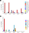
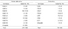
The genotypes in the 249 isolates from the HAdV-infected patients were identified mainly as HAdV-8 (183, 73.5%), HAdV-37 (14, 5.6%), HAdV-3 (9, 3.6%), and HAdV-4 (9, 3.6%). HAdV-4 (3/66, 4.5%) in 2012, HAdV-3 (8/96, 8.3%) in 2013, HAdV-37 (6/24, 25%) in 2014, HAdV-37 (2/19, 9.0%) in 2015, HAdV-37 (6/41, 14.6%) in 2016 (Fig. 1). Furthermore, the total number of genotypes of HAdV isolated also varied each year as follows: 3 genotypes in 2012, 5 in 2013, 4 in 2014, 3 in 2015, 5 in 2016. We also detected HAdV-22 (n=2) in 2016 and HAdV-55 (n=2) in 2014. The 19 isolates from enterovirus-infected patients were mainly identified as CVA24 (8, 42.0%) and CVB1 (4, 21.0%) (Table 1).
Epidemiological features
The annual distribution of the HAdV and enterovirus strains from 268 samples collected over a period of 5 years were analyzed (Fig. 1). A total of eight genotypes were detected in the HAdV isolates. Among the HAdV isolates collected over the years, the percentage of HAdV-8 was the highest. Characteristically, HAdV-3 (n=8) and HAdV-4 (n=6) isolates in 2013, HAdV-37 in 2014 (n=6) and 2016 (n=6) had a high percentage (Fig. 1, panel A). A total of seven genotypes were detected in the enterovirus isolates. Of these, the highest percentage was of CVA24 in 2012 and CVB1 in 2014, with 8 and 4 isolates, respectively.
Phylogenetic analysis of the hexon gene in HAdV-8 and the VP1 regions in CVA24
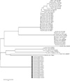
The phylogenetic relationships between the isolates were inferred by the neighbor-joining method (Fig. 2 and 3). Sequences from the HAdV-8 and CVA24 South Korean isolates were compared to those available in GenBank. Phylogenetic analysis of the hexon gene of HAdV-8 was performed using the 17 isolates from South Korean and 43 isolates from different parts of the world that were available in GenBank (Fig. 2). The nucleotide sequences of the 17 South Korean isolates analyzed from 2014 to 2016 were identical. All the hexon sequences from 17 strains clustered together with that of HAdV-8 isolated in USA, UK, Sweden and Singapore.
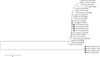
Similarly, phylogenetic analysis of the VP1 gene of CVA24 was performed with the 8 isolates from South Korean and 19 isolates from different parts of the world that were available in GenBank (Fig. 3). The nucleotide sequences of the 8 South Korean isolates analyzed from 2014 to 2016 were identical at 3 and 4 cases, respectively. Phylogenetic analysis of the VP1 region revealed that South Korean strains isolated during 2014-2016 had highest nucleotide identity with CVA24 strains isolated from China.
DISCUSSION
Viral conjunctivitis can be caused by the adenoviruses, rubella virus, rubeola (measles) virus, herpesviruses, and picornaviruses. Among viral conjunctivitis, EKC is a highly contagious disease that mainly affects the surface of the eye (13). It is known that EKC occurs around the world in all age groups and at all times of year (14). AHC, also known as pinkeye, is an inflammation of the conjunctiva. EV70 and CVA24 are the main agents responsible for AHC (15). Furthermore, AHC outbreaks caused by CVA24 have been reported around world: Uganda, Southern Sudan, Pakistan, Mexico and Guangdong in China in 2010 (16). Therefore, the epidemiological data are vital to the prevention and better management of the EKC and AHC outbreaks in South Korea as well as around the world. Our long-term molecular epidemiological studies on EKC and AHC are the first of its kind in South Korea. In this study, we investigated the molecular epidemiology of EKC and AHC in Gwangju in the southwest area of South Korea for 5 years (2012–2016). HAdV-8 (183/268, 73.5%) in EKC was found to be the dominant genotype in this region. This observation is identical to that made in other countries, for instance: Vietnam in 2006, and Saudi Arabia in 2002–2007. HAdV-8 was found to be the predominant genotypes for each year (Fig. 1). However, the second-most common genotype detected varied for each year. Thus, we found that the infection patterns were different for each year. It is noteworthy that in 2016 the most number of genotypes were detected in relatively fewer samples. We detected HAdV-56 in 2 samples each in 2013 and 2016. To the best of our knowledge, this is the first report of EKC associated with HAdV-56 genotype in South Korea. HAdV-56 has only been observed sporadically in France, Japan and Thailand. The first outbreak associated with HAdV-56 worldwide was reported from China in 2012 (17).
In the case of AHC, CVA24 (8/19, 42.1%) was detected in 2012 and found to be the dominant genotype in the region during the year. However, CVA24 was not detected in any of the other years surveyed. CVB1 (4/8, 50%) was the second most common genotype detected. Other genotypes detected in AHC: detected include CVA21, CVB3, CVA9, CVA16, CVB1, and EV71. Of note, these other genotypes are not known to be the causative agents for AHC, and at present we do not fully understand their contribution to AHC in patients from our study. Importantly, we also found cases of coinfections with the HAdV and enterovirus genotypes: 3 cases were detected in the total 268 EKC- or AHC-positive samples. The genotypes detected were as follows: HAdV-8 and CVA10; HAdV-8 and CVB1; HAdV-8 and EV71.
In conclusion, our study detected a variety of genotypes in EKC and AHC patients during the 5 year surveillance in South Korea, suggesting a changing pattern of molecular diversity in the causative viruses each year from 2012 to 2016. We were the first to detect HAdV-56 in EKC cases in South Korea. Due to the continually changing molecular features of HAdVs and enteroviruses causing EKC and AHC, we think that a continued surveillance of viral conjunctivitis is needed for the better management of these diseases in South Korea.
 XML Download