

 PDF
PDF ePub
ePub Citation
 Print
Print


INTRODUCTION
Antibody-mediated rejection (AMR) is one of the most common causes of allograft dysfunction and allograft failure in kidney transplant recipients (KTRs) because it is often unresponsive to the current conventional treatments(1). The Kidney Disease: Improving Global Outcomes recommendations recognize that no definitive treatment is available for AMR and that steroids, plasma exchange, intravenous immunoglobulin (IVIG), anti-CD20 monoclonal antibodies, and T-cell depleting agents have all been used with some success(2). Although some strategies for AMR treatment have been studied in controlled clinical trials, no treatments are currently recommended by the international guidelines(345). Thus, AMR remains a therapeutic challenge for physicians.
The conventional treatments for AMR have focused on antibody removal and B-cell depletion; however, these treatments do not target the plasma cells that produce the antibodies. Recently, bortezomib, a proteasome inhibitor, has been used as a plasma cell-targeting agent during transplantation(67) and has been shown to reduce antibodies by depleting antibody-producing plasma cells and blocking anti-human leukocyte antigen (HLA) immunoglobulin G secretion. In previous studies, the use of bortezomib for the treatment of refractory AMR and primary treatment of AMR has also been reported(48910). These previous case series using bortezomib therapy as a treatment for refractory and primary AMR show that bortezomib can be an alternative treatment for such recipients.
Advanced immunosuppressive agents have reduced acute rejection rates in KTRs. However, some infectious diseases caused by more intensive immunosuppression could lead to complications and make the choice of treatment more difficult. BK virus-associated nephropathy (BKVAN) is one of the most important causes of allograft loss in KTRs. BKVAN still remains a major challenge in KTRs due to the high percentage of allograft loss and the paucity of effective therapeutic agents. In fact, poor clinical outcomes have been reported in several studies when AMR is associated with BKVAN(11). The incidence of BKVAN associated with acute allograft rejection was reported as 1% to 24%(1213). However, no reports have indicated that bortezomib could be used for AMR complicated with BKVAN.
Herein, we report the use of bortezomib for a patient who developed both AMR and BKVAN in the second kidney transplantation (KT), with recurrent glomerular disease.
CASE REPORT
A 38-year-old man with allograft dysfunction after a second KT was admitted to our hospital. He was initially diagnosed as having membranoproliferative glomerulonephritis (MPGN) on the basis of a renal biopsy finding in 1996. We reviewed only his pathology report, which described cellular proliferation and the lobular accentuation of the glomeruli and electron dense deposits at the mesangium and subendothelium. Immunofluorescence (IF) findings were positive only for C3 antibody and positivity for anti-immunoglobulins. He was commenced on hemodialysis because of end-stage renal disease in 2000. He received a living-donor KT from his father in 2004 and was diagnosed as having recurrence of the primary disease at 1 year after the first KT. Allograft failure developed 2 years after the first KT, and he received a B-cell cross-match-positive deceased KT in 2014. Flow cytometry cross-matching result was positive for B-cells. There were HLA-AB 2 and HLA-DR 1 mismatches and donor-specific anti-HLA antibodies (DSA) against DQ7 (mean fluorescence intensity [MFI], 19899). Considering the presence of DSA, he received desensitization therapy. He received 600 mg of rituximab before transplantation, and three cycles of plasmapheresis and immunoglobulin therapy (0.5 g/kg) postoperatively. The desensitization therapy decreased the MFI of DQ7 DSA to 3199. He also received induction therapy with basiliximab and maintenance immunosuppressive therapy with methylprednisolone, tacrolimus, and mycophenolate mofetil. The graft showed good function with a decrease in serum creatinine level and a high urinary output. valacyclovir, cotrimoxazole, and nystatin were administered for infection prophylaxis. His serum creatinine level was 1.2 mg/dL at discharge.
During follow-up, we performed allograft biopsy at 4 months after KT, as his allograft function had decreased. His serum creatinine level increased to 1.52 mg/dL without any clinical symptoms. The biopsy examination revealed microvascular inflammation (g3, ptc1, and C4d0) with recurrence of the primary disease that was reclassified as C3 glomerulonephritis (C3GN). He was also diagnosed as having AMR with recurrence of the primary disease and received steroid pulse (methylprednisolone 250 mg every 12 hours for 3 days), 600 mg of rituximab, four times of plasmapheresis, and IVIG therapy (0.5 g/kg).
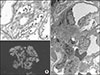
The second biopsy at 10 months after KT showed sustained microvascular inflammation and C3GN (Fig. 1). Thereafter, steroid pulse (methylprednisolone 250 mg every 12 hours for 3 days), and IVIG therapy (0.5 g/kg) were repeated once. Cyclophosphamide was added for the treatment of C3GN.
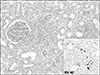
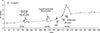
The third biopsy at 11 months after KT (Fig. 2) demonstrated that stage 1 BKVAN was complicated with the previous diagnoses. Glomerular changes considerably pro gressed, and cellular crescents developed. When a polymerase chain reaction (PCR) assay for BK virus DNA in plasma was performed, the BK virus PCR titer was 10,000 copies/mL, and the tacrolimus dose was adjusted 3.5 to 5 ng/mL. When the diagnosis of BKVAN was made, a reduction of immunosuppressive agents was initiated by discontinuing the tacrolimus therapy. As the creatinine level was increased to 3.58 mg/dL even after repeated administration of the conventional therapies for AMR. One cycle of bortezomib (1.3 mg/m2×4 doses for 2 weeks) was injected. Methylprednisolone and sirolimus were used as maintenance immunosuppressants. Thereafter, allograft function was stabilized and BK viremia became undetectable after 6 months. At present, the patient has been followed up carefully at our outpatient clinic, his serum creatinine level at the latest follow-up was 3.4 mg/dL (Fig. 3).
DISCUSSION
With the progress of immunosuppressive agents and improved graft survival rates after KT, indications for KT have been further expanded(14). Thus, more high-risk recipients are receiving KT. These high-risk recipients include patients with second transplantation, multiple transfusions, long-term hemodialysis, and pregnancy(15), and these conditions can sensitize recipients and induce the production of DSA. Thus, this highly sensitized KTRs are a high-risk group for AMR. AMR is one of the most common causes of allograft failure in highly sensitized KTRs(16). Generally, AMR is treated with steroids, plasma exchange, IVIG, and rituximab. However, the efficacy of these conventional treatments is limited and has not been confirmed(17). Unlike previous studies, recent studies found that rituximab therapy has no additional benefit to graft survival(18). The efficacy of newer agents such as bortezomib and complement inhibitors in the treatment of AMR is still under research. Thus, plasma exchange and IVIG therapy have become standard treatments for AMR therapy, even though their efficacies are uncertain(19). Despite the advances in the understanding of AMR mechanisms, some aspects are still, including the role of B-cells and plasma cells.
The limitations of the conventional treatments for AMR may be that they have no direct effect on plasma cells. As a selective proteasome inhibitor, bortezomib can cause plasma cell apoptosis in the bone marrow with subsequent inhibition of antibody production and treat AMR effectively. Several transplant centers have used bortezomib to treat AMR with various success rates(792021). In these successful cases, treatment with bortezomib therapy showed reversal of graft rejection, improved renal allograft function, and decreased DSA levels. Bortezomib therapy might be an effective treatment to target plasma cells. In a recently published randomized, placebo-controlled trial, bortezomib failed to prevent glomerular filtration rate loss, improve histological or molecular disease features, or reduce DSA level. However, this randomized trial compared the effect of a single course of bortezomib treatment. Some authors have proposed that the effect of bortezomib could be enhanced by combination therapy(192223). Therefore, the effect of bortezomib for the treatment for chronic AMR should be determined after the ongoing trial to determine its efficacy, in association with steroids, plasma exchange, and polyclonal IVIG therapy.
High-intensity immunosuppression, especially after treatment of AMR, may increase the risk of infection by weakening the immune functions against pathogens. BKVAN remains a major cause of infectious diseases that can lead to allograft failure in KT(24). The cause of BKVAN is well known to be related to excessive immunosuppression(2526). Therapeutic reduction of maintenance immunosuppression can restore immunity, control viral replication, and prevent progression of BKVAN. However, the therapeutic reduction of immunosuppression can increase the risk of developing de novo DSA and acute rejection episodes and chronic AMR(27). BKVAN was considered as a complication that resulted in poor graft survival.
As in our case, BKVAN occasionally develops concurrently with acute rejection. The incidence of BKVAN associated with acute allograft rejection was reported to range from 1% to 24%(1213). Generally, acute rejection is considered to be treated first. Once the acute rejection has subsided, it is advisable to reduce the dose of immunosuppressive agents to prevent the progression of BKVAN(1228). Bortezomib therapy has been used to treat AMR in transplant recipients, and a case of BKVAN disease with plasma cell-rich infiltrates treated with bortezomib has been reported(2930). In such case, the therapeutic effect is thought to be mediated through the reduction of plasma cells and CD20+ cells. BK virus replication remained stable during the follow-up periods(29). Therefore, we speculate that bortezomib may be beneficial for refractory AMR with BKVAN because it does not activate BK virus replication. No histological examination was performed during the follow-up period, but the BK virus PCR titer was maintained at <100 copies/mL. It may indicate that bortezomib administration did not activate BK virus replication in the KTRs.
In this report, we describe our experience with bortezomib therapy for the salvage treatment of a patient who developed AMR combined with BKVAN, which was refractory to a combination of plasmaphereses with IVIG, steroid pulse, and rituximab therapy. In conclusion, our report suggests that bortezomib represents a rescue therapy for AMR refractory to conventional treatments, even in cases with concurrent BKVAN. However, during the follow-up period, the C3 level was consistently low. Approximately 15% of death-censored graft failures are due to recurrent glomerulonephritis(31). Recent data show that monoclonal- related type I MPGN has an early recurrence rate of 60%, and polyclonal-Ig-related MPGN recurrence is more variable. Low C3 and C4 levels could predict a higher risk of recurrence(3132). C3GN is the result of dysregulation in the alternative pathway of complement and is characterized by dominant staining for complement factor C3 with minimal or no staining for immunoglobulins on IF microscopy(33). C3GN recurs in 66.7% of cases with a high risk of early graft loss(34). However, no treatment has been established for C3GN, neither for the prevention of its recurrence(32). Although bortezomib may be effective in the treatment of transient rejection, it is unlikely to be effective for inhibiting C3GN progression. Future prognosis depends on the clinical effort to prevent rejection and the progression of unresolved C3GN.
Considering the limitations of our short-term follow-up period and limited experience, larger, well-designed, randomized controlled trials are essential to confirm these results and ensure the long-term clinical efficacy of bortezomib therapy for graft survival. Moreover, further evaluation is required regarding the safety and efficacy profiles of bortezomib, including its dosage and timing of administration.
 XML Download