

 PDF
PDF Citation
 Print
Print


INTRODUCTION
Orofacial dyskinesia is stereotyped movement, consisting of smacking and pursing of the lips, lateral deviation and protrusion of the tongue, and occasionally lateral deviation and protrusion of the jaw. And, it can lead to orofacial pain, speech difficulty, dysphagia, and the inability to wear dentures, causing problems in everyday life. Several biochemical mechanisms have been proposed as causes, and the disease is diversely categorized according to the cause or type of movement [1].
Drugs with different action mechanisms, including haloperidol, estradiol, clonazepam, and antioxidants, have been used to treat orofacial dyskinesia, and research results have demonstrated that they had some effects [234]. But, treatment of orofacial dyskinesia is largely with medications that, unfortunately, are not highly successful. Less than 10% of patients experienced sustained benefits from anticholinergic agents and, although neuroleptic medication may have better efficacy, the side effects and the risk of tardive manifestations prevented their general use [5].
The occurrence of orofacial dyskinesia is accepted as due to the abnormality of various neurotransmitters, and it is known that supersensitivity of the dopamine receptors is a major cause [6]. We have also reported complete remission in a 27-year-old male patient who had developed oral dyskinesia after a traumatic brain injury following the administration of metoclopramide [7]. With this theoretical background and experience, we attempted to treat patients with orofacial dyskinesia using metoclopramide.
Below, we report seven cases in which an improvement in the symptoms was observed by using metoclopramide, a dopaminergic receptor blocking agent, in patients who had developed orofacial dyskinesia after suffering brain damage.
CASE REPORT
Patient 1
An 84-year-old female patient was receiving outpatient department follow-up for left hemiplegia which had developed from a right middle cerebral artery (MCA) infarction. The patient had two old cerebral vascular attacks in her medical history, and magnetic resonance imaging (MRI) taken at that time seem like a recent small infarction in the right parietal lobe, a chronic infarction in the bilateral basal ganglia (BG) and left cerebellar hemisphere (Fig. 1A). She was diagnosed with diabetes mellitus (DM) and hypertension (HTN), for which she was receiving oral medication, and had worn dentures for 5 years. She came to the hospital complaining about orofacial dyskinesia symptoms, as her jaw had been moving 2–3 Hz side-to-side for a month. She also suffered from an orofacial cavity ulcer, as the dentures were not fixed due to the dyskinesia, and from a related eating disorder. The dopaminergic agents; ropinirole (Requip®; GlaxoSmithKline, Research Triangle Park, NC, USA) 2 mg #2 and amantadine (PK-merz®; Ursapharm Arzneimittel GmbH Industriestrasse, Saarbrücken, Germany) 200 mg #2 were prescribed and taken for a week with no improvement. After explaining the treatment to the patient and guardians and obtaining their consent, metoclopramide (Macperan®; Hospira, Inc., Lake Forest, IL, USA) 5 mg 6T #3 was administered. Two hours after administration, the symptoms started to improve and after taking the medication for approximately two weeks, the dosage was reduced to 3T #3. However, the symptoms returned (although at a decreased frequency), and the dosage was therefore raised back to 6T. The frequency of the symptoms has now significantly decreased, and the patient and guardians report that her meal intake has increased as the dentures are now well secured. There have been no particular complications.
Fig. 1
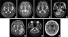
T2-weighted axial MRI for case 1–6 and CT image of case 7. (A) T2-weighted axial MRI of the case 1 patient shows chronic infarctions in bilateral BG. (B) T2-weighted axial MRI of the case 2 patient shows recent infarction in right MCA territory with hemorrhagic transformation and chronic infarctions in bilateral BG. (C) T2-weighted axial MRI of the case 3 patient shows chronic infarction in left BG. (D) T2-weighted axial MRI of the case 4 patient shows recent infarction in the right PICA territory. (E) T2-weighted axial MRI of the case 5 patient shows recent infarction in the left MCA territory. (F) T2-weighted axial MRI of the case 6 patient shows low signal intensity on bilateral pontine. (G) CT image of case 7 patient shows chronic infarctions in the left frontal lobe and bilateral BG.
CT, computed tomography; MRI, magnetic resonance imaging; BG, basal ganglia; MCA, middle cerebral artery; PICA, posterior inferior cerebellar artery.
Patient 2
A 64-year-old male patient had been receiving follow-up in the clinic after hospitalization in the Rehabilitative Medicine Department for left hemiplegia caused by the right MCA infarction four years prior. Brain MRI taken at that time seemed like a recent infarction in right MCA territory and chronic infarctions in bilateral BG, periventricular white matter, cerebellar hemispheres and left pons (Fig. 1B). The patient came to the hospital as he started experiencing orofacial dyskinesia symptoms, with the jaw moving 2–3 Hz side-to-side, and a loss of appetite for two months. His medical history revealed that he had HTN and DM and had been using dentures since 2008. He had been taking risperidone (Risperdal®; Ortho-McNeil-Janssen Pharmaceuticals, Inc., Titusville, NJ, USA) 0.5 mg for three years for behavioral problems and after emerging oral dyskinesia, stopping risperidone. Having explained the treatment to the patient and guardians and obtained their consent, we started using metoclopramide (Macperan®; Hospira, Inc.) 5 mg 6T #3. The symptoms began to improve two hours after administration, and prescription of the medication has been maintained for five months.
Patient 3
An 84-year-old male patient had been receiving follow-up in the clinic after hospitalization in the Rehabilitative Medicine Department for quadriplegia caused by “top of the basilar” syndrome, which had occurred two years prior. He had HTN, DM, old tuberculosis, hyperlipidemia, hepatocellular carcinoma, chronic renal failure, and benign prostatic hyperplasia in his medical history, and had been using dentures for five years. For three months, he had been experiencing symptoms of orofacial dyskinesia by which his jaw moved from side to side, which had caused an eating disorder and subsequent poor food intake, resulting in a weight loss of 5 kg. Brain MRI revealed chronic infarctions in bilateral posterior cerebral artery territories, cerebellums, frontal lobes, left BG, and right midbrain (Fig. 1C). And, he had been taking risperidone 1 mg for unstable mood three months before orofacial dyskinesia began. Having explained the treatment to the patient and guardian and obtained their consent, we started to use metoclopramide (Macperan®; Hospira, Inc.) 5 mg 6T #3. The symptoms of orofacial dyskinesia began to improve two hours after administration of the medication. After taking the medication for two weeks, the patient is now free of symptoms and his meal intake has increased, without particular complications from the medication (Supplementary Video 1).
Patient 4
A 78-year-old female patient received decompressive craniectomy from the Neurology Department for a right cerebellar infarction with hemorrhagic transformation which had appeared 1 month prior. She was transferred to the Rehabilitative Medicine Department and received rehabilitation treatment. Brain MRI revealed a recent infarction in the right posterior inferior cerebellar artery territory and chronic infarctions in the right periventricular white matter and right cerebellar hemisphere (Fig. 1D). She had a history of taking medication for HTN and atrial fibrillation, and not taking dopamine-related medication such as antipsychotics, and had been using dentures for five years. After transferring to the Rehabilitative Medicine Department, symptoms of orofacial dyskinesia, with the jaw moving about 3–4 Hz side-to-side, were observed in the daytime, although they disappeared when the patient slept. When feeding orally, most of the food was spilled out and she had difficulty swallowing because of the dyskinesia symptoms; hence, nasogastric tube feeding was applied. When a videofluoroscopic swallowing study (VFSS) was performed, it was observed that due to the continuous orofacial dyskinetic movement of the mandible and tongue, the bolus did not move to the pharyngeal phase from the oral phase, causing an anterior spill. Metoclopramide (Macperan®; Hospira, Inc.) 5 mg 6T #3 was used, and 1 hour later the symptoms had decreased and a symptom-free state maintained from the next day on (Supplementary Video 2). Following discharge from the hospital, the abnormality in the oral phase disappeared on the VFSS, and the patient is now taking general diet orally.
Patient 5
A 75-year-old female patient had experienced bilateral cerebellar infarction in October 2011, and after that she had experienced left MCA territory infarction 3 years later (Fig. 1E). Since then, she had received rehabilitation treatment in several hospitals. She had a history of receiving total abdominal hysterectomy, cholecystectomy, and left renal mass removal, and was taking warfarin for a diagnosis of atrial fibrillation. She had no history of using dentures and dopamine related medication. Following hospitalization, orofacial dyskinesia symptoms, with the jaws moving left to right, were observed in the daytime, although they disappeared when the patient slept. In the VFSS test, the bolus did not move from the oral phase due to the continuous dyskinetic movement, and the patient was therefore fed by nasogastric tube feeding. Having explained the treatment to the patient and guardian and obtained their consent, metoclopramide (Macperan®; Hospira, Inc.) 5 mg 6T #3 was administered. However, there was no improvement in the symptom, and the dosage was reduced to 5 mg 4T #2. After two days, the oral dyskinetic movement had been significantly reduced, and the patient later became-symptom free, so that an oral diet was slowly resumed. The dyskinetic movement of the tongue and jaws had disappeared but the movement of the head from left to right was observed. There were no other observations of complications from the medication.
Patient 6
A 48-year-old male patient was diagnosed with quadriplegia from pontine intracerebral hemorrhage (Fig. 1F). He had a history of HTN, hepatitis B virus carrier, and liver cirrhosis, and had not used dentures before. He also did not take antipsychotics, antiparkinsonism agents, antiemetics, anticonvulsants, etc. which are known to be associated with orofacial dyskinesia. Two weeks before the hospitalization, he began to manifest symptoms of orofacial dyskinesia in the daytime, which disappeared when he slept. The dyskinesia symptoms usually worsened with increased emotional stress. The patient was on a gestational diabetes diet, and orofacial dyskinesia was not observed while feeding. Having explained the treatment to the patient and guardian and obtained their consent, metoclopramide (Macperan®; Hospira, Inc.) 5 mg 3T #3 was administered. Afterward, the orofacial dyskinesia symptoms observed in the daytime decreased significantly, and there were no particular side effects.
Patient 7
An 88-year-old female patient came to the rehabilitative medicine clinic complaining about poor oral intake and gait disturbance. The patient had developed right hemiplegia from the stroke 20 years prior and brain computed tomography revealed chronic infarctions in the left frontal lobe and bilateral BG (Fig. 1G). She had been able to walk despite a lower extremity equinovarus deformity, however, due to recent general deterioration, she was only able to sit alone. When the patient was awake, she thrust her tongue out 1–2 times per second and spat out more food than she swallowed when eating due to the tongue thrust movement. Because of the orofacial dyskinesia, her dentures were not secured, so that she had to continually re-insert them with her hands. She had taken risperidone 0.5 mg and escitalopram (Lexapro®; Forest Laboratories LLC, St. Louis, MO, USA) 10 mg about for 4 years in the psychiatric department, but stopped risperidone after emerging orofacial dyskinesia. Her symptoms started to improve after the first administration of metoclopramide (Macperan®; Hospira, Inc.) 5 mg 3T #3, so that the irregular tongue thrusting was reduced to once every 3–4 seconds, and she was able to keep her mouth closed when instructed. According to reports from her guardian, the amount of spilled food had decreased and the meal intake increased. Later, complete remission of the symptoms was observed after the dosage was increased to 5 mg 4T #2.
DISCUSSION
Oral dyskinesia consists of abnormal, involuntary, uncontrollable movements predominantly affecting the tongue, lips, and jaw. And, they are classified according to the phenomenology of the abnormal movements or causes; bucco-linguo-mandibular dyskinesia, oral dyskinesia, oromandibular dystonia, diurnal bruxism, tics, perioral tremor. They may go unnoticed or cause social embarrassment, oral traumatic injury, speech difficulty, chewing and eating disorders, inability to wear prosthetic devices [1].
Prior to the seven cases described above, we had already reported the cases of a 51-year-old patient with subarachnoid hemorrhage, and a 36-year-old patient with involuntary diurnal bruxism from a traumatic brain injury, whose symptoms had been successfully managed with the administration of metoclopramide [8]. We had also reported complete remission in a 27-year-old male patient who had developed oral dyskinesia after a traumatic brain injury following the administration of metoclopramide [7].
In this case reports, we tried metoclopramide in seven patients who developed oral dyskinesia after stroke. And, we have described demographic characteristics and clinical information of seven case patients in Table 1.
Table 1
Demographic characteristics and clinical information of case series

The causes of orofacial dyskinesia are currently unclear. However, in order to discuss why we used metoclopramide, we need to see why orofacial dyskinesia has been developed.
Among orofacial dyskinesia, Tardive dyskinesia (TD) is known to be caused by a drug. It is an iatrogenic condition that results from the long-term use of dopaminergic antagonist medication [9]. The pathophysiology of TD is not understood but striatal dopamine receptor supersensitivity has been the traditional and oversimplistic explanation [10]. And, the minimum length of antipsychotic drug exposure required to propose a causal relation between the dyskinesia and the offending drug, which is typically at least three months in younger individuals, can be as short as 1 month in elderly people [1].
Second, edentulous status is thought to be a cause of oral dyskinesia, although the number of studies on this association is surprisingly low. Unlike drug-induced dyskinesia, the movements were always confined to the oral region and never dystonic, and no tongue movements were recorded when the mouth was open. Oral dyskinesia appeared only after a long period of edentulous status, an average of 12 years separating tooth extraction and the onset of oral movements. It is probable that, with time, progressive changes occurred in the mouth. Inadequate dento-oral prostheses may cause disruption of dental proprioception, resulting in dyskinetic searching movements of the oral cavity [11].
Finally, it is not known exactly which brain lesion causes oral dyskinesia, but several lesions are known to be associated with oral dyskinesia. Motor control is a complex process that is governed by sophisticated motor circuits involving both pyramidal (cortical) and extrapyramidal (basal ganglionic and cerebellar) circuits. Motor commands are generated in the motor cortex, but BG and cerebellum closely refine these signals by acting as feedback loops to allow for smooth, accurate, coordinated movements [12]. Neuroleptic drugs chronically block dopamine receptors in the BG. The result would be a chemically-induced denervation supersensitivity of the dopamine receptors which leads to excessive movement [6]. And, deep brain stimulation in thalamus, thalamotomy and pallidotomy are performed for the treatment of orofacial movements disorders [13]. Previous studies revealed that dopamine is the major neurotransmitter involved in balance of the motor output of the prefrontal cortex by maintaining an inhibitory tone, so, the hypoperfusion of the frontal lobe is closely related to orofacial dyskinesia [14].
We have summarized the potential causes of orofacial dyskinesia in Table 2 according the following categories: brain lesions, medication, and denture durations. First, when reviewing these cases, patients 2, 3, and 7 used risperidone, orofacial dyskinesia may have been caused by drugs when considering the duration of use. And, patients 1, 2, 3, 4, and 7 used dentures, but considering that it takes about 12 years for orofacial dyskinesia to occur after edentulous status, the likelihood is not high. Among the brain structures, BG is known to be highly related to orofacial dyskinesia, and lesions in BG have been found in most of our patients. From these findings, we believe BG lesions are likely to be associated with orofacial dyskinesia. Nonetheless, we cannot exclude the possibility of other potential causes as other brain regions are closely linked by numerous circuits [12].
Table 2
Several factors related to the development of orofacial dyskinesia

Our patients responded well to metoclopramide, selective dopamine 2 receptor (D2R) antagonist. Metoclopramide readily crosses the blood-brain barrier and is equal in potency to chlorpromazine in dopamine receptor blocking affinity. In mesocortical neurons, a preferential binding to presynaptic D2R rather than postsynaptic D2R by D2R antagonists is noted [15]. Metoclopramide binds to D2R in a biphasic manner; to presynaptic D2R at low doses and to postsynaptic D2R with increasing doses [16]. A selective blockade of the hypersensitive presynaptic D2R by low dose metoclopramide recovers the dopaminergic flow and attenuates bruxism or oral dyskinesia. The absence of extrapyramidal side effects during metoclopramide therapy may be due to a higher sensitivity of mesocortical D2R to low dose metoclopramide than that of mesolimbic or nigrostriatal neurons [17].
Metoclopramide may also exert its effect through other mechanisms. The prefrontal cortex and ventral tegmental area have abundant noradrenergic connections. Destruction of noradrenergic fibers innervating the ventral tegmental area would result in a decrease in dopamine utilization in the prefrontal cortex, probably due to an impairment of the stimulatory a1-aderenoceptor effects on postsynaptic dopaminergic neurons. Metoclopramide may thus act through an increase in noradrenaline [14].
In previous reports, the effect of metoclopramide appeared 3–4 hours after dosing, and in these case patients, the effect appeared 1 to 2 hours after taking pills. The degree of improvement in dyskinesia is subjective, so the difference in the time may be probably due to measurement error of time. Therefore, it will be necessary to quantitatively divide the degree of improvement and objectively suggest the time to act.
We used metoclopramide in dosages of 15–30 mg/day in seven patients who had developed orofacial dyskinesia following a brain injury and observed an improvement in the symptoms within a short time frame. This supports the hypothesis that dopamine receptors play an important role in orofacial dyskinesia, and calls for more research into unraveling the exact mechanisms. The use of metoclopramide for the treatment of orofacial dyskinesia patients is relatively safer than the administration of haloperidol. However, as metoclopramide can cause extrapyramidal symptoms, close monitoring is required.

 XML Download