

 PDF
PDF Citation
 Print
Print


INTRODUCTION
Stroke survivor regularly receives physiotherapy (PT) training with the aim to make progress as the outcome of functional rehabilitation. In some programs, the transformation between the functional outcomes after the training cannot be noticed [1234]. Thus, to a significant degree, the success of neurorehabilitation depends on the amount and effectiveness of rehabilitative training [567]. Effective rehabilitation therapies are needed for stroke patients showing long-term deficits in upper-arm function by increasing force and reduce spasticity in muscle. The primary objective of rehabilitation is to further improve motor function and the impaired limb in daily activity of life [234].
Non-invasive brain stimulation procedures, such as transcranial direct current stimulation (tDCS) and repetitive transcranial magnetic stimulation (rTMS), as an assistant to other managements, may expand therapeutic effects by increase ipsilesional or decrease contralesional excitability [89]. Previous studies showed the anodal tDCS stimulation depolarizes, while cathodal stimulation produces hyperpolarization of the resting membrane potential of neurons [91011]. Recent studies showed that brain stimulation priming with motor training might share synergistic effects on synaptic and network plasticity, which provide larger behavioral effects in animal and human [10111213].
Proven on this concept, it is practical to explore the effect of excitability diminishing by cathodal tDCS of the unaffected hemisphere priming with cycling training on the motor function in chronic stroke patients.
MATERIALS AND METHODS
Patients
Three male patients (71, 63, and 52 years) had left-sided effected with ischemic capsular and ischemic sylvian stroke (30 months before, Table 1) participated in this study. Each of them had given his written informed consent. The computerized tomography shown no patient had more than one cerebral lesion. Inclusion criteria were; 1) to be capable to take part for at least 20 minutes of exercise using a hand ergometer and 2) to have no problems of understanding the study procedure. Patients with aphasia, shoulder pain or serious neuropsychological deficits were excluded.

Experimental protocol
Soterix medical 1 × 1 device with two 35 cm2 (5 × 7 cm) saline-soaked sponges' electrode was used for stimulation with intensity of 1 mA tDCS for 20 minutes. The objective of the study design was achieved by cathode was placed over M1 of the unaffected hemisphere to decrease in excitability in circuits controlling the left hand and the anode over the contralateral supraorbital area.
MOTOmed Movement Therapy System®
The commercial motorized arm-ergometer (MOTOmed® viva RECK, Tampa, FL, USA), was used for optimal training by gently moved and loosened with motor and software assistance of the system. The previous work described in detail of the MOTOmed system® and procedure [14].
Experimental protocol
An A-B-A protocol was tested during 4 sessions with; 1) at T0, the beginning of the base line phase A; 2) at T1, a beginning of training phase B of 1 weeks; 3) at T2, the end of phase B; and 4) at T3, the end of the follow-up phase A of 1 week was used.
The patients, who were sited in front of the ergometer in their wheelchair or on an armless chair, completed the arm training for 20 minutes daily, 5 days a week. The performed tests involved clinical and behavioral assessment at T0, T1, T2, and T3.
Clinical assessment
Physiotherapists used Rivermead Motor Assessment (RMA) to quantity gross motor task, upper, lower limb, and trunk control [15]. In this study all 3 sections were assessed, but only data concerning the upper limb control were presented in result section.
The Motricity Index (MI) tools used to tested manually the force of the elbow, shoulder flexors and extensors in the patients [1516]. Medical Research Council provides the grades ranging from 0 (no movement and no contraction can be palpated) to 5 (movements with normal power). The patients' performance was evaluated on the affected side by taking the sum of the quotation on the elbow flexors, extensors, shoulder extensors, and flexors.
The maximum cycling force was measured on the affected side by which the patient was able to cycle for 10 seconds at a constant frequency. The resistance to cycling might be accustomed on the scientific ergometer.
Behavioral measurement
Maximal pinch force (specific dynamometers, Saehan Hydraulic Hand Evaluation, model SH5003; Saehan Corporation, Masan, Korea) were used to measure the manual force. Maximum pinch and grip forces were measured in 2 efforts using both hands with the patient seated and the elbow at 90° of flexion and a neutral position of the wrist. This method is sound consistent and showed high test-retest reliability.
The 9-hole peg test (9HPT) is a simple task to test the hand dexterity. Patients performance was measured by insert 9 pegs into 9 holes of a board in the time to complete the task using a chronometer.
Statistical analysis
The Statistical Package for the Social Science (SPSS Inc., Armonk, NY, USA) was used for Statistical analysis. The muscular force was measured by 1) the RMA, 2) the MI, and 3) the cycling force, while spasticity was measured by 1) the Ashworth Scale, 2) the maximum active extension of the biceps, and 3) the minimum torque on the affected side during arm cycling. Data were summarized in terms of their mean and standard deviation.
The 3 data sets cannot be combined directly to test the effect of training since they are measure in different units. Therefore, they were transformed. The mean of all observations was subtracted from each observation and the resulting values were divided by their standard deviation (z-transformation) [14]. The data sets, which have then a mean of zero and a standard deviation of one, were pooled and a t-test was performed for pre- and post-testing.
RESULTS
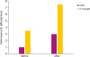
The RMA, MI, and cycling force was assessed for muscular force and the data were transformed (z-transformation), pooled and a t-test was performed. The numerical data with the confidence limits are presented in Fig. 1. Due to the data transformation, the average of all values is 0, which remained constant during the base line, but increased significantly (p = 0.01) over the period of training priming with tDCS. All the patients increased force during the training priming with tDCS and this force increase remained during the following 3 weeks. Further the Ashworth Scale and the maximum extension of the biceps, the minimum torque on the lesioned side through arm cycling was careful as a constraint related to spasticity. Since the patients are not capable to decline the flexor muscles of the spastic arm during the delay phase, these contracted flexor muscles can even result in a negative torque on the spastic side. Before the training spasticity was greater than after training priming with tDCS (p = 0.03).
 | Fig. 1Average arm force before and after cycling training priming with cathdol transcrinal direct current stimulation of the unaffected hemisphere. Due to the normalization, values can be negative. Due to the z-transformation, values are positive and negative and the grand mean is 0. There was a significant increase of force (A) and spasticity (B).
|
Behavioral testing showed favorable results on all the tests (9HPT; maximal pinch force test) for the affected hand. The 9HPT showed an improvement in the lesion hand following tDCS and training. Post-training the patient was able to place 4 pegs (p = 0.01, Fig.2) in the same time period. Both the finger tapping task and 9HPT showed greater affects (p = 0.02) on after training priming with stimulation application.
DISCUSSION
The finding that cathodal stimulation of the contralateral hemisphere could yield motor improvements of the affected hemisphere in combined therapy with ergometer is a suitable device for training stroke patients with unilateral paralysis. Since all aspects of muscle force improved might be valuable for patient in daily life as assessed in qualitative approaching by RMA, MI, and quantitative by maximum cycling force for an adequate motor control [14].
Previous studies shown the therapy increased the force and decrease spasticity in chronic stroke patients with severe upper limb dysfunction [3411]. The present results clearly showed that these effects were due to both the demanding, repetitive practice of cycling and the stimulation of neurons by cathodal tDCS in unaffected hemisphere from possible excessive transcallosal inhibition, potentially allowing some functional improvement.
These results regarding the effect of repetitive training by an arm ergometer on spasticity were corroborated by other studies [3461114], where we evaluated spasticity by Ashworth Scale and the maximum extension of the biceps, the minimum torque on the lesioned side during arm cycling.
Our findings were similar to those reported by Fregni et al. [17] that mean improvement of motor performance with cathodal tDCS of the unaffected hemisphere was higher when the left hemisphere was inhibited in chronic stroke patients.
CONCLUSION
To confirm the present outcomes, next large multi-center clinical trials would study the possibility of other types of stimulation (anodal or dual), in order to examine altered likely interactions between neuroplasticity and achievable functional outcome by priming with training. In summary, noninvasive cathodal tDCS mutual with cycling training did enhance the function of affected upper arm in chronic stroke patients.
 XML Download