Journal List > Ann Surg Treat Res > v.95(4) > 1101700

Kim and Hong: Focal change of the pancreatic texture using a direct injection mixture of N-butyl cyanoacrylate and lipiodol in the pig model: a strategy for preventing pancreatic leakage during pancreatic surgery

Abstract

Purpose

A soft texture of the pancreas is one of the most important predisposing factors for a pancreatic fistula. Thus, in a porcine model, we investigated a method to harden the pancreas locally by directly injecting an artificial material.

Methods

During the laparotomy, 51 samples from 17 pigs, including 13 survival models, were randomly divided into 3 groups and either received a direct injection into the pancreas of MHL (1:4 mixture of histoacryl [n-butyl cyanoacrylate] and lipiodol) (group E) or saline (group C) or only received a pinprick into the pancreas without injecting a substance (sham). We measured the change in the pancreatic hardness after the injection using a durometer and examined the histological change of the pancreas using the fibrosis grade in the survival model.

Results

The postinjection hardness of the pancreas was significantly increased in group E compared to group C and the sham group (P < 0.001). Pathologically, all cases in group E showed a severe fibrotic change, whereas the other groups demonstrated mild to no fibrosis (P < 0.001). The fibrosis in group E was localized to the area of the injection, while the surrounding areas were preserved.

Conclusion

The direct injection of MHL could induce focal hardening and fibrotic changes in the pancreas of the porcine model.

INTRODUCTION

Despite technical improvements and advancements in operative skills and perioperative care, the postoperative pancreatic fistula (POPF) after a pancreatectomy remains a conundrum to pancreatic surgeons [1]. Although the operative mortality after pancreaticoduodenectomy has dramatically decreased to less than 5% in the past 40 years [2], the morbidity rate remains high, claiming 30%–50% of the patients, and most of the serious morbidities are related to POPF [3]. To reduce this noticeably high incidence of POPF, various novel surgical techniques for the pancreatoenterostomy (or the handling of the pancreas stump following a distal pancreatectomy) have been introduced and steadily refined to ensure a safe surgery and acquire a firm pancreas [456]. However, to the best of our knowledge, no definite solution exists to date.
Furthermore, the identification and prevention of the risk factors related to POPF has recently been a key issue. The various risk factors for developing POPF includes a high fat content in the pancreas, a small main pancreatic duct, and an especially soft texture of the pancreas [78]. Among the various risk factors, a soft pancreas is a key factor for increasing the risk of a pancreatic fistula, as shown in several large-sized prospective trials [910]. In fact, pancreatic leakage following a pancreaticoduodenectomy most often occurs in the presence of the soft, fragile pancreas often seen with a benign tumour or borderline malignancy of the pancreas that is not associated with chronic inflammatory changes. Therefore, two trials have reported a method that seeks a histomorphological change of the pancreatic texture using octreotide [511].
The aim of this study was to investigate a novel technique using an artificial material that could harden the pancreas and evoke focal fibrosis at the pancreatic anastomosis site or at the resected stump site. We assessed the feasibility and effectiveness of this method using the porcine experimental model.

METHODS

The present study was approved by the Institutional Animal Care and Use Committee of the institution (IRB No. 2017-0017-01). During the study period, a total of 17 female, 3-month-old domestic pigs, weighing 31–41 kg, underwent the operation, and all procedures were performed at the animal laboratory centre under the supervision of a specialized veterinarian. All pigs underwent a bowel enema at midnight prior to the operation and were fasted for 24 hours before the surgery for the preoperative bowel preparation.
A total of three different groups were analysed in our study: (1) the experimental group was injected with a mixture of histoacryl and lipiodol (MHL) into the pancreas (group E, 51 samples), (2) the control group was injected with saline (group C, 51 samples), and (3) the sham group did not received an injection but only received a pancreatic pinprick (sham, 51 samples). We selected 3 different areas of the pancreas for each pig, and each of the 2 substances (MHL and saline) was injected into the 2 different areas in a random order, whereas the third pancreatic area was pricked with a needle. A working solution of MHL was generated by mixing Histoacryl (n-butylcyanoacrylate, Braun GmbH, Kronberg im Taunus, Germany) with lipiodol (Guerbert, Roissy, France) at a ratio of 1:4, and 1 mL of MHL was injected into each pancreas. Histoacryl, which is the commercial name of N-butyl-2-cyanoacrylate (NBCA), is widely used as an embolic agent for the haemostasis of bleeding or for a pseudoaneurysm [12]. In the saline control group, 1 mL of saline was injected using the same method as for MHL. The sham treatment included a needle pinprick without injecting any substances. We used the sham model in addition to the saline control because the pinprick could result in iatrogenic pancreatitis without any injection, which could also occur when injecting the artificial substances. To estimate the hardness of the pancreatic parenchyma before and after the injection, we used a commercially available durometer using the technique described below. The baseline characteristics and surgical outcomes, including the postoperative complications and the existence of each case after surgery, were also recorded and retrospectively analysed.

Surgical procedure

On the operation day, each pig was moved to the operation room in the animal laboratory of Seoul St. Mary's Hospital following the preoperative preparation. Preanaesthesia was performed with a combination of 1-mg/kg xylazine, 3-mg/kg azaperone, and 3-mg/kg alfaxalone. General anaesthesia was maintained with isoflurane gas via an endotracheal tube. Respiratory and cardiac parameters were closely monitored during the whole procedure.
The pig was placed in the supine position under general anaesthesia, and a midline incision was made. Initially, the posterior wall of the stomach was lifted, the peritoneum that covers the surface of the viscera was opened, and the distal part of the pancreas was dissected from its posterior attachments. The upper and lower margins of the pancreatic tail were dissected and isolated from the mesenteric attachments. Next, the tail and distal portions of the pancreatic body were mobilized to the junction of the splenic vein, superior mesenteric vein and portal vein. The head of the pancreas, which has a C-shape with respect to the duodenum, was also dissected to expose the full anterior aspect of the pancreas. The pig pancreas is composed of the following three different lobes with a nodular surface: (1) the splenic lobe, (2) the duodenal lobe, and (3) the connecting lobe. After sufficient exposure, the lateral side of the pig pancreas, including the splenic and duodenal lobes for each pig, was marked for the three different areas by clipping the connective tissue attached to the border of the pancreas that are collinear to each injected area. Next, we injected one of the 2 substances (MHL or saline) or administered the pinprick in a random order as shown in Fig. 1.
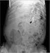
Among the 17 pigs, 4 were sacrificed immediately after the initial operation (sacrifice model). The other 13 pigs were kept alive for 1 week after the initial operation (survival model) to perform a second laparotomy with a total pancreatectomy on postoperative day 7 to investigate the delayed histological change to the pancreas. For the survival model, an X-ray examination of the abdomen occurred before the second laparotomy to assess the location of the injected substances and to confirm whether the substance was absorbed or diffused (Fig. 2).

Hardness measurement

To measure the pancreatic hardness, a Check-line DD-100 durometer (Electromatic Equipment Co., Cedarhurst, NY, USA) was used, and the pancreatic hardness was measured by 1 investigator who received the pancreatic tissue in a random order and did not participate in the operation, which enabled the investigator to be blinded to the treatment. The device was positioned on the surface of specimen as shown in Fig. 3. Once the working surface of the durometer was placed on the tissue, the indention body was pressed, and the spring measuring the force of the indention body could be converted into the indicator of tissue hardness [11]. Before the measurement, the repeated hardness tests were performed to acquire an accurate testing pressure while eliminating the human error. For each case, 3 measurements were taken each time by placing the working face of the durometer on the exact single area of the specimen, and the results were calculated as a mean value. Once the pancreas was exposed after the laparotomy, the investigator initially measured the baseline hardness of the tissue for every 3 measurements in three different places of the pancreas from each animal. Next, the treatment (injection or pinprick) was administered as previously described. For the 4 pigs in the sacrifice model, the change in hardness was measured immediately following the pancreatic harvest using the same technique as the baseline hardness measure. However, only the baseline hardness was measured during the first laparotomy for the survival model. After a week, those in the survival model underwent the re-laparotomy, and we measured the pancreatic hardness at the previous experimental sites, which was marked at the exact location with the clipping. After the pancreatic harvest, all of the animals were sacrificed.

Histopathological examination

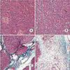
For the survival model, the pancreatic tissue was fixed in formalin for histological examination and later transferred to the clinical research support centre of the Catholic University of Korea. Slide preparation was performed using the Masson trichrome staining method (Sigma-Aldrich, Inc., St. Louis, MO, USA). To estimate the pancreatic fibrosis, the fibrosis grade was estimated using the four-stage scoring system described by Wellner et al. [1]. Briefly, the 4 grades include a normal pancreatic parenchyma without fibrotic changes (grade 0), mild fibrosis with thickened periductal fibrous tissue (grade 1), moderate fibrosis with marked sclerosis of the interlobular septa without evidence of architectural changes (grade 2), and severe fibrosis with detection of architectural destruction or acinar atrophy (grade 3) (Fig. 4).

Statistical analysis

All statistical analyses were conducted using IBM SPSS Statistics ver. 23.0 (IBM Co., Armonk, NY, USA). Continuous data are presented as the mean ± standard deviation. For the continuous data, overall differences were analysed using Student t-test and analysis of variance, and multiple comparison tests were used for further analysis. The categorical variables were analysed using Fisher exact test or chi-square test. The descriptive statistics are described as the mean ± standard deviation, and differences were considered to be statistically significant when P < 0.05.

RESULTS

During the study period, a total of 17 female pigs were assessed. The mean operation time and anaesthesia time was 31.7 ± 3.9 minutes (range, 25–35 minutes) and 126.2 ± 15.4 minutes (range, 90–145 minutes), respectively. We classified the pigs into the 2 groups according to the type of experimental model (4 sacrificed and 13 survival) and analysed them according to the type of the injected substance. The ratio of injected lobes was definitely the same in each group.
In each of 4 cases in the sacrificed model, the measurements were performed 3 times per case. Regarding the hardness of pancreatic texture (Table 1), the preinjection hardness showed no significant difference among the three groups (P = 0.457). Contrarily, there was a significant difference between the postinjection hardness of the pancreas, which was measured in the procured pancreas immediately after injection of the substances, among the three groups (P < 0.001). The pancreas texture of group E was harder than that of group C (94.1 ± 3.8 vs. 22.6 ± 10, respectively, P < 0.001) and group E was also harder than group S (94.1 ± 3.8 vs. 14 ± 0.7, respectively, P < 0.001), and there was no difference between groups C and S.
Additionally, in each of the 13 survival models, three measurements per case were taken in the same way as in the sacrificed model. Regarding the hardness of the pancreatic texture (Table 1), the preinjection hardness of the pancreas showed no significant difference among the three groups (P = 0.955). However, there was a significant difference in the postinjection hardness of the pancreatic texture that was measured on the seventh day postinjection among the three groups (P < 0.001). The hardness of group E exceeded that of group C (85 ± 17.6 vs. 15.5 ± 14.2, respectively, P < 0.001), and group E was greater than group S (85 ± 17.6 vs. 14.2 ± 22.3, respectively, P < 0.001). There was no difference between groups C and S. Regarding the histopathological change (Table 2), the distribution of fibrosis grade was significantly different among the three groups (P < 0.001). In group E, 13 cases (92.3%) showed a severe histological change with a grade of 3, and the others (7.7%) had a moderate change with a grade of 2. All cases in groups C and S presented no fibrotic change (grade 0; 6 cases [46.2%] in group C and 11 cases [84.6%] in group S) or mild fibrotic change (grade 1; 7 cases [53.8%] in group C and 2 cases [15.4%] in group S). Fig. 5 shows the focal change of the pancreas glossly at the cross-section (Fig. 5A) and the fibrotic change at the injection area upon histopathological examination (Fig. 5B) after the MHL injection. Contrarily, there was no histological change in the pancreas beyond the MHL injection site, and the pancreas demonstrated normal parenchymal findings. Additionally, the abdomen X-ray image, which was taken before the second laparotomy, presented the injected lipiodol obviously seen at the injected area locally in all survived models. No mortalities were observed during the experimental period in the survival models.

DISCUSSION

The incidence of POPF occurs 30%–50% of the time following pancreatic surgery, and the incidence should be higher with a fragile pancreas that has a soft texture of the parenchyme. Belyaev et al. [2] reported that the soft texture of the pancreas' parenchyme is a significant predisposing factors for POPF from a histomorphological aspect. Several reasons may explain this impressive pancreatic texture factor on the development of POPF. First, the soft pancreas is inevitably accompanied by the technical difficulty of a pancreaticoenteric anastomosis, a consensus generally accepted by most pancreatic surgeons. This finding is attributable to the histological characteristics of the soft pancreas that decreases the suture holding capacity of the pancreatic tissue and due to the easy distortion of the pancreatic gland or tissue by a needle penetration or suturing. These properties could aggravate the parenchymal injury caused by the tangential shear stress of the needle and make the anastomosis more fragile, which consequently results in POPF. Second, the exocrine function of the remnant pancreas could be another clue to the occurrence of POPF in the soft pancreas. The exocrine function of the pancreas that contributes to the development of POPF is the pancreatic juices exceeding the ductal boundary, which is significantly associated with the soft texture of the pancreas [31314]. The exocrine function of a hard texture pancreas accompanied by chronic inflammation should be minimized because of the ductal atrophy and parenchymal fibrotic change contrary to a soft pancreas [5]. These fibrotic changes in a hard pancreas usually obstruct the opening of the minor ducts at the cut-surface of the pancreas, which would prevent POPF by blocking the secretions from these ducts.
Therefore, several techniques have been recently developed to modify the texture of the pancreas into a hard pancreas through parenchymal fibrosis or ductal atrophy. Konstadoulakis et al. [5] applied an intra-arterial injection of octreotide, which is expected to harden the pancreas and inhibit the exocrine secretion of the pancreas [15]. However, this method, which uses an infusion of octreotide via arterial circulation, includes the concern of unnecessary, systemic adverse effects including vasoconstriction or reduction of splanchnic blood flow [16]. Additionally, Ahmadi et al. [17] reported a mouse model of chronic pancreatitis with fibrotic changes induced by the intraperitoneal injection of ethanol and cerulean. However, this method is limited for use during actual pancreatic surgery because its effect is neither localized nor does it occur immediately.
The MHL used in the current study is made by mixing histoacryl and lipiodol. Cyanoacrylate in histoacryl has a characteristic feature that could rapidly sclerose tissue when a hydrate is added, such as water or alcohol. Polymerization of NBCA in the MHL could be promoted by mixing the solution with ethanol, which is initiated by contact with a positively charged ion such as the sodium (Na+) in the serum or water of the cellular tissue. Since its first introduction in 1984 [18], histoacryl has been used in various clinical fields without significant complications. Notably, histoacryl is generally mixed with lipiodol, a sterile injectable radio-opaque agent that contains iodine organically combined with ethyl esters of fatty acids from poppy seed oil [19]. Because of these characteristics, histoacryl is usually used with a mixture of lipiodol during the embolization of a blood vessel or for tissue visualized during a diagnostic CT or other imaging technique. MHL also enables one to accurately localize the injection site of the embolic agent or to measure the quantity of the injected agents [20].
In our experiments, MHL acts as a potent, effective sclerosing agent through the rapid polymerization of NBCA at the injection site. Moreover, MHL facilitated the secondary fibrotic change in the tissue as shown in our histopathologic examination of the injected specimen. The injection of MHL makes the acute and chronic inflammatory change primarily due to the foreign body reaction, which could lead to the fibrotic change in the tissue around the injection site. This histological change would result in the texture of the pancreatic parenchyma hardening.
There are other characteristic properties of our method that could be advantageous for its clinical use. First, as shown in the sacrificed model, this histological change occurred immediately after the MHL injection. The time spent procuring the pancreas was approximately 15–20 minutes on average and seems sufficient to induce the inflammatory change to harden the pancreas. The prompt effect of the MHL might be attributed to the rapid polymerization of the histoacryl when contacting the water in the tissue, and this property of the MHL would make this method useful during the operation under direct vision. This polymerization could help to inject the substance more accurately to the target site with the exact amount suitable for the intraoperative method. Additionally, the hardness value of the injected sites in the survival models have been preserved for at least a week following the MHL injection, and it would support the stability and persistency of the tissue change induced by the MHL.
Second, confining the histological change to the area of injection is very important. One serious caution for the artificial degeneration of the pancreas might be the excessive suppression of the physiological function in the remnant pancreas resulting from the diffuse sclerosing change. The artificial degeneration could lead to iatrogenic diabetes mellitus or even malnutrition associated with the degenerated exocrine function. However, our method seems safe from this concern because the sclerosing change only occurred in a limited area around the MHL injection site in the histopathologic exam, whereas the other site of the remnant pancreas demonstrated normal pancreas tissue or glandular structure even after a week. We believe that the MHL use could induce a focal hardening of the pancreas without the inevitable functional loss of the normal pancreatic tissue.
Additionally, the local, direct MHL injection into the pancreatic parenchyme would reduce the risk of morbidities such as the extravasation of drugs or the systemic embolization that could occur after the intravascular injection of histoacryl [2122]. Additionally, the lipiodol in the MHL could be useful as an indicator or marker to confirm and follow the exact site or status of the injection area using a CT scan or a simple X-ray image serially.
In conclusion, the authors suggest that the direct injection of MHL into the pancreas could be an effective and feasible method for preventing pancreatic leakage through the focal hardening and the fibrotic change of the pancreas at the injection site. A large case study using MHL with a long-term follow-up should be conducted to assess the feasibility of the procedure before application to human pancreas surgery.

Figures and Tables

Fig. 1

View of the operation field. (A) The entire anterior surface of the pancreas was exposed, including the splenic and duodenal lobes. (B) After the injection of each substance into the pancreas (mixture of histoacryl and lipiodol or saline) or the pinprick, we marked the 3 different areas of the pancreas by clipping the connective tissue that was collinear to each treated area (arrows).

astr-95-175-g001
Fig. 2

Presence of the mixture of histoacryl and lipiodol was markedly revealed by X-ray film (arrow), which was taken on the 7th day following the injection for the survival model.
astr-95-175-g002
Fig. 3

Hardness of the pancreatic parenchyme was measured using a durometer in vivo (A) and ex vivo (B).

astr-95-175-g003
Fig. 4

Slide preparation was performed using the Masson trichrome staining method (original magnification x400). Fibrosis grade of the pancreas was estimated using the 4-stage scoring system as described by Wellner et al. [4] Briefly, the 4 grades include a normal pancreatic parenchyma without fibrotic changes (grade 0, A), mild fibrosis with thickened periductal fibrous tissue (grade 1, B), moderate fibrosis with marked sclerosis of the interlobular septa without evidence of architectural changes (grade 2, C), and severe fibrosis with detection of architectural destruction or acinar atrophy (grade 3, D).
astr-95-175-g004
Fig. 5

Focal change of the pancreatic parenchyme was confined to the mixture of histoacryl and lipiodol injection site (arrow) and not in the surrounding area, which was preserved to the normal structure of the pancreas (arrowhead), as shown in a cross-section (A) and during the histopathologic examination (B). Slide preparation was performed using the Masson trichrome staining method (original magnification x400).

astr-95-175-g005
Table 1

Comparative analysis of the hardness of harvested pancreatic tissue according to the injected substances

astr-95-175-i001

Values are presented as mean ± standard deviation.

a)A mixture of histoacryl and lipiodol (MLH) which was generated by mixing the Histoacryl (n-butylcyanoacrylate, Braun GmbH, Kronberg im Taunus, Germany) with lipiodol (Guerbert, Roissy, France) at a ratio of 1:4. b)Shore unit (S.U.) which reprents the tissual hardness checked by the durometer.

Table 2

Comparative analysis of the fibrosis grade of harvested pancreatic tissue according to the injected substances in the case of survival models

astr-95-175-i002

a)A mixture of histoacryl and lipiodol (MLH) which was generated by mixing the Histoacryl (n-butylcyanoacrylate, Braun GmbH) with lipiodol (Guerbert, Roissy, France) at a ratio of 1:4. b)The histopathologic examination was peformed only in the case of survival models (n = 13) because the sacrificed model that killed immediately after the laparotomy was not suitable to reflect the fibrotic change on pancreatic tissue. c)The fibrosis grade of pancreas was estimated using the 4-stage scoring system described by Wellner et al. [1].

Notes

CONFLICTS OF INTEREST No potential conflict of interest relevant to this article was reported.

References

1. Wellner UF, Kayser G, Lapshyn H, Sick O, Makowiec F, Hoppner J, et al. A simple scoring system based on clinical factors related to pancreatic texture predicts postoperative pancreatic fistula preoperatively. HPB (Oxford). 2010; 12:696–702.
crossref
2. Belyaev O, Munding J, Herzog T, Suelberg D, Tannapfel A, Schmidt WE, et al. Histomorphological features of the pancreatic remnant as independent risk factors for postoperative pancreatic fistula: a matched-pairs analysis. Pancreatology. 2011; 11:516–524.
crossref
3. Sato N, Yamaguchi K, Chijiiwa K, Tanaka M. Risk analysis of pancreatic fistula after pancreatic head resection. Arch Surg. 1998; 133:1094–1098.
crossref
4. Wellner U, Makowiec F, Fischer E, Hopt UT, Keck T. Reduced postoperative pancreatic fistula rate after pancreatogastrostomy versus pancreaticojejunostomy. J Gastrointest Surg. 2009; 13:745–751.
crossref
5. Konstadoulakis MM, Filippakis GM, Lagoudianakis E, Antonakis PT, Dervenis C, Bramis J. Intra-arterial bolus octreotide administration during Whipple procedure in patients with fragile pancreas: a novel technique for safer pancreaticojejunostomy. J Surg Oncol. 2005; 89:268–272.
crossref
6. Torer N, Ezer A, Nursal TZ. Mattress sutures for the modification of end-to-end dunking pancreaticojejunostomy. Hepatobiliary Pancreat Dis Int. 2013; 12:556–558.
crossref
7. Hashimoto Y, Traverso LW. Incidence of pancreatic anastomot ic fai lure and delayed gastric emptying after pancreatoduodenectomy in 507 consecutive patients: use of a web-based calculator to improve homogeneity of definition. Surgery. 2010; 147:503–515.
8. Gaujoux S, Cortes A, Couvelard A, Noullet S, Clavel L, Rebours V, et al. Fatty pancreas and increased body mass index are risk factors of pancreatic fistula after pancreaticoduodenectomy. Surgery. 2010; 148:15–23.
crossref
9. Yeo CJ, Cameron JL, Maher MM, Sauter PK, Zahurak ML, Talamini MA, et al. A prospective randomized trial of pancreaticogastrostomy versus pancreaticojejunostomy after pancreaticoduodenectomy. Ann Surg. 1995; 222:580–588.
crossref
10. Suzuki Y, Fujino Y, Tanioka Y, Hori Y, Ueda T, Takeyama Y, et al. Randomized clinical trial of ultrasonic dissector or conventional division in distal pancreatectomy for non-fibrotic pancreas. Br J Surg. 1999; 86:608–611.
crossref
11. Foitzik T, Gock M, Schramm C, Prall F, Klar E. Octreotide hardens the pancreas. Langenbecks Arch Surg. 2006; 391:108–112.
crossref
12. Yonemitsu T, Kawai N, Sato M, Sonomura T, Takasaka I, Nakai M, et al. Comparison of hemostatic durability between N-butyl cyanoacrylate and gelatin sponge particles in transcatheter arterial embolization for acute arterial hemorrhage in a coagulopathic condition in a swine model. Cardiovasc Intervent Radiol. 2010; 33:1192–1197.
crossref
13. Sato N, Yamaguchi K, Yokohata K, Shimizu S, Morisaki T, Mizumoto K, et al. Preoperative exocrine pancreatic function predicts risk of leakage of pancreaticojejunostomy. Surgery. 1998; 124:871–876.
crossref
14. Uchida E, Tajiri T, Nakamura Y, Aimoto T, Naito Z. Relationship between grade of fibrosis in pancreatic stump and postoperative pancreatic exocrine activity after pancreaticoduodenectomy: with special reference to insufficiency of pancreaticointestinal anastomosis. J Nippon Med Sch. 2002; 69:549–556.
crossref
15. Williams ST, Woltering EA, O'Dorisio TM, Fletcher WS. Effect of octreotide acetate on pancreatic exocrine function. Am J Surg. 1989; 157:459–462.
crossref
16. McCormick PA, Seifalian AM, Stansby G, McGann G, Collins P, Chin J, et al. Superior mesenteric artery blood flow in man measured with intra-arterial Doppler catheters: effect of octreotide. J Hepatol. 1993; 17:20–27.
17. Ahmadi A, Nikkhoo B, Mokarizadeh A, Rahmani MR, Fakhari S, Mohammadi M, et al. An optimised mouse model of chronic pancreatitis with a combination of ethanol and cerulein. Cent Eur J Immunol. 2016; 41:54–63.
crossref
18. Rajoriya N, Forrest EH, Gray J, Stuart RC, Carter RC, McKay CJ, et al. Long-term follow-up of endoscopic Histoacryl glue injection for the management of gastric variceal bleeding. QJM. 2011; 104:41–47.
crossref
19. Yue J, Sun X, Cai J, Yin FF, Yin Y, Zhu J, et al. Lipiodol: a potential direct surrogate for cone-beam computed tomography image guidance in radiotherapy of liver tumor. Int J Radiat Oncol Biol Phys. 2012; 82:834–841.
crossref
20. Hawkins MA, Brock KK, Eccles C, Moseley D, Jaffray D, Dawson LA. Assessment of residual error in liver position using kV cone-beam computed tomography for liver cancer high-precision radiation therapy. Int J Radiat Oncol Biol Phys. 2006; 66:610–619.
crossref
21. Lee GH, Kim JH, Lee KJ, Yoo BM, Hahm KB, Cho SW, et al. Life-threatening intra-abdominal arterial embolization after histoacryl injection for bleeding gastric ulcer. Endoscopy. 2000; 32:422–424.
crossref
22. Fan CS, Soon MS. Portal vein embolization as a complication of Histoacryl injection for a bleeding giant gastric ulcer. Endoscopy. 2007; 39:Suppl 1. E110.
crossref
TOOLS
ORCID iDs

Tae Ho Hong
https://orcid.org/0000-0003-3864-8104

Similar articles