This article has been
cited by other articles in ScienceCentral.
Abstract
Persistent right umbilical vein (PRUV) is a common anomaly of the venous system. Although candidates for future PRUV were expected to occur more frequently in earlier specimens, evaluation of serial horizontal sections from 58 embryos and fetuses of gestational age 5–7 weeks found that only two of these embryos and fetuses were candidates for anomalies. In a specimen, a degenerating right umbilical vein (UV) joined the thick left UV in a narrow peritoneal space between the liver and abdominal cavity, and in the other specimen, a degenerating left UV joined a thick right UV in the abdominal wall near the liver. In these two specimens, the UV drained into the normal, umbilical portion of the left liver. These results strongly suggested that, other than the usual PRUV draining into the right liver, another type of PRUV was likely to consist of the right UV draining into the left liver.
Keywords: Persistent right umbilical vein, Right paramedian sector, Umbilical portion of the left liver, Gallbladder, Human embryos
Introduction
Persistent right umbilical vein (PRUV) is a common anomaly of the venous system: ultrasound anatomic surveys of 150,017 fetuses of gestational age 16–24 weeks found PRUV in 313 (0.21%) of these fetuses [
1]. Of these 313 fetuses, 217 (69.3%) had normal neonatal outcomes, 69 (22.0%) were lost to follow-up, and 19 (6.1%) had congenital heart defects. Other ultrasound studies of midterm fetuses also showed normal outcomes in those with PRUV [
23].
The association of PRUV with intrahepatic anatomy had been described in detail previously. Examination of 8,050 adults showed that 35 (0.43%) had PRUV, excluding those with situs inversus [
4]. In all 35 patients, the PRUV was connected to the right paramedian portal pedicle (intrahepatic portal vein to Couinaud's segments 5+8 or the right anterior sector); and the gallbladder was always located on the “left” side of the ligamentum teres containing the PRUV. Other studies have also reported a left-sided gallbladder in patients with PRUV [
56]. However, a detailed line-drawing of a 79-year-old cadaver showed that the PRUV was connected to the right anterior sector, but the gallbladder was on the “right” side of the ligamentum teres [
7]. Factors associated with gallbladder laterality during fetal development of the PRUV have not yet been examined.
To better understand the development of the PRUV, this study sought to identify intermediate morphologies during degeneration of the left umbilical vein in serial sections of human embryos and fetuses.
Case Report
In almost all of the 58 specimens, the left umbilical vein (UV) was connected to the left liver, while the right UV was degenerated or degenerating in the abdominal body and/or umbilical cord (
Fig. 1), irrespective of stages or sizes. Candidate intermediate morphologies suggestive of PRUV were detected in two of the 58 specimens (3.4%): a 13 mm-specimen (
Fig. 2) and a 14 mm-specimen (
Fig. 3).
These two specimens had common features or intermediate morphologies. First, the left UV drained into the normal umbilical portion of the left liver; and second, the right UV merged with the left UV near the liver. The umbilical portion of intrahepatic portal veins was consistently normal, being located on the left side of the gallbladder. The merging sites of umbilical vein differed in the two specimens. In one specimen (crown-rump length [CRL], 14 mm), the merger sites were located in a narrow peritoneal cavity between the abdominal wall and liver (
Fig. 3B). In the other specimen (CRL, 13 mm), the merger sites were located in the abdominal wall at levels below the umbilical portion of the left portal vein (
Fig. 2C). In the former specimen, the right UV was degenerating in the abdominal wall and disappeared in the proximal part of the umbilical cord. Conversely, in the latter specimen, the left UV was degenerating and disappeared in the umbilical cord. Therefore, the right UV was likely to drain into the normal umbilical portion of the left liver on the left side of the gallbladder. In contrast to the morphology reported in patients with PRUV, we did not observe a “usual right UV” draining into the right paramedian portal vein. Similarly, we did not detect a gallbladder along or on the left side of the UV draining into the left liver.
Discussion
We identified two specimens carrying a likely candidate for a future PRUV draining into the normal umbilical portion of the left liver. Such a paradoxical drainage pattern, in which the right UV is connected to the left liver, has not been reported in many patients of PRUV. Likewise, we identified two specimens, in which a degenerating right UV merged with the left UV in a narrow peritoneal cavity between the liver and abdominal wall. Because the site of merging was fragile, the right vein was liable to be bent or twisted at a possible sliding of the liver along the body wall in association with respiratory movement in late fetal age. Although a left-sided gallbladder has been hypothesized in PRUV, the ligamentum teres of liver, rather than the UV itself, was observed in the previous studies (e.g., Shindoh et al. [
4]). Usually, the adult ligamentum teres of liver does not contain the UV but does contain other, secondarily-developed veins that drain into the internal thoracic or inferior phrenic vein [
8910]. Moreover, a peritoneal fusion was likely with the PRUV. Therefore, it may be difficult to identify the laterality of embryonic UV according to whether the ligament is on the left or right side of the gallbladder in adults.
Figures and Tables
Fig. 1
A degenerating right umbilical vein (RUV) in a specimen of crownrump length 13 mm: normal morphology. Panels A and F represent the most superior and inferior sites, respectively, in the figure. The intervals between panels are 0.08 mm (A–B), 0.1 mm (B–C, C–D), and 0.05 mm (D–E, E–F). The RUV is degenerating and restricted in planes of almost 0.15 mm thickness (panels C–E). The left umbilical vein (LUV) drains into the liver in planes 0.4 mm superior to panel A. SMA, superior mesenteric artery; UA, umbilical artery. Scale bar=1 mm.
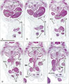
Fig. 2
A degenerating left umbilical vein (LUV) joining the right umbilical vein (RUV) near the liver in a specimen of crown-rump length 13 mm. Panels A and F represent the most superior and inferior sites, respectively, in the figure. Intervals between panels are 0.1 mm (A–B), 0.05 mm (B–C, C–D, D–E), and 0.1 mm (E–F). The RUV is thicker than the LUV; the latter is degenerating and disappears in planes almost 1.0 mm below panel F. Following fusion of these bilateral umbilical veins (C), a common thick vein drains into the umbilical portion of the left liver (UP in panel A). As the configuration of intrahepatic portal veins is normal, the gallbladder (GB) is located in the right side of the umbilical portion. CA, celiac artery; CBD, common bile duct; CL, caudate lobe of the liver; P2, P5+8 or P6+7, segmental portal vein to Couinaud's segment 2, 5+8 (i.e., anterior sector) or 6+7 (i.e., posterior sector). Scale bar=1 mm.

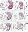
Fig. 3
A degenerating right umbilical vein (RUV) joining the left umbilical vein (LUV) near the liver in a specimen of crown-rump length 14 mm. Panels A and F represent the most superior and inferior sites, respectively, in the figure. The intervals between panels are 0.1 mm (A–B, B–C), 0.05 mm (C–D), 0.1 mm (D–E) and 0.2 mm (E–F). The RUV is degenerating and restricted in planes of almost 0.35 mm thickness (A–E). However, the right vein joins the LUV near the liver (B). A configuration of intrahepatic portal veins is normal. P2 or P3, segmental portal vein to Couinaud's segment 2 or 3; SMA, superior mesenteric artery; UP, umbilical portion of the left liver; VV, vitelline vein. Scale bar=1 mm.
Acknowledgements
This research was supported by Basic Science Research Program through the National Research Foundation of Korea (NRF) funded by the Ministry of Education (grant number: 2016R1D1AB03935784).
All specimens were part of the large collection kept at the Embryology Institute of the Universidad Complutense, Madrid, Spain. The study protocol was approved by the Ethics Committee of University (B08/374).
References
1. Canavan TP, Hill LM. Neonatal outcomes in fetuses with a persistent intrahepatic right umbilical vein. J Ultrasound Med. 2016; 35:2237–2241.

2. Kirsch CF, Feldstein VA, Goldstein RB, Filly RA. Persistent intrahepatic right umbilical vein: a prenatal sonographic series without significant anomalies. J Ultrasound Med. 1996; 15:371–374.
3. Yagel S, Cohen SM, Valsky DV, Shen O, Lipschuetz M, Messing B. Systematic examination of the fetal abdominal precordial veins: a cohort study. Ultrasound Obstet Gynecol. 2015; 45:578–583.
4. Shindoh J, Akahane M, Satou S, Aoki T, Beck Y, Hasegawa K, Sugawara Y, Ohtomo K, Kokudo N. Vascular architecture in anomalous right-sided ligamentum teres: three-dimensional analyses in 35 patients. HPB (Oxford). 2012; 14:32–41.
5. Ricklan DE, Collett TA, Lyness SK. Umbilical vein variations: review of the literature and a case report of a persistent right umbilical vein. Teratology. 1988; 37:95–100.
6. Gupta R, Miyazaki A, Cho A, Ryu M. Portal vein branching pattern in anomalous right-sided round ligament. Abdom Imaging. 2010; 35:332–336.
7. Kawai K, Koizumi M, Honma S, Tokiyoshi A, Kodama K. Right ligamentum teres joining to the right branch of the portal vein. Anat Sci Int. 2008; 83:49–54.
8. Martin BF, Tudor RG. The umbilical and paraumbilical veins of man. J Anat. 1980; 130(Pt 2):305–322.
9. Lin G, Lunderquist A, Hägerstrand I. Umbilical and paraumbilical veins in ligamentum teres. Their significance as collaterals in portal hypertension. Acta Radiol Diagn (Stockh). 1984; 25:1–5.
10. Ibukuro K, Tanaka R, Fukuda H, Abe S, Tobe K. The superior group of vessels in the falciform ligament: anatomical and radiological correlation. Surg Radiol Anat. 2008; 30:311–315.


 PDF
PDF ePub
ePub Citation
 Print
Print


 XML Download