

 PDF
PDF ePub
ePub Citation
 Print
Print


Introduction
Gallbladder is a pear-shaped organ that belongs to extrahepatic biliary apparatus. It is situated under the right lobe of the liver. It is involved in the storage, concentration, and ejection of the bile. Some of the reported variations of the gallbladder include its agenesis, duplication, the presence of phrygean cap, floating gallbladder with a mesentery and intrahepatic gallbladder. These variations have been reported from cadaveric dissections, autopsies, or surgical procedures. Most of the variations of gallbladder either hinder the biliary flow or predispose it to cholecystitis or cholelithiasis. These variations may also pose challenges to the diagnosing radiologist and operating surgeons. We report here, a unique variation of double pouched gallbladder that can lead to diagnostic dilemmas. A similar case has not been reported yet in the literature.
Case Report
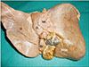
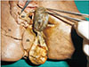
During dissection classes for medical undergraduates, we observed a double pouched sigmoid gallbladder in a male cadaver aged approximately 60–65 years. The gallbladder had two pouches; anterior and posterior. The anterior pouch was covered by peritoneum, but the posterior pouch was just covered by extrahepatic connective tissue. There was a narrow isthmus between the two pouches. At the constriction, the diameter of the gallbladder was 1 cm. It was folded at the constriction two times, thus getting a sigmoid shape. The posterior pouch was situated in a posterior extension of the fossa for gallbladder. When unfolded, it measured 13 cm. The cystic duct was short and straight and measured 2 cm in length. Due to the folding and pouching of the gallbladder, the quadrate lobe was narrow, and the porta hepatis was very congested. The interior of the bladder showed normal folds of the mucosa. There was no evidence of any inflammation or cholelithiasis. The variations are shown in Figs. 1 and 2.
Discussion
Gallbladder develops from the foregut through the hepatic diverticulum. Many of its congenital anomalies are well known. In rare cases, the gallbladder undergoes atresia. It may be partially or completely duplicated [12], or may be septate [3] or may possess a phrygean cap [4]. In rare cases, the gallbladder may be suprahepatic in position [5]. ‘Left sided gallbladder’ is another congenital anomaly of the gallbladder where the bladder is situated on the left side of the ligamentum teres [6]. In a condition called ‘floating gallbladder’, the gallbladder is suspended by mesentery and is free to move [7]. Most of these congenital anomalies may remain asymptomatic for some time and may be incidental findings. However, they may predispose the bladder for cholecystitis and cholelithiasis. They may also cause hindrance in the free flow of bile from in and out of the gallbladder.
The current case that we are presenting differed from all the previously presented congenital anomalies of gallbladder. The present case is not of duplication but a single gallbladder with two pouches. This type of variation has not been published before. Additionally, in all previous reported cases there were two cavities in gallbladder with a septum in between but in the present case the gallbladder did not have septum, just one cavity which was constricted in the middle. We presume that this case is also a congenital anomaly as there were no obvious signs of pathology in the walls or at the constriction. Also, there were no gall stones in the lumen. The constriction found in the gallbladder would have appeared early during development of the cystic bud from the original hepatic diverticulum. The two pouches were located in two separate parts of gallbladder fossa and there was associated narrowing of the quadrate lobe.
Meticulous dissection of the callot's triangle or tubular structures in the subhepatic region, identification of anatomical details prior to surgery along with a high degree of awareness of gallbladder anomalies could possibly prevent catastrophic consequences [8]. Complete pre-operative evaluation of anatomy with special attention to the biliary ductal anatomy is must to avoid potential damage to the ductal system. This anatomical variation might lead to diagnostic dilemmas to the radiologists and might pose difficulties during the laparoscopic cholecystectomy. Hence, laparoscopic cholecystectomy with intraoperative cholangiography is the appropriate treatment in such conditions.
 XML Download