Dear Editor,
Internuclear ophthalmoplegia (INO) results in the slowing, limitation, or inability of adduction of one eye, which is associated with nystagmus in the abducting eye and is caused by a lesion in the medial longitudinal fasciculus.1
A 39-year-old woman presented with sudden-onset vertigo, diplopia, and bilateral INO. Five years earlier she had developed subacute left-body numbness. Brain MRI at that time showed several small punctate T2-weighted hyperintensities in the periventricular and subcortical white matter, which were probably related to clinically and serologically suspected neurocysticercosis. She was treated with albendazole and corticosteroids. She developed symptomatic bradycardia during that hospitalization and a pacemaker was implanted. Four years prior to her current presentation she was admitted to our hospital with a 1-month history of new-onset headache, and was found to have a subarachnoid hemorrhage. Vascular imaging identified an intracranial dural arteriovenous fistula (DAVF) at the right sigmoid and transverse sinus junction. The DAVF was treated endovascularly and surgically.
On examination she had impaired adduction of both eyes on lateral gaze, with horizontal nystagmus of the abducting eye and intact convergence, which was compatible with bilateral INO. She also had mild ataxia on the left side in both finger-nose-finger and heel-to-sheen testing, as well as difficulty performing tandem gait. The findings of neurological and general physical examinations were otherwise unremarkable. A vascular etiology was suspected due to the acuity of onset and her prior history of a DAVF after the endovascular and surgical interventions. The initial noncontrast head CT produced unremarkable findings. A digital subtraction cerebral angiogram showed occlusion of the right transverse and sigmoid sinuses and no new intracranial vascular lesion.
Stroke, multiple sclerosis (MS), and infectious etiologies are the most common causes of bilateral INO.23 Brain MRI is the best tool for differentiating between ischemic stroke and MS, but the prior implantation of a non-MRI conditional pacemaker precluded MRI from initially being performed in our patient. An acute brainstem stroke manifests as areas of restricted diffusion in the arterial territory or a lacunar pattern. On the other hand, patients with MS usually have T2-weighted hyperintense (and possibly gadolinium-enhanced) lesions in the subcortical white matter, periventricular white matter, brainstem, and spinal cord. Cysticercosis usually presents in MRI as cystic or calcified lesions in the cortex or brainstem. Other diagnostic modalities that can aid the diagnosis of MS (e.g., CSF analysis for oligoclonal bands, visual evoked potentials, and somatosensory evoked potentials) are not as sensitive or specific as MRI. Nevertheless, we found that the opening pressure, cell counts, protein level, glucose level, veneral disease research laboratory results, and cultures were all within normal limits for the CSF of the present patient.
MRI is often contraindicated for patients with standard pacemakers. The radiofrequency, static magnetic, and gradient magnetic fields can potentially cause movement of the device, fracture or dislodgment of the leads, lead tip heating, current induction, oversensing, undersensing, or permanent device damage.4 Most MRI protocols state that the information obtained from a scan needs to be important, the pacemaker should be interrogated before and after the scan, the scan time should be minimized, and personnel trained in advance cardiac life support be present during the scan.4 Using an MRI device with magnets stronger than 1.5 Tesla (T) is not recommended. Strict adherence to basic screening and scan protocols is also required.567 The recommended safety protocol includes considering alternative imaging modalities, the timing of lead placement, the presence of abandoned or fractured leads, device interrogation, pacemaker dependency of the patient, presence of a defibrillator, and post-MRI interrogation and device reprograming.5
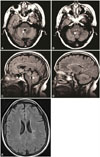
The cardiac electrophysiology and radiology departments were consulted to help with performing brain MRI. The pacemaker was interrogated, and it was determined that the patient was not pacemaker-dependent. After obtaining informed consent from the patient, she underwent brain MRI with contrast agent in a device with a 1.5-T magnet, which revealed a demyelinating lesion in the posterior pons and other lesions compatible with a diagnosis of MS (Fig. 1).
The CSF oligoclonal bands of the patient became positive after discharge, and applying the 2017 McDonald criteria led to a diagnosis of relapsing-remitting MS.8 She had three different unrelated conditions affecting the central nervous system: DAVF, neurocysticercosis, and MS. Many neurologists assume that having a cardiac pacemaker is an absolute contraindication to performing MRI. Here we demonstrate that MRI can be performed in selected patients with cardiac pacemakers when the information obtained from the MRI could change the clinical management.
Figures and Tables
Fig. 1
Brain MRI of our patient with a pacemaker who presented with bilateral internuclear ophthalmoplegia. FLAIR sequences are shown. Arrows indicate lesion. A and B: Consecutive axial views through the pons revealing bilateral T2 hyperintensities within the posterior pons that involve both medial longitudinal fasciculi. C: Sagittal view through the brainstem. The demyelinating lesion extends approximately 7 mm. D: Sagittal view demonstrating multiple periventricular T2 hyperintensities consistent with demyelination. E: Axial FLAIR image showing several periventricular lesions.

References
1. Karatas M. Internuclear and supranuclear disorders of eye movements: clinical features and causes. Eur J Neurol. 2009; 16:1265–1277.

2. Bolaños I, Lozano D, Cantú C. Internuclear ophthalmoplegia: causes and long-term follow-up in 65 patients. Acta Neurol Scand. 2004; 110:161–165.

3. Keane JR. Internuclear ophthalmoplegia: unusual causes in 114 of 410 patients. Arch Neurol. 2005; 62:714–717.
4. Cronin EM, Mahon N, Wilkoff BL. MRI in patients with cardiac implantable electronic devices. Expert Rev Med Devices. 2012; 9:139–146.

5. Korutz AW, Obajuluwa A, Lester MS, McComb EN, Hijaz TA, Collins JD, et al. Pacemakers in MRI for the neuroradiologist. AJNR Am J Neuroradiol. 2017; 38:2222–2230.

6. Padmanabhan D, Jondal ML, Hodge DO, Mehta RA, Acker NG, Dalzell CM, et al. Mortality after magnetic resonance imaging of the brain in patients with cardiovascular implantable devices. Circ Arrhythm Electrophysiol. 2018; 11:e005480.


 PDF
PDF ePub
ePub Citation
 Print
Print


 XML Download