

 PDF
PDF ePub
ePub Citation
 Print
Print


INTRODUCTION
Chronic periodontitis (CP) is a common disease worldwide that has a bacterial etiology and is characterized by an inflammatory process, resulting in the destruction of the soft and hard tissues that support the teeth. The severity of the disease process can be altered by a variety of factors. Recently, the role of reactive oxygen species (ROS) has been established in the pathogenesis of periodontitis. Despite providing an important function in normal metabolic reactions, ROS are highly toxic and destructive in nature. Phagocytic cells, predominantly polymorphonuclear leucocytes (PMNLs), are their potential source. It has been suggested that as a result of stimulation by bacterial antigens, PMNs produce and release a large quantity of ROS, culminating in heightened oxidative damage to gingival tissue, periodontal ligament, and alveolar bone [1]. ROS are active in the depolymerization of extracellular matrix components, lipid peroxidation, oxidation of enzymes such as antiproteases, induction of proinflammatory cytokines, and DNA damage [2,3].
All organisms possess a range of enzymatic and nonenzymatic antioxidant (AO) systems, which are a biological counterfoil to ROS-mediated harmful oxidative reactions. One key AO enzyme implicated in the regulation of ROS-mediated tissue damage is superoxide dismutase (SOD). It removes damaging ROS by catalyzing the dismutation of two superoxide radicals to hydrogen peroxide and oxygen [4,5] and can be detected in extra- and intracellular compartments. The SOD family includes cytosolic Cu, Zn-SOD, mitochondrial Mn-SOD, and extracellular Cu, Zn-SOD (EC-SOD). EC-SOD shows some sequence homology to the cytosolic Cu, Zn-SOD but has a glycosylated structure. It is found in the extracellular matrix of tissues and is ideally suited to prevent cell and tissue damage initiated by extracellularly produced ROS [6].
Oxidative stress has been implicated in the pathogenesis of many systemic diseases, which include rheumatoid arthritis, chronic obstructive pulmonary disease, acquired immune deficiency syndrome, and atherosclerosis [7,8,9,10]. Iron deficiency anemia (IDA), the most common nutritional deficiency worldwide, is also associated with enhanced oxidative stress. The deficiency of iron causes tissue hypoxia and affects the production of iron-containing AO proteins, which tilts the balance to the oxidative side [11]. In an anemic state, a relative decrease in oxygen into the tissues has been suggested to act as a modifying factor in the response of the periodontium to local irritation [12]. A number of environmental, physical, and psychosocial factors have the potential to alter the periodontal tissues and host immune response, resulting in relatively severe periodontal disease expression. It is important to appreciate that these disorders and conditions do not initiate periodontitis, but they may predispose, accelerate, or increase its progression. Moreover, periodontal diagnosis aims to identify the high-risk groups for a destructive periodontal disease. While our understanding of risk factors associated with periodontitis has expanded, the identification of groups and individuals at risk for periodontal disease progression still represents one of the greatest challenges in the management of periodontal patients.
The aims of this study were twofolds: (1) to clinically investigate the extent to which the presence of IDA affects the severity of CP and (2) to compare the local (saliva) and systemic (serum) SOD activity in patients with IDA, CP, and IDA and CP with that of healthy individuals.
MATERIALS AND METHODS
Study groups
The study was performed as a joint collaboration between the Department of Periodontics and Oral Implantology of the Post Graduate Institute of Dental Sciences, Rohtak, and the Department of Medicine of the Post Graduate Institute of Medical Sciences, Rohtak. The duration of study was from July 2011 to October 2012. The study was conducted in agreement with the principles embodied in the 1964 Declaration of Helsinki, as revised in 2008, and was approved by the Institutional Review Board (Department of Periodontics and Oral Implantology, Pandit Bhagwat Dayal Sharma University of Health Sciences, Rohtak).
Out of the 92 IDA patients referred from the Department of Medicine, 23 patients in the iron deficiency anemic periodontally healthy (IDA-PH) group and 22 patients in the iron deficiency anemia with periodontitis (IDA-CP) group were found to be eligible on the basis of the inclusion and exclusion criteria. Three eligible patients of the IDA-PH group and two patients of the IDA-CP group refused to participate; therefore, 20 patients each from the IDA-PH group and the IDA-CP group were enrolled in the study. 22 periodontally and systemically healthy controls and 20 patients with chronic periodontitis were selected from the Outpatient Department of Periodontics and Oral Implantology.
The study included 82 female patients, consisting of 22 periodontally and systemically healthy individuals (control group, CG) (mean±standard deviation [SD], 33.13±6.38 years), 20 IDA-PH patients (mean±SD, 33.60±3.08 years), 20 CP patients (mean±SD, 35.90±4.14 years), and 20 IDA-CP patients (mean±SD, 34.45± 6.57 years).
The CG participants had no evidence of interproximal attachment loss, no probing pocket depth (PPD) of ≥3 mm at any sites on any teeth, and whole-mouth bleeding scores of ≤10% [13]. The patients with chronic periodontitis had at least two or more interproximal sites with an attachment loss of ≥4 mm, or two or more interproximal sites with PPDs of ≥5 mm, not on the same tooth [14]. The description criteria for IDA were hemoglobin<12 g/dL, serum iron<30 mg/mL, serum ferritin<15 ng/mL, total iron binding capacity>400, and red blood cell (RBC) morphology microcytic/hypochromic [15]. It was ensured that the total number of teeth in the mouth was ≥20.
The exclusion criteria included a course of nonsteroidal anti-inflammatory drugs or antimicrobial drugs within a 3-month period before the commencement of study, pregnancy or lactating mothers, use of mouthwashes or vitamin supplements within the previous 3 months, and patients with other medical conditions that could affect the results, like diabetes mellitus, hypertension, rheumatoid arthritis, and chronic lung diseases. All participants had a negative history of current or previous smoking or recreational drug use and special dietary requirements.
In order to ensure investigator blinding, patients were recruited into each of the four groups by one investigator (S.T.), while the oral examination was carried out by another investigator (S.C.). Prior written informed consent was taken from each patient after explaining the procedure along with the risks and benefits in their own language. After enrollment, all patients were reappointed for the collection of baseline saliva and venous blood samples before recording clinical measurements.
Clinical measurements
The periodontal status of all individuals was detected by the measurement of PPD, clinical attachment level (CAL), gingival index (GI) [16], sites with bleeding on probing (BOP), and plaque index (PI) [17]. PPD, CAL, and BOP% were measured on six sites of teeth (mesial, median, and distal points at buccal and palatal aspects) with a calibrated periodontal probe (UNC-15, Hu-Friedy, Chicago, IL, USA). The bleeding sites were registered in a dichotomous way, and the scores were expressed as the percentage of positive sites per subject (BOP %). A clinical periodontal examination was carried out by the same trained examiner (S.C.) to preclude interexaminer variability. The examiner reproducibility was determined by carrying out double clinical periodontal data recording on ten patients. Reproducibility of the data collection was determined for each site by calculating the percentage of the sites examined where the scores were matching or within ±1 mm. An assessment of the mean difference in the scores (with 90% accuracy) indicated that there was no systematic bias in the measurements.
Collection of samples
All the samples, saliva, and blood at the baseline were obtained in the morning following an overnight fast. The subjects were asked not to drink (except water) or chew gum for the same period, and abstention was checked prior to biological sample collection. Unstimulated whole saliva samples were used in this study. Seated patients were instructed to allow saliva to pool at the bottom of the mouth and passively flow into disposable, sterile, and clean tubes in an area away from the main clinic. Two milliliters of whole saliva was collected and centrifuged immediately to remove cell debris (1,000×g for 10 minutes at 4℃). The supernatant was removed and stored in small aliquots in a deep freezer at -80℃ until analysis.
To avoid circadian rhythm changes, early morning venous blood samples were obtained from each patient for biochemical and hematologic screening tests. Venous blood was collected in vacutainer tubes without an additive and was centrifuged at 3,500×g for 5 minutes to obtain the serum. Serum aliquots were stored in a deep freezer at -80℃ until analysis.
Laboratory assessment
The SOD activity was evaluated using an SOD Assay Kit (Sigma Aldrich, St. Louis, MO, USA) according to the manufacturer's instructions, and an enzyme-linked immunosorbent assay (ELISA) reader (Robonik India Private Limited, Maharashtra, India) at 450 nm. The SOD activity was obtained from the manufacturer's formula using the values from ELISA. The unit of measurement for the SOD activity was percentage.
Statistical analysis
All statistical analyses were carried out using SPSS ver.17.0 (SPSS Inc., Chicago, IL, USA) with a two-tailed P-value of 0.05 used as a threshold for significance. A minimum sample size of 15 per group was required for the detection of a significant difference in BOP% with 80% power and a two-sided 0.05 level of significance [18]. The normality of the data distribution was examined using the Kolmogorov-Smirnov test. Data were found to be nonnormally distributed. A Kruskal-Wallis test was used for a comparison of groups, and post hoc tests were performed using the Mann-Whitney U test with a Bonferroni correction. To avoid spurious significance among multiple inferences (type-1 error), the Bonferroni adjustment was used to interpret the significance of the P-values. Therefore, P-values <0.008 were regarded as statistically significant test results. The statistical significance of correlations among clinical and biochemical variables was determined using the Spearman rank correlation coefficients.
RESULTS
Clinical findings
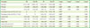
Demographic data and mean±SD of periodontal parameters and biochemical parameters are presented in Table 1. There was no significant difference between the mean ages of the individuals in the four groups (P>0.0008). A comparison among all four groups showed a significant difference in periodontal parameters and biochemical parameters (P>0.008). The clinical periodontal parameter scores (PI, GI, BOP, PPD, and CAL) were statistically higher in groups of patients with CP and IDA-CP than in the periodontally healthy individuals (CG and IDA-PH) (P<0.008).
Further, Table 1 demonstrated that the IDA-CP patients exhibited significantly higher GI, BOP%, PPD, and percentage of sites with CAL of ≥6 mm than CP patients (P<0.008), in spite of the same plaque score for both the groups (P>0.008).
Laboratory findings
Saliva findings
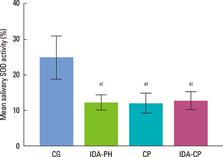
Salivary SOD activity was significantly lower in the IDA-PH, CP, and IDA-CP patients than in the CG group (P<0.008). No statistically significant difference could be detected among the IDA-PH, CP, and IDA-CP groups with respect to the salivary SOD activity (P>0.008) (Fig. 1).
Serum findings
The serum SOD activity of the CG was higher than that of the IDA-PH, CP, and IDA-CP groups (P<0.008). In addition, no difference could be found in the serum SOD activity among the IDA-PH, CP, and IDA-CP groups (P>0.008) (Fig. 2).
Correlations of SOD activity and clinical periodontal parameters
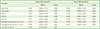
Spearman's rank correlation coefficients among salivary, serum SOD activity, and clinical periodontal parameters in iron deficiency anemia patients (pool data of the CG, IDA-PH, and IDA-CP groups)
The data of Table 2 reveal a significant positive correlation between the salivary and the serum SOD activity (P≤0.05). Salivary SOD activity was found to be negatively and significantly correlated with all periodontal parameters (P≤0.05) except CAL and the percentage of sites with CAL of 4-5 mm (P≥0.05). Serum SOD activity was found to be negatively and significantly correlated with all periodontal parameters including the percentage of sites with CAL of 4-5 and ≥6 mm (P≤0.05).
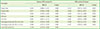
Spearman's rank correlation coefficients among salivary, serum SOD activity, and clinical periodontal parameters in chronic periodontitis patients (pool data of the CG and CP groups)
Table 3 shows that no correlation was found between the salivary and the serum SOD activity (P≥0.05). The salivary SOD activity was found to be negatively and significantly correlated with all the periodontal parameters including the percentage of sites with CAL of 4-5 and ≥6 mm (P≤0.05), whereas the serum SOD activity showed a significant and negative correlation with all periodontal parameters (P≤0.05) except PPD (P≥0.05).
DISCUSSION
Considerable activity of reactive oxygen radicals may lead to the destruction of normal cell functions and the integrity of cell structures. Oxidative stress in biological systems can be induced by the consumption of AOs and/or by an overload of oxidant species, such that AO levels become deficient. Oxidative stress has been determined by the estimation of the products of oxidative damage to lipids, proteins, and DNA or by assaying a single AO compound in isolation or in groups, or to measure the total AO capacity. By detecting a particular AO enzyme activity, we evaluated its importance to a given pathogenic process, for example, the detection of SOD enzyme activity in our study, which represents an important AO defense against excess ROS production in both CP and IDA.
Perusal of the available literature reveals only a single study wherein IDA and CP have both been taken into consideration [19]. To the best of our knowledge, this is the first reported study that evaluated the relationship between chronic periodontitis and iron deficiency anemia with respect to salivary and serum SOD activity levels.
Thus far, numerous studies have been conducted on the association of the serum and salivary SOD activity levels with periodontitis, and the results are equivocal. Kim et al. [20] reported a decrease in the salivary SOD activity in chronic periodontitis patients. Studies by Baltacioglu et al. [21], Canakci et al. [22], and Akalin et al. [23] have also endorsed the decreased SOD activity in periodontitis. Conversely, Wei et al. [24] demonstrated a higher activity of SOD in the saliva, serum, and gingival crevicular fluid of chronic periodontitis patients. In our study, the serum and salivary SOD activity levels in the CP patients were significantly lower than those of systemically and periodontally healthy individuals. Lower salivary SOD activity could be interpreted as a result of the suppression of SOD production in saliva/PMNLs because of oxidative damage caused by increased ROS/O2- generation [25], while a decrease in the serum SOD activity can be attributed to hyperreactive peripheral neutrophils with respect to the production of ROS in response to Fc gamma-receptor stimulation in patients with chronic periodontitis [26].
A significantly lower value of both salivary and serum SOD activity was also observed in IDA-PH patients than in the CG at baseline. Studies by Amirkhizi et al. [27], Isler et al. [28], and Kurtoglu et al. [29] have also endorsed the decreased activity of SOD in patients with IDA. In contrast, studies by Acharya et al. [30] and Hafez et al. [31] found increased erythrocytic Cu-Zn SOD activity in IDA patients. In the present study, the lower SOD activity in anemic patients may be linked to increased hemoglobin auto-oxidation owing to hypoxia resulting in the increased RBC production of superoxide and hydrogen peroxide. These RBC-derived ROS can damage the cell membrane and leak out of the RBC [32]. It is well known that ROS, particularly hydrogen peroxide, inhibit SOD activity [27].
Although there was no significant difference in the salivary and serum SOD activity in the IDA-CP patients and the CP patients, significantly higher baseline PPD, GI, and BOP% were observed in the IDA-CP group than in the CP group in spite of similar PI values. Moreover, the present study showed that the IDA-CP patients had worse periodontal conditions, expressed as the percentage of sites with a CAL of ≥6 mm, than the CP group. Higher periodontal destruction in the IDA-CP group can be attributed to the synergistic effect of both ROS-induced oxidative stress and other mechanisms augmenting inflammation owing to iron deficiency anemia. ROS can directly cause periodontal tissue damage by the degradation of the extracellular matrix components of periodontal tissues or can play an indirect role in potentiating extracellular matrix degradation by matrix metalloproteinase, via the activation of latent enzymes, such as collagenase and gelatinase [33], and via the inactivation of enzyme inhibitors, such as tissue inhibitor of metalloproteinase and α 1-antiproteinase [34]. In addition, the production of ROS and the subsequent disturbance in the tissue redox status can modulate the expression of a variety of immune and inflammatory molecules via redox-sensitive transcription factors (e.g., nuclear factor κB), thereby causing indirect tissue damage and exacerbating inflammation [35].
Furthermore, IDA has been reported to augment inflammation through a number of mechanisms. Hypersegmented neutrophils found in IDA can cause immune suppression by directly suppressing T-cell responses by the creation of an immunological synapse and the direct delivery of hydrogen peroxide resulting in tissue damage. Inhibition of the activity of iron-dependent myeloperoxidase, which mediates the bactericidal activity of macrophages [36], may be another associated mechanism. Moreover, alteration in the gingival blood flow due to the reduced erythrocyte count might decrease the oxygen content in the gingival tissues [12]. Hypoxia produced as a result, induced the activation of the transcription factor hypoxia-inducible factor-1, triggering the expression of proinflammatory genes [37] and increased production of mitochondrial ROS, which activate the endothelial secretion of leukocyte adhesion receptors, thereby promoting the inflammatory response [38].
We also analyzed the relationship between the serum and the salivary SOD activities in iron deficiency anemia patients and found a significant positive correlation, while the CP group did not demonstrate any correlation between the salivary and the serum SOD activity. However, a comparison of the test groups (IDA-PH, CP, and IDA-CP) did not show a significant difference in the salivary and serum SOD activity.
The study was carried out with female patients because IDA in males is rare and is generally related to neoplastic diseases [39]. This was the limitation of our study, as we could not evaluate the effects in men.
Being a preliminary study, this study led to the following major conclusions: (1) Higher periodontal inflammation and severe destruction was observed in the IDA-CP patients than in the CP group. (2) The serum and salivary SOD activities in all test groups were lower than those in the CG. (3) A significant positive correlation between the salivary and the serum SOD activity was observed in the iron deficiency anemia group. Furthermore, the serum SOD activity was significantly and negatively correlated with all periodontal parameters as well as the percentage of sites with CAL of 4-5 mm and ≥6 mm, while salivary SOD did not show significant correlation with CAL and the percentage of sites with CAL of 4-5 mm. (4) In the CP patients, no correlation was observed between the serum and the salivary SOD activity. The salivary SOD activity was significantly and negatively correlated with all periodontal parameters as well as the percentage of sites with CAL of 4-5 mm and ≥6 mm, while the serum SOD activity was significantly and negatively correlated with PI, GI, and mean CAL as well as the percentage of sites having CAL of 4-5 and ≥6 mm. (5) IDA influences the serum SOD activity to a greater extent but does not appear to affect the salivary SOD activity in patients with both IDA and CP than in patients having only CP.
The results of our study indicate that IDA patients may be at increased risk of periodontal destruction and have lower SOD defense levels. However, studies of larger groups with an interventional nature will be required to gain a clearer understanding of the relationship between these two disease entities.
 XML Download