Abstract
Arthroscopy has become an attractive modality in the diagnosis and treatment of joint diseases in toy breed dogs. However, the application of arthroscopy is limited by small joint space. Our objective was to evaluate the efficacy of a stifle lever for joint distraction during stifle arthroscopy in toy breed dogs. Paired stifles (n = 32 each) collected from 16 cadavers of toy breed dogs were randomly assigned to one of two groups: the stifle lever group or the external manipulation group. All stifles underwent arthroscopic cranial cruciate ligament transection, and the visualization of the medial meniscus was evaluated. Medial meniscal release (MMR) was then performed. Following arthroscopic examination, the success rates of MMR and damages of tibial and femoral cartilages were evaluated. Visualization of the medial meniscus was significantly better, and meniscal probing was significantly easier, in the stifle lever group than in the external manipulation group (p = 0.001). There were no significant differences between groups for MMR success or articular cartilage damage. Using the stifle lever on arthroscopic examination improved visualization and probing on the medial meniscus in toy breed dogs. The stifle lever can be used as a good modality in assessing medial meniscal pathology in toy breed dogs.
Cranial cruciate ligament (CrCL) rupture is a major cause of orthopedic diseases leading to lameness and stifle osteoarthritis in dogs [6]. It results in cranial translation of the tibia. Tearing of the medial meniscus most commonly occurs secondary to tibial translation because of the impingement of the medial meniscus between the femoral condyle and the tibia condyle during weight-bearing activity. The incidence of a meniscal tear with CrCL rupture in dogs has been reported to be as high as approximately 52% to 78%; tears at the caudal horn of the medial meniscus occur commonly [8912]. Accurate evaluation of the medial meniscus is important because a meniscal tear can lead to pain, lameness, and osteoarthritis and may necessitate a second surgery.
Arthrotomy and arthroscopy have been most commonly used for the diagnosis and treatment of CrCL rupture and meniscal tear in veterinary orthopedics [11011]. Although arthrotomy is a relatively simple approach, it does not provide adequate visualization of the caudal horn of the medial meniscus and can also cause pain and lameness [3]. Arthroscopy is a minimally invasive approach with low morbidity and provides better visualization of and accessibility to intra-articular structures through illumination and magnification. Thus, arthroscopy has higher sensitivity and specificity than arthrotomy for the assessment of the caudal horn of the medial meniscus and the detection of medial meniscal injury [1911].
However, arthroscopy with probing may be difficult in dogs, specifically toy breed dogs, due to the small joint space. Joint distraction techniques, such as external manipulation of the joint, the use of an intra-articular stifle distractor, and the use of an externally applied joint distractor, have been described to increase joint space and lead to good visualization of and accessibility to a meniscal tear during arthroscopy [5101314].
To the authors' knowledge, the use of a stifle lever for intra-articular joint distraction during stifle arthroscopy in toy breed dogs has not been reported. The only previous study focused on stifle arthroscopy in toy breed dogs investigated the use of an external joint distractor for medial meniscal release (MMR) in toy breed dogs [7]. The use of an external joint distractor for stifle arthroscopy has some disadvantages, such as the possibility of incorrect pin position or interference with the tibial plateau leveling osteotomy plate, especially in toy breed dogs [27].
The objective of this study was to evaluate the efficacy of a stifle lever for joint distraction during stifle arthroscopy in toy breed dogs. We hypothesized that visualization and accessibility of the caudal horn of the medial meniscus would increase with the use of a stifle lever and that arthroscopic MMR with a stifle lever would result in a high success rate with less iatrogenic articular damage than that from arthroscopic MMR with external manipulation.
Thirty-two stifle joints from 16 toy breed dogs euthanized for reasons unrelated to this study were obtained. The body weight, body condition score, and breed were recorded for each cadaver. The physical and radiographic examinations of the pelvic limbs revealed no pre-existing orthopedic diseases. The pelvic limbs were clipped, and the cadavers were stored at −20℃ until used for this study.
The paired stifles (n = 32) from the 16 cadavers were assigned to one of two groups using a computer software program (Microsoft Excel 2016; Microsoft, USA): arthroscopy with a stifle lever or arthroscopy with external manipulation.
Cadavers were thawed for 24 h at room temperature. A single surgeon (H.B. Lee) performed all arthroscopic procedures. Surgeon's experiences with arthroscopy included approximately 200 stifle arthroscopies in toy breed dogs. Arthroscopic examination was performed as previously described [47]. Briefly, a cadaver was positioned in dorsal recumbency and secured with a bean bag positioner. A lateral subpatellar stab incision was made lateral to the patellar ligament by using a No. 11 surgical blade. A medial suprapatellar egress portal was established and fluid ingress flow was maintained with a pressurized fluid pump (AR-6475; Arthrex, USA) set at 25 mmHg [4]. A 1.9 mm, 30° fore-oblique arthroscope (Stryker, USA) was placed at the lateral subpatellar portal. The CrCL of the stifles in the external manipulation group was transected by using a meniscus hook knife (Veterinary Instrumentation, UK) through a medial subpatellar instrument portal. A meniscal probe having a 1.5 mm tip (BS Corem, Korea) was used for assessment of the medial meniscus, and visualization of the medial meniscus was scored by using a defined evaluation protocol. Transection of the caudal meniscotibial ligament was performed with a meniscus hook knife. In the stifle lever group, the CrCL was transected in the same manner. Next, an additional lateral subpatellar portal was established, and a Ventura Stifle Thrust Lever (VSTL; IMEX Veterinary, USA) was applied. The joint was distracted with the VSTL (Fig. 1), and the visualization of the medial meniscus was evaluated. Transection of the caudal meniscotibial ligament was performed in the same manner.
During the arthroscopic examination, the time to place the VSTL and the time to complete arthroscopy after CrCL transection were measured in seconds. The visualization, assessed by measurement of the minimal distance between the medial femoral condyle and medial tibial articular cartilage with a meniscal probe having a 1.5 mm tip, was scored by applying the following grading scale: 0 = excellent, greater than 1 times the tip length (> 1.5 mm); 1 = good, 0.5 to 1 times the tip length (0.75–1.5 mm); 2 = fair, 0 to 0.5 times the tip length (0–0.75 mm); and 3 = poor, less than 0 times the tip length (0 mm). The meniscal probing difficulty score was assigned by applying the following grading scale: 0 = easy, 1 = moderate, 2 = difficult, and 3 = not possible (panel A in Fig. 2).
After the arthroscopy was completed, the length of all stifle incisions, including the lateral subpatellar incision, the medial subpatellar incision, and the additional lateral incision for the VSTL, was measured in millimeters; the total incision length in each stifle was then calculated. Next, disarticulation of the stifle was performed. The completeness of the CrCL transection, the damage to the medial collateral ligament, the damage to the caudal cruciate ligament (CdCL), and the damage to the caudal part of the tibial intercondylar area were evaluated as previously described [47]. Briefly, the amount of release of the medial meniscus was evaluated in 5% increments by using computer software (Photoshop CS6; Adobe Systems, USA). After removal of the meniscus, the percentage articular cartilage damage (% ACD) of the femur and tibia was evaluated by using India ink staining and digital image software (ImageJ ver. 1.50; National Institutes of Health, USA). The India ink staining in the articular cartilages images was blindly evaluated (B.S. Jeong) (panels B and C in Fig. 2).
All data analysis was performed using a statistical software program (IBM SPSS Statistics ver. 22.0; IBM, USA). Each outcome measure was expressed as the mean ± SD. The scores for both visualization and difficulty of meniscal probing were compared between groups by using Fisher's exact test. The Fisher's exact test was also used to evaluate significant differences in MMR success between the two groups, and the odds ratio (OR) for the likelihood of MMR success was determined. The effect of body side (left or right) of the stifles on cartilage damage was also evaluated. Based on the normality of the data, either the two-sample t-test (for total incision length) or the Mann-Whitney U test (for percentage transected medial meniscus, femoral % ACD, tibial % ACD, and time required for arthroscopic procedures) was used to compare the groups. A p value < 0.05 was considered statistically significant.
Sixteen canine cadavers included the following breeds: mixed (9 dogs), Poodle (4), Maltese (2), and Shih Tzu (1). Their body weights ranged from 2.8 to 5.1 kg (mean ± SD, 3.84 ± 0.68 kg). The body condition scores ranged from 3 to 6 out of a maximum score of 9 (mean ± SD, 4.44 ± 0.89).
The scores for visualization of the medial meniscus during arthroscopic examination were significantly lower in the stifle lever group than in the external manipulation group (p = 0.001; Table 1). Overall, the degree of visualization was rated from grade 0 to grade 1 in the stifle lever group and from grade 1 to grade 2 in the external manipulation group (Fig. 3). The scores for difficulty of meniscal probing showed the same result as the visualization scores for both groups (p < 0.001).
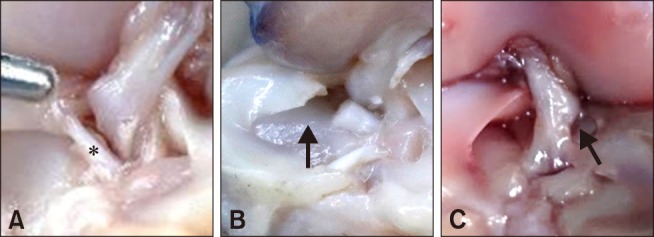
Complete transection of the CrCL was observed in all stifles. The proportion of transected caudal meniscotibial ligaments was 92.5 ± 13.3% in the stifle lever group and was 99.2 ± 2.9% in the external manipulation group (panels A and B in Fig. 4). This difference was not significant (p = 0.272). The MMR was successfully performed in 87% in the stifle lever group and in 93% in the external manipulation group. These results were not significantly different (p = 0.212). The OR results indicated that the use of a VSTL did not affect the complete transection of the caudal meniscotibial ligament (p = 0.179).
Most of the damage to the femoral cartilage was detected at the medial femoral condyle in the area involved in MMR. Most of the damage to the tibial cartilage was also detected adjacent to the caudal meniscotibial ligament.
India ink-staining results revealed no significant difference in either femoral or tibial ACD between the stifle lever group and the external manipulation group.
The mean % ACD for the distal femoral cartilage was 0.57 ± 1.19% in the stifle lever group and 0.77 ± 0.65% in the external manipulation group (p = 0.064). The mean % ACD for the proximal tibial cartilage was 0.22 ± 0.31% in the stifle lever group and 0.26 ± 0.48% in the external manipulation group (p = 0.477). There were no significant differences between the left and right stifles (p = 0.515).
Approximately 8% of the width of the CdCL was compressed by the VSTL in 2 stifles in the stifle lever group (panel C in Fig. 4), but compression did not occur in any stifles in the external manipulation group. No damage to either the posterior intercondylar area or the medial collateral ligament occurred in any of the stifles.
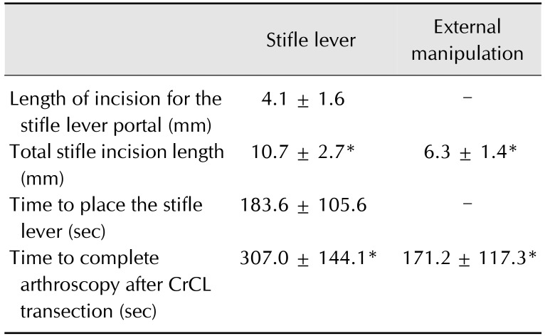
The total incision length in the stifle was significantly greater in the stifle lever group (10.7 ± 2.7 mm) than in the external manipulation group (6.3 ± 1.4 mm; p < 0.001) (Table 2). The mean length of the additional lateral incision to accommodate the stifle lever was 4.1 ± 1.6 mm.
The time spent on placement of the stifle lever was 183.6 ± 105.6 sec. The procedure for stifle lever placement, meniscal probing, and MMR were included in the time to complete arthroscopy after CrCL transection; the stifle lever placement procedure was not performed in the external manipulation group. The time to complete arthroscopy after CrCL transection was significantly longer in the stifle lever group (307 ± 144.1 sec) than in the external manipulation group (171.2 ± 117.3 sec; p = 0.004) (Table 2).
Our study showed that the use of a stifle lever resulted in a significant improvement in visualization of the medial meniscus during stifle arthroscopy compared with that during external manipulation. The surgeon also reported greater convenience in performing arthroscopic procedures such as meniscal probing in the stifle lever group. The improved visibility provided by the stifle lever may aid in the evaluation of the meniscal status and the diagnosis of meniscal injury.
The arthroscopic MMR model was used to evaluate visibility and accessibility during stifle arthroscopy in toy breed dogs and to evaluate the efficacy of a stifle lever in arthroscopic procedures. The MMR success rate was 87% in the stifle lever group and was 93% in the external manipulation group. Despite the improved visualization of the medial meniscus in the stifle lever group, the lower success rate of MMR with a stifle lever likely resulted from instrumental fighting between the stifle lever and the meniscus hook knife when the stifle lever was placed close to the caudal meniscotibial ligament. However, unsuccessful MMR did not occur in the late stages of this study as appropriate placement of the stifle lever had improved.
Damage to both the distal femoral and the proximal tibial cartilage did not differ significantly between the stifle lever group and the external manipulation group. The mean % ACD in the stifle lever and the external manipulation groups were 0.57% and 0.77%, respectively, in the distal femur and were 0.22% and 0.26%, respectively, in the proximal tibia. Most of the cartilage damage to the femoral and tibial condyle was observed at the area of the caudal meniscotibial ligament. These results were similar to those of MMR in toy breed dogs during stifle arthroscopy in which an external joint distractor was used [7]. This cartilage damage could be caused by manipulation of meniscus hook knife. There were no significant differences between groups for MMR success or the damage of articular cartilage in this study.
In this study, we used normal cadaver dogs without CrCL disease. Those with CrCL disease have periarticular fibrosis and synovial proliferation. This situation makes joints difficult to open. If stifle arthroscopy is performed on dogs with CrCL disease, we speculate that the clinical efficiency of the stifle lever may be significantly higher than that from external manipulation. Further clinical studies, however, are needed.
The performance of MMR on the right stifle seemed more convenient for the right-handed surgeon, but no significant difference in cartilage damage was observed between the right-sided stifles and the left-sided stifles.
A compressed CdCL was detected in 2 of the 16 stifles in which a stifle lever was used during stifle arthroscopy. Incidences of compressed CdCL did not occur in the late stages of this study. Thus, the early results may be the results of a lack of familiarity with the placement of the stifle lever. Further studies are needed to assess the clinical significance of a compressed CdCL. No iatrogenic damage to the caudal part of the intercondylar area or to the medial collateral ligament, both of which were evaluated in this study, was observed.
The group in which a stifle lever was used during stifle arthroscopy needed an additional incision for the stifle lever portal and thus had a significantly longer total incision length than the external manipulation group. The mean time to place the stifle lever was approximately 3 minutes (183.6 sec). A significant difference in the total time to complete arthroscopy after CrCL transection was detected between the stifle lever group and the external manipulation group; the time difference was approximately 2 minutes. In some stifles of the stifle lever group, stifle lever placement was attempted repeatedly due to inappropriate placement of the stifle lever with respect to the caudal aspect of the CdCL. Moreover, additional debridement of the fat pad was needed because stifle lever-induced movement of the fat pad disturbed visualization in all stifles of the stifle lever group. The difference in the time to complete arthroscopy may have been associated with the appropriate placement of the stifle lever and the additional debridement of the fat pad. However, if stifle arthroscopy is performed on a CrCL-deficient stifle with osteoarthritis, we speculate that the total time for arthroscopic examination may be longer when external manipulation is used than when a stifle lever is used because of decreased visualization during meniscal probing and increased difficulty of externally manipulating a stifle with periarticular fibrosis. Further clinical studies, however, are needed.
Arthroscopy is very challenging in toy breed dogs due to their small joint spaces. Using the stifle lever on arthroscopic examination can improve visualization and probing of the medial meniscus in toy breed dogs. Thus, it can be a good modality in assessing medial meniscal pathology.
References
1. Beale BS, Hulse DA. Arthroscopy versus arthrotomy for surgical treatment. In : Muir P, editor. Advances in the Canine Cranial Cruciate Ligament. 1st ed. Ames: Wiley-Blackwell;2010. p. 145–158. .
2. Böttcher P, Winkels P, Oechtering G. A novel pin distraction device for arthroscopic assessment of the medial meniscus in dogs. Vet Surg. 2009; 38:595–600. PMID: 19573060.
3. Case JB, Hulse D, Kerwin SC, Peycke LE. Meniscal injury following initial cranial cruciate ligament stabilization surgery in 26 dogs (29 stifles). Vet Comp Orthop Traumatol. 2008; 21:365–367. PMID: 18704244.

4. Cha JG, Lee HB, Cheong HY, Heo SY, Ragetly GR. Evaluation of a Veress needle for the fluid egress system of stifle arthroscopy in toy dog breeds. Vet Comp Orthop Traumatol. 2016; 29:149–155. PMID: 26846402.

5. Gemmill TJ, Farrell M. Evaluation of a joint distractor to facilitate arthroscopy of the canine stifle. Vet Surg. 2009; 38:588–594. PMID: 19573059.

6. Innes JF, Bacon D, Lynch C, Pollard A. Long-term outcome of surgery for dogs with cranial cruciate ligament deficiency. Vet Rec. 2000; 147:325–328. PMID: 11058021.

7. Kim K, Lee H, Ragetly GR. Feasibility of stifle medial meniscal release in toy breed dogs with and without a joint distractor. Vet Surg. 2016; 45:636–641. PMID: 27357273.

8. Luther JK, Cook CR, Cook JL. Meniscal release in cruciate ligament intact stifles causes lameness and medial compartment cartilage pathology in dogs 12 weeks postoperatively. Vet Surg. 2009; 38:520–529. PMID: 19538675.

9. Mahn MM, Cook JL, Cook CR, Balke MT. Arthroscopic verification of ultrasonographic diagnosis of meniscal pathology in dogs. Vet Surg. 2005; 34:318–323. PMID: 16212585.

10. McKee WM, Cook JL. The stifle. In : Houlton JEF, Cook JL, Innes JF, Langley-Hobbs SJ, editors. BSAVA Manual of Canine and Feline Musculoskeletal Disorders. 1st ed. Gloucester: Wiley;2006. p. 350–395.
11. Pozzi A, Hildreth BE 3rd, Rajala-Schultz PJ. Comparison of arthroscopy and arthrotomy for diagnosis of medial meniscal pathology: an ex vivo study. Vet Surg. 2008; 37:749–755. PMID: 19121170.

12. Ralphs SC, Whitney WO. Arthroscopic evaluation of menisci in dogs with cranial cruciate ligament injuries: 100 cases (1999–2000). J Am Vet Med Assoc. 2002; 221:1601–1604. PMID: 12479333.

13. Whitney WO. Arthroscopically assisted surgery of the stifle joint. In : Beale BS, Hulse DA, Schulz KS, Whitney WO, editors. Small Animal Arthroscopy. 1st ed. Philadelphia: Saunders;2003. p. 137–140.
14. Winkels P, Pozzi A, Cook R, Böttcher P. Prospective evaluation of the Leipzig stifle distractor. Vet Surg. 2016; 45:631–635. PMID: 27357272.

Fig. 1
Application of the arthroscopic stifle lever (arrow) to the caudal aspect of the tibia with cranial tibial thrust.
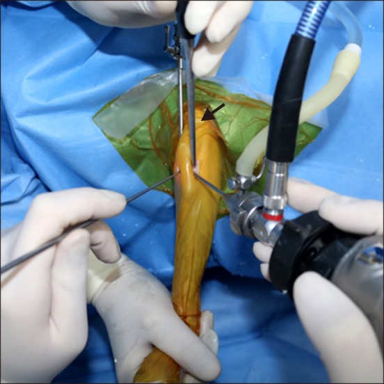
Fig. 2
(A) Arthroscopic view of a stifle documenting the measurement of the distance between the medial femoral condyle (mFC) and the tibial articular cartilage. Visualization was scored by using the tip of the probe. (B and C) Digital imaging and a computer software program were used to trace the entire surface of the articular cartilage, the area of damaged articular cartilage (which was stained with India ink), and to calculate the percentage area of cartilage damage for both the femoral and tibial surfaces. mM, medial meniscus; MP, meniscal probe; pTAC, proximal tibial articular cartilage.
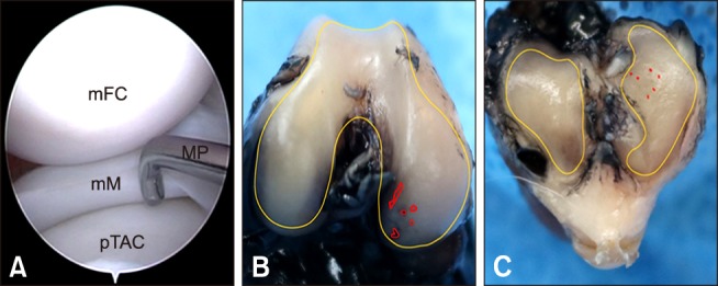
Fig. 3
Arthroscopic view during medial meniscal release (MMR). (A) Visualization of the medial meniscus was better when a stifle lever was used, and the stifle lever protected the caudal cruciate ligament (CdCL) during arthroscopic MMR. (B) Arthroscopic view when MMR was performed using external manipulation. mFC, medial femoral condyle; mM, medial meniscus; pTAC, proximal tibial articular cartilage; MHK, meniscus hook knife.
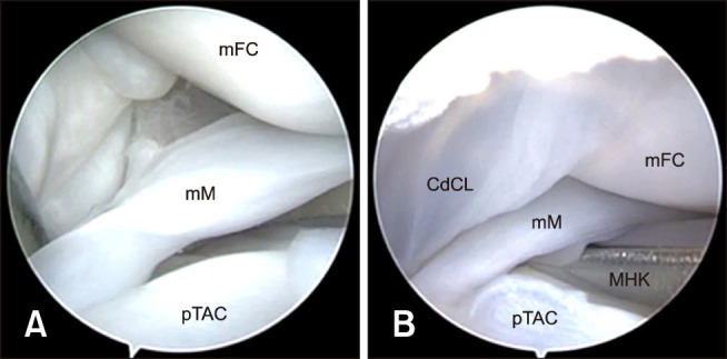
Fig. 4
Gross observations on disarticulation. (A) Incompletely transected caudal meniscotibial ligament in a stifle from the stifle lever group. The remaining caudal meniscotibial ligament is denoted by the asterisk. (B) The arrow points to a completely transected caudal meniscotibial ligament in a stifle from the external manipulation group. (C) A compressed caudal cruciate ligament (arrow) caused by the placement of the stifle lever.


 PDF
PDF ePub
ePub Citation
 Print
Print


 XML Download