

 PDF
PDF ePub
ePub Citation
 Print
Print


Introduction
Variant angina commonly manifests as chest pain and transient ST elevation by coronary spasm, and generally follows a benign clinical course.1) Rarely, syncope induced by ventricular arrhythmia associated with transient myocardial ischemia can be developed by coronary spasm.2)3) If the cause of syncope is not correctly diagnosed, patients are at risk of recurrent syncope.4) In particular, syncope without chest pain is an uncommon manifestation of variant angina. Thus, as in this case, it can be missed as the cause of syncope.
Case
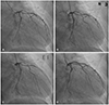
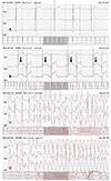
A 68-year-old man visited our emergency room due to recurrent syncope. He had experienced four episodes of syncope with dizziness and chest discomfort during the prior 2 months. The episodes were evoked when he was preparing his boat for sailing in early morning. On admission, his mentality was clear. Vital signs showed blood pressure (BP) of 120/80 mmHg, pulse rate of 60 beats/min, respiratory rate of 20 breaths/min and body temperature of 36.5℃. He had a 10-year history of diabetes mellitus with application of oral hypoglycemic agents. He had stopped smoking 40 years previously. He drank 1 bottle of soju per week. Physical examination, chest radiography and laboratory findings were unremarkable. The initial electrocardiogram (ECG) showed sinus rhythm with T inversion in the V5 and V6 leads. Transthoracic echocardiogram revealed left ventricular hypertrophy, mildly decreased ejection fraction, and hypokinetic left ventricular wall motion. Twenty four hour Holter monitoring showed normal sinus rhythm and infrequent premature ventricular complexes (PVCs). Coronary angiography revealed 70% stenosis at proximal left anterior descending artery (LAD) (Fig. 1A). Stenosis persisted after intracoronary nitrates administration (Fig. 1B). We planned medical treatment instead of coronary angioplasty. The tilt table test and brain magnetic resonance angiography were unremarkable. The patient was discharged and regarded as an unexplained syncope. After 4 days, he was readmitted because of severe dizziness and we performed 48-hour Holter monitoring. During his second admission, syncope occurred for about 3 minutes. Fortunately, he completely recovered without any injury. The Holter monitoring during the syncope episode revealed ST-segment elevation followed by PVCs (Fig. 2B). Polymorphic ventricular tachycardia (VT) started with the PVCs (Fig. 2C) and degenerated into ventricular fibrillation (Fig. 2D). We suspected that the ventricular arrhythmia resulted from transient myocardial ischemia by variant angina, because STsegment elevation was seen before VT and syncope occurred in the morning. A repeat coronary angiography was carried out for a spasm provocation test. Right coronary artery was negative in the provocation test. In contrast, after intracoronary ergonovine injection, nearly total occlusion of LAD developed (Fig. 1C). There was no chest pain and no change of BP or ECG during the provocation test. Coronary vasospasm was rapidly relieved by intracoronary nitroglycerin. We speculate that it may have caused no changes of ECG during the provocation test. Intravascular ultrasonography revealed that minimum lumen area was 2.3 mm2 after intracoronary nitroglycerine injection. We performed percutaneous coronary intervention (PCI) at proximal LAD (Fig. 1D). The patient was discharged with medications including calcium channel blockers. At present, he has been regularly followed in the outpatient clinic without symptoms.
Discussion
Variant angina was first described a specific entity of angina pectoris characterized by angina pain occurring at rest and associated with ST-segment.1) Variant angina generally has a good prognosis; the 10-year survival rate is over 80%. However, variant angina patients with severe coronary artery disease have poor prognosis.4) Rarely, vasospasm-induced ischemia causes life-threatening ventricular arrhythmia, syncope, and sudden cardiac death.5) Syncope associated with VT can be caused by myocardial ischemia due to severe coronary artery stenosis in ischemic heart disease.6) In the present case, since the patient had no chest pain or evidence of ischemic heart disease on routine workup, we presumed that he had no significant coronary lesion that could cause ischemia that would lead to ventricular arrhythmia. However, the patient had coronary spasm, which can cause transient myocardial ischemia leading to lethal arrhythmia and syncope. The frequency of variant angina presenting as syncope is not uncommon, but only a few cases of variant angina without chest pain were reported.2)3)789)
Holter monitoring of the patient revealed ST-segment elevation followed by VT during syncope indicating that the probable cause of syncope was variant angina. Ergonovine provocation test with coronary angiography confirmed coronary spasm. After the syncope episode, we reanalyzed the previous 24-hour Holter monitoring that was performed during the first admission, and observed that ST-segment elevation was present before PVC development. If we had recognized abnormal ST-segment elevation on the first Holter monitoring and suspected variant angina, we could have avoided the re-development of syncope. We therefore emphasize detailed interpretation of Holter monitoring.
In this case, total occlusion of LAD developed during ergonovine injection, however, we did not identify the ST elevation and polymorphic VT during the provocation test. Polymorphic VT in Holter monitoring might develop only in prolonged coronary vasospasm. A reported case of variant angina showing polymorphic VT with resting chest pain was not associated with ST elevation during ergonovine provocation test10) is similar to our case.
The main therapy for variant angina is medical treatment using nitrate and calcium channel blockers. Additional coronary intervention in variant angina patients with fixed coronary stenosis is controversial. In one study, coronary spasm after PCI was induced in 76.7% of cases in follow-up provocation testing.11) The results suggest that medical therapy with calcium channel blockers is important and should be continued regardless of whether PCI was performed or not. Coronary intervention is helpful in patients with discrete proximal fixed stenosis, and it may be indicated in patients refractory to optimal pharmacologic management.12) VT can disappear upon revasularization.13) In patients with recurrent life-threatening ventricular arrhythmia in spite of maximal medical treatment, implantable cardioverter defibrillator insertion can be indicated.14) For our case, we performed coronary stenting in the patient, because severe eccentric plaques were shown on intravascular ultrasonography and uncontrolled ventricular arrhythmia occurred even on medications. After coronary stenting, the previous symptoms and ventricular arrhythmia were not represented in the patient. Thus, we suggest that coronary stenting along with optimal medical therapy may be the most appropriate option in variant angina with severe fixed lesion in proximal coronary artery.
Our case demonstrates that syncope and ventricular arrhythmia could be related to coronary vasospasm. We missed the diagnosis of variant angina during the first admission because the patient did not complain of chest pain and we did not identify abnormal ST-segment elevation. Thus, syncopal patient of variant angina without typical chest pain may be misdiagnosed as in our case. Therefore, in the event of an unexplained syncope, it is important to interpret Holter monitoring carefully and to consider coronary spasm provocation test for the diagnosis of variant angina, if clinically indicated.
 XML Download