

 PDF
PDF ePub
ePub Citation
 Print
Print


INTRODUCTION
Autogenous bone was once considered to be the gold standard for sinus augmentation, as well as in other types of defects. However, autogenous bone from intraoral sources may not provide a sufficient amount of augmentation for pneumatized and atrophic maxillary sinuses to allow for the placement of implants of adequate lengths in many cases [1]. Moreover, it was believed that placing a long implant (i.e., ≥13 mm long) with a machined surface would ensure appropriate function in augmented sinuses [1]. This prompted the use of extra-oral sources, such as iliac bone, but these have been associated with a wide range of resorption rates and donor-site morbidity [2]. However, bone substitutes have been demonstrated to be as effective as autogenous bone in recent systematic reviews [34]. This finding has led to a growing number of bone substitutes being used for sinus augmentation. While there is long-term evidence for the efficacy of some of these substitutes, the evidence supporting others is currently weak.
One of the most popular bone substitutes is biphasic calcium phosphate (BCP), whose efficacy in sinus augmentation has been widely demonstrated [5678]. BCP consists of hydroxyapatite (HA) and β-tricalcium phosphate (β-TCP), and it is known that rates of resorption and bone formation vary with the HA:β-TCP ratio [9]. In contrast with stable HA, β-TCP is highly resorptive and replaced by newly formed bone. As a result, the bone-forming characteristic of BCP is mainly facilitated by the presence of β-TCP.
The behavior of β-TCP has been demonstrated to be similar to that of autogenous bone [10]. This has led to it being called a temporary bone replacement. However, it has also been found that the rate of bone formation sometimes does not always keep up with the rate of β-TCP resorption [11]. A sinus augmented with β-TCP was demonstrated to exhibit more than 50% resorption of its grafted height [12]. Thus, BCP with a high proportion of β-TCP may exhibit long-term volume instability. In contrast, BCP with a low proportion of β-TCP may result in only a small amount of newly formed bone, which impairs vital bone-to-implant contact.
An augmented sinus consisting of various components including new bone tissue, a residual bone substitute, and fibrovascular tissue is subjected to dynamic stimuli such as air pressure [13] and occlusal forces [14]. Re-pneumatization sometimes occurs, which can result in an implant apex protruding into the sinus cavity [1315]. This is problematic because the stability of the sinus-to-graft height is important for the long-term success of implants [13]. The ratio of tissue components in an augmented sinus may also affect the stability of sinus augmentation.
Lim et al. [16] recently observed that BCPs with low and high proportions of β-TCP had similar effects on bone formation and similar space-maintaining capabilities when present in a rabbit sinus for up to eight weeks. However, different tissue compositions in augmented sinuses have been found depending on the HA:β-TCP ratio, and their effects on long-term stability are yet to be determined. Furthermore, it has been demonstrated that an augmented sinus containing BCP with an HA:β-TCP ratio of 60:40 exhibits some resorption over time [617]. Therefore, an extension of the healing period is required in studies of BCPs with different HA:β-TCP ratios in order to confirm their volume stability and to examine whether these ratio differences eventually influence new bone formation.
The aim of the present pilot study was to determine the outcomes of augmented sinuses when applying BCPs at different ratios in the standardized rabbit sinus model over an extended healing period. In addition, fluorochrome staining was performed to evaluate bone-forming activity.
MATERIALS AND METHODS
This study was conducted after receiving approval from the Institutional Animal Care and Use Committee, Yonsei Medical Center. Five adult New Zealand white rabbits weighing 2.5–3.5 kg were used. Each sinus cavity in each rabbit was assigned to one of two experimental groups according to the HA:β-TCP ratio in the BCP: group 1, TCP30, in which the sinus was grafted with Osteon I (Genoss, Suwon, Korea) characterized by HA:β-TCP=70:30, porosity=77%, and pore size=300-500 μm; or group 2, TCP70, in which the contralateral sinus was grafted with Osteon II (Genoss, Suwon, Korea) characterized by HA:β-TCP=30:70, porosity=70%, and pore size=250 μm.
Surgical procedure
The entire surgical procedure followed that used by Lim et al. [16]. In brief, the animals were anesthetized by administering ketamine hydrochloride (Ketalar, Yuhan, Seoul, Korea) and xylazine (Rompun, Bayer Korea, Seoul, Korea). The surgical area was then shaved and disinfected before applying local anesthesia (lidocaine HCl, Huons, Seoul, Korea). A mid-sagittal skin incision was made over the nasal bone, and the lateral antral bone was exposed. Bony windows were made using a trephine bur, and the sinus membrane was carefully elevated without tearing. Each sinus was grafted with 0.2 cc of each type of BCP (Figure 1). No membrane coverage was performed, and the flaps were sutured with 4-0 monosyn (B-Braun, Aesculap, Center Valley, PA, USA).
Fluorochrome staining
Bone-forming activity was evaluated by injecting a fluorochrome calcein green (10 mg/kg, Sigma-Aldrich, St. Louis, US) five days before euthanizing the animals at four months post-surgery.
Micro-computed tomography analysis
Block sections of the grafted sites were harvested and fixed in 10% buffered formalin solution. Before histologic processing, micro-computed tomography (micro-CT; SkyScan 1173, SkyScan, Kontich, Belgium) was performed at a resolution of 8.88 μm (achieved using 130 kV and 60 μA). A 1.0-mm-thick aluminum filter was used. The total exposure time was 500 ms, and the frame averaging was five at each projection. The acquired data were reconstructed using NRecon software (version 1.6.8.0, SkyScan, Kontich, Belgium). The region of interest (2,240×2,240 pixels) was established and analyzed. The grayscale threshold values for the graft particles and newly formed bone (NB) were standardized; the corresponding grayscale values were within ranges of 90–225 and 55–90, respectively (Figure 2). The augmented sinus cavity surrounded by the Schneiderian membrane and adjacent native bone was measured, indicating the total augmented volume (TV). In the TV, the volume of the newly formed mineralized bone (NV), residual graft material (RV), and fibrovascular tissue (FV) were separately measured. The microstructural parameters, the trabecular thickness (t.Th), number (n.Th), and separation (s.Th) were also measured.
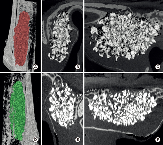
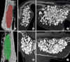 | Figure 2Micro-CT views of the TCP30 (A–C) and TCP70 groups (D–F). (A, D), Three-dimensional reconstructions; (B, E), coronal views; (C, F), sagittal views. Dome-shaped augmentation consisting of new bone and residual particles was observed in both groups. New bone and residual particles were well blended.
|
Images of the sections that had been stored in the DICOM format were used to produce three-dimensional images with commercially available software (On-Demand3D, CyberMed, Seoul, Korea), and the augmented space was color-coded to facilitate visualization.
Histologic and histomorphometric analysis
After performing micro-CT, the blocks were embedded in methylmethacrylate resin and polymerized (Exakt, Apparatebau, Norderstedt, Germany). The resin block was cut at the center of the sinus in an anteriorposterior direction, with final sections that were 15 μm thick. All of the sections were first examined with the aid of immunofluorescence microscopy (Leica DM LB, Leica Microsystems, Wetzlar, Germany), and the histologic images were captured and saved (cellSens Standard 1.11, Olympus Corporation, Center Valley, PA, USA). The sections were then stained with hematoxylin-eosin, and images of these stained sections were also saved. Histomorphometric analysis was performed with an automated image-processing system (Photoshop CS5, Adobe systems, CA, USA). The total augmented area (TA) surrounded by the Schneiderian membrane and adjacent native bone was identified. In the TA, the area containing newly formed mineralized bone (NA), residual graft material (RA), and fibrovascular tissue (FA) were separately measured. The perimeters of all the residual particles as well as the contact length between the new bone and the residual particles were measured. The percentage of NB-to-graft particle contact (%NPC) was also calculated.
The following five areas of interest (AOIs: 1.5×1.5 mm) were established for regional analysis: anterior area of the sinus, posterior area of the sinus, area adjacent to the bony window, central area, and area adjacent to the Schneiderian membrane. NA, RA, and FA were also measured in these five AOIs.
Statistics
All the data are presented as the mean and standard deviation values. Due to the smallness of the samples, the nonparametric Wilcoxon signed-rank test was used to evaluate the intergroup difference between the TCP30 and TCP70 groups. The Friedman test was used for the intra-group difference in AOI recordings made at the five areas of each sinus. Dunn’s correction was then utilized for a post-hoc pairwise comparison. The statistical significance was determined at a P<0.05 (SPSS 20.0, IBM Corporation, Armonk, NY, USA).
RESULTS
Clinical observations
Perforation of the Schneiderian membrane was not observed during surgery in any of the samples. After four months of healing, no animal showed signs of inflammation.
Micro-CT analysis
Dome-shaped augmentation consisting of NB and residual particles was observed in the sinus cavity. The bony window was almost healed by NB, and corticalization was evident with some depression also being present. NB and residual particles looked well blended regardless of their location in the augmented sinus (Figure 2).
The results of the volumetric and microstructural analyses are presented in Tables 1 and 2. TV and NV did not differ significantly between the two groups. RV was significantly smaller and FV was significantly larger in the TCP70 group than the TCP30 group (P<0.05). The values of t.Th, n.Th, and s.Th did not differ significantly between the two groups.
Table 1
Results of the volumetric analysis

Values are presented as mean±standard deviation (median).
TV, total volume of augmentation; NV, volume of new bone; RV, volume of residual bone material; FV, volume of fibrovascular tissue.
a)Significantly different from the TCP30 group (P<0.05).
![]()
Table 2
Results of the microstructural analysis

| Group | t.Th (mm) | n.Th (1/mm) | s.Th (mm) |
|---|---|---|---|
| TCP30 (n=5) | 0.06±0.01 (0.06) | 4.15±0.34 (4.27) | 0.24±0.02 (0.23) |
| TCP70 (n=5) | 0.06±0 (0.06) | 5.11±0.95 (4.68) | 0.24±0.04 (0.27) |
Values are presented as mean±standard deviation (median).
t.Th, trabecular thickness; n.Th, trabecular number; s. Th, trabecular separation.
There was no statistical difference between the two groups (P≥0.05).
![]()
Histologic and histomorphometric analysis
The Schneiderian membrane was elevated into a dome shape in both groups, and the bony window had mostly healed with NB. Almost uniform NB formation was found in all locations of the augmented sinus. The NB fragments were larger in the TCP30 group than the TCP70 group. Almost all particles were in close contact with NB. Some particles adjacent to the Schneiderian membrane had not been incorporated into NB. Instead, they were only in contact with the membrane. The resorption of the graft particles showed distinct intergroup differences. Like the appearance of NB, the graft particles in the TCP30 group were much larger and bulkier than those in the TCP70 group, and these particles were also more scattered in the TCP70 group (Figure 3 and 4).
 | Figure 3Histologic observations of the TCP30 (A) and TCP70 (B) groups. (A) Relatively large particles closely contacting new bone were observed. (B) Small and scattered particles were in close contact with new bone. Hematoxylin-eosin stain, original magnification ×12.5.
|
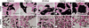 | Figure 4Histologic observations of the AOIs in groups TCP30 (A–E) and TCP70 (F–J). Regardless of the groups and areas, new bone formation was abundant and in close contact with the residual particles. Hematoxylin-eosin stain, original magnification ×40.
|
The histomorphometric results for the entire augmented sinus are presented in Table 3. The TA, NA, RA, FA, and %NPC did not differ significantly between the two groups. The histomorphometric results for the AOIs are shown in Figures 5, 6, and 7. Within each group, only FA in the TCP70 group differed significantly between the anterior and membrane areas. NA in the anterior, center, window, and membrane areas did not differ significantly between the groups, but the posterior area was significantly larger in the TCP70 group than the TCP30 group. RA was larger in all of the AOIs in the TCP30 group than those in the TCP70 group with statistically significant differences found in the posterior, center, and membrane areas. The increase in FA in all of the AOIs between the TCP30 and TCP70 groups was insignificant.
Table 3
Results of the histomorphometric analysis

Values are presented as mean±standard deviation (median).
TA, the total augmented area; NA, area of new bone; RA, area of residual material; FA, area of fibrovascular tissue; %NPC, percentage of new bone to graft particle contact.
There was no statistical difference between the two groups (P≥0.05).
![]()
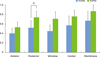 | Figure 5Values of new bone area (NA) in the AOIs. For the intergroup difference between the TCP30 and the TCP70 groups, the Wilcoxon signed-rank test was used. For the intragroup difference among the five AOIs (anterior, posterior, window, center, and membrane), the Friedman test with post-hoc Dunn’s correction was used. *Statistically significant difference (P< 0.05).
|
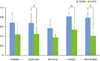 | Figure 6The values of the residual material area (RA) in the AOIs. For the intergroup difference between the TCP30 and the TCP70 groups, the Wilcoxon signed-rank test was used. For the intragroup difference among the five AOIs (anterior, posterior, window, center, and membrane), the Friedman test with post-hoc Dunn’s correction was used. *Statistically significant difference (P<0.05).
|
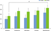 | Figure 7Fibrovascular tissue area (FA) values in the AOIs. For the intergroup difference between the TCP30 and the TCP70 groups, the Wilcoxon signed-rank test was used. For the intragroup difference among the five AOIs (anterior, posterior, window, center, and membrane), the Friedman test with post-hoc Dunn’s correction was used. *Statistically significant difference (P<0.05).
|
Fluorochrome staining observations
The fluorescence of the NB tissue and graft particles varied in both groups, with no clear intergroup difference detected. A few luminous streaks were observed near the particles in both groups, and the fluorescence was frequently brighter in the Haversian canal (Figure 8).

DISCUSSION
Long-term stability is currently one of the greatest concerns in the implant dentistry. The stability of sinus augmentation is influenced by consolidation of graft material and sinus pneumatization [18]. Therefore, a chosen graft material should facilitate as much NB formation as possible and concomitantly provide space maintenance, which requires long-term evaluation. The influence of BCPs with different HA:β-TCP ratios on osteoconductivity and volume stability was previously found to be minimal after short-term healing (i.e., two and eight weeks [16]), but this was not confirmed over a more-extended healing period. The use of a different HA:β-TCP ratio may gradually lead to distinct changes in an augmented sinus as time passes. Therefore, the present study evaluated the outcomes of BCPs with HA:β-TCP ratios of 70:30 (TCP30) and 30:70 (TCP70) after four months of healing.
The rabbit sinus is widely considered to be an appropriate model for simulating a human sinus due to the anatomical similarity and almost equivalent air pressure [19]. However, some differences should be taken into account. Firstly, the graft material after augmentation is positioned on the Schneiderian membrane, which may result in a larger amount of micromotion relative to the graft compared to a clinical situation. Secondly, considering that a rabbit sinus can accommodate less graft material compared to a human sinus, the air pressure over the area of a graft may be larger in rabbits than in humans. Thirdly, the bone remodeling period is known to be shorter in rabbits than in humans (e.g., six weeks versus 17 weeks) [20], which indicates that any change in the bone tissue of a rabbit at a certain point in time is equivalent to that in a human over a longer period.
The ratio of HA to β-TCP in BCP is known to be a major factor influencing bone formation and graft resorption [9]. Ideally, all of the bone substitute should be replaced by NB, and the replacement should occur at a balanced rate between material resorption and new bone formation. However, a high rate of resorption in sinus augmentation may be undesirable considering the constancy of air pressure. β-TCP was demonstrated to show similar behavior to autogenous bone in a canine model, but its rapid resorption did not coincide with NB formation in some specimens [21]. Thus, it can be speculated that BCP with a high proportion of β-TCP may result in inferior volume stability, especially in conditions exposed to constant pressure such as a maxillary sinus. However, the present study found no significant volume difference between the TCP30 and TCP70 groups after four months of healing. This finding is consistent with a previous study that evaluated the results at two and eight weeks of healing [16]. Volumetrically, the initial amount of grafts (0.2 cc per sinus) did not diminish throughout the healing period—the resulting volume (i.e., TA) after four months was more than 200 mm3 in both groups. This result may indicate that BCPs with high and low proportions of β-TCP have similar space-maintaining capabilities. The findings from the present animal study may be in line with retrospective studies using TCP30 [522] with graft height resorptions of 0.88 mm and 0.79–1.00 mm being observed after mean follow-ups of 42 months and one year, respectively. However, there has been no clinical study evaluating resorption of augmented sinuses using BCP with a high proportion of β-TCP.
Previous studies have evaluated dimensional changes when using various bone materials for sinus augmentation in humans [26122324252627]. All of the investigated bone materials showed volume decreases over time, which occurred mainly during the initial healing period [18]. The reported amount of resorption varied depending on the origin [6] and type [2] of materials. The resorption of BCP with an HA:β-TCP ratio of 60:40 was reportedly 15%–20% after six months [617]. Therefore, the notable resistance of TCP30 and TCP70 to volume constriction in the present study needs to be interpreted carefully due to the difference in healing potential between humans and experimental animals.
The stability of an augmented sinus is affected by the quality and quantity of NB. An increase in NB formation is expected when using BCP with a high proportion of β-TCP. The histologic results obtained in this study indicated that the amount of NB was significantly greater in the TCP70 group than in the TCP30 group only in the posterior AOI, while %NBC was comparable in the two groups. In micro-CT, NV and the microstructural indices did not differ significantly between the two groups. In fluorochrome staining, both TCP30 and TCP70 showed varying degrees of fluorescence with a few bright streaks near the particles; it was difficult to determine which type of BCP provided more-sustained and stronger bone-forming activity. These findings may indicate that a larger amount of β-TCP does not necessarily result in greater bone formation. However, it should be noted that this experiment was performed using a contained-type defect in the maxillary sinus. Furthermore, these results need to be confirmed in a larger number of samples than our pilot study.
In contrast to the results of the volumetric analysis, comparable amounts of residual material were observed in the histologic sections of the TCP30 and TCP70 groups. Previous histologic studies of rabbit calvaria using BCPs with varying HA:β-TCP ratios found similar resorption rates among the BCPs [2829]. This may reflect a limitation of histologic investigations. A single histologic section might not be representative of the tissue composition of an entire harvested tissue, which is why volumetric analysis using micro-CT has been employed in recent studies of bone tissue engineering.
It therefore remains to be determined whether augmented bone can furnish an implant with sufficient stability. Stability is improved when vital bone forms a tight network with residual material and is anchored to as much of the implant surface as possible. In this study, the contact ratio of the two BCPs with NB was around 80%. Histologically, the TCP30 group showed relatively larger fragments of NB and residual material compared to the TCP70 group. Two scenarios are possible: (1) bulkier NB may produce greater primary stability when an implant is installed, or (2) a network of smaller fractions of NB and residual material may achieve a larger degree of bone-to-implant contact.
Within the limitations of this pilot study, the osteoconductivity and dimensional stability of the BCPs with high and low proportions of β-TCP were comparable after four months of healing. The two BCPs tested in the present study were successfully used in sinus augmentation. However, these findings need to be confirmed using a larger number of samples and in a prospective clinical study.
 XML Download