

 PDF
PDF ePub
ePub Citation
 Print
Print


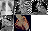
A 28-year-old female presented with shortness of breath and cyanosis over a long period of time. Chest radiography revealed situs inversus with levocardia, right heart enlargement, and engorged right interlobar pulmonary arteries (Fig. 1A). Transthoracic echocardiography (TTE) detected a large ventricular septal defect (VSD), right ventricular hypertrophy, pulmonary atresia, and a sinus venosus atrial septal defect (ASD). Persistent right-sided superior vena cava and sinus venosus ASD were detected on contrast TTE. Besides, adequate biventricular performance and trivial tricuspid regurgitation were noted. The actual anatomy of this congenital heart disease (CHD) could not totally be explored by echocardiography. Cardiac computed tomography (CT) was arranged for advance evaluation.
Cardiac CT showed the bilateral superior vena cava and the single left side inferior vena cava, which drained into the left-sided morphologic right atrium (Fig, 1B). The right pulmonary artery was supplied by the patent ductus arteriosus, and narrowed at the proximal part, and caused post-stenotic dilatation of the interlobar portion (Fig. 1C). Left pulmonary artery was supplied by multiple major aorto- pulmonary collateral arteries. The right-sided aorta was from the right ventricle with prominent trabeculation and ventricular hypertrophy (Fig. 1D). An axial view of cardiac CT showed D-loop ventricle, right ventricular hypertrophy with prominent trabeculation and no papilla muscle, large in-let type VSD, tricuspid aortic valve without coaptation defect, sinus venosus ASD. Volume rending images showed the single coronary artery (Fig. 1E). Complex CHD was diagnosed as congenitally corrected transposition of great arteries (CCTGA). Few CCTGAs were isolated, but the association with situs inversus, levocardia, ASD, VSD, bilateral superior vena cava and absence of pulmonary trunk were rare. CCTGAs may not easily be identified during obstetric ultrasound survey and its clinical presentation depend on the severity of associated cardiac malformation. If the patient has balanced systemic and pulmonary circulation, long term survival is possible1 and conservative management is better than surgical intervention. The risk factors for mortality of CCTGAs are tricuspid regurgitation, right ventricular dysfunction, and complete atrioventricular block.2 CTs are particularly useful in the assessment of adults with operated or unoperated complex malformations. Our patient has free of symptoms since her childhood because of balanced shunts. Although most adult with complex CHD will need surgery, our patient was treated conservatively because of the absence of risk factors of CCTGAs' mortality.
All procedures performed in studies involving human participants were in accordance with the ethical standards of the institutional and/or national research committee and with the 1964 Helsinki declaration and its later amendments or comparable ethical standards.
 XML Download