Abstract
We investigated predictors of major adverse cardiac events (MACE) with two years after medical treatment for lesions with angiographically intermediate lesions with intravascular ultrasound (IVUS) minimum lumen area (MLA) <4 mm2 in non-proximal epicardial coronary artery. We retrospectively enrolled 104 patients (57 males, 62±10 years) with angiographically intermediate lesions (diameter stenosis 30–70%) with IVUS MLA <4 mm2 in the non-proximal epicardial coronary artery with a reference lumen diameter between 2.25 and 3.0 mm. We evaluated the incidences of major adverse cardiovascular events (MACE including death, myocardial infarction, target lesion and target vessel revascularizations, and cerebrovascular accident) two years after medical therapy. During the two-year follow-up, 15 MACEs (14.4%) (including 1 death, 2 myocardial infarctions, 10 target vessel revascularizations, and 2 cerebrovascular accidents) occurred. Diabetes mellitus was more prevalent (46.7% vs. 18.0%, p=0.013) and statins were used less frequently in patients with MACE compared with those without MACE (40.0% vs. 71.9%, p=0.015). Independent predictors of MACEs with two years included diabetes mellitus (odds ratio [OR]=3.41; 95% CI=1.43–8.39, p=0.020) and non-statin therapy (OR=3.11; 95% CI=1.14–6.50, p=0.027). Long-term event rates are relatively low with only medical therapy without any intervention, so the cut-off of IVUS MLA 4 mm2 might be too large to be applied for defining significant stenosis. The predictors of long-term MACE were diabetes mellitus and statin therapy in patients with angiographically intermediate lesions in non-proximal epicardial coronary artery.
The cut-off value of intravascular ultrasound (IVUS) minimum lumen area (MLA) 4 mm2 is currently being used for the prediction of future clinical events in patients with proximal epicardial coronary artery disease.123 However, the MLA cut-off value of 4 mm2 cannot be used in lesions in non-proximal epicardial coronary artery because the vessel size is smaller in the non-proximal epicardial coronary artery compared with proximal epicardial coronary artery. Currently, little is known about risk factors of clinical outcomes for non-proximal epicardial coronary arteries, which are particularly common in Asian patients.45 We investigated predictors of two-year major adverse cardiac events (MACE) after medical treatment for the lesions with angiographically intermediate lesions with IVUS MLA <4 mm2 in non-intervened, non-proximal epicardial coronary arteries.
The study population consisted of 104 patients who underwent IVUS examinations of angiographically intermediate lesions (diameter stenosis 30–70%) with IVUS MLA <4 mm2 in the non-intervened non-proximal epicardial coronary artery (Fig. 1) who were clinically followed up on after twoyears. The protocol was approved by the institutional review board, and written informed consent was obtained from all patients. Hospital records of patients were reviewed to obtain information on clinical demographics.
Peripheral blood samples were obtained before coronary angiography using direct venipuncture. The blood samples were centrifuged, and serum was removed and stored at −70℃ until the assay could be performed. Absolute creatine kinase-MB levels were determined by radioimmunoassay (Dade Behring Inc., Miami, Florida). Cardiac-specific troponin I levels were measured by a paramagnetic particle, chemiluminescent immunoenzymatic assay (Beckman, Coulter Inc., Fullerton, California). The serum levels of total cholesterol, triglyceride, low-density lipoprotein cholesterol, and high-density lipoprotein cholesterol were measured by standard enzymatic methods. High-sensitivity, C-reactive proteins were analyzed turbidimetrically with sheep antibodies against human C-reactive protein; this has been validated against the Dade-Behring method.6
Coronary angiograms were analyzed with validated quantitative coronary angiography (QCA) system (Phillips H5000 or Allura DCI program, Philips Medical Systems, the Netherlands). With the outer diameter of the contrast-filled catheter as the calibration standard, the minimal lumen diameter, reference diameter, and lesion length were measured in diastolic frames from orthogonal projections. Perfusion was evaluated according to Thrombolysis In Myocardial Infarction (TIMI) criteria.7
All IVUS examinations were performed after intracoronary administration of 300 µg nitroglycerin using a commercially available IVUS system (Volcano Corp, Rancho Cordova, CA, USA). The IVUS catheter was advanced distally to the target lesion, and imaging was performed retrograde to the aorto-ostial junction at an automatic pullback speed of 0.5 mm/sec.
Qualitative analysis was performed according to the American College of Cardiology Clinical Expert Consensus Document on Standards for Acqusition, Measurement and Reporting of Intravascular Ultrasound Studies.8 Using plannimetry software (Echoplaque 3.0, INDEC Systems Inc., Santa Clara, CA), the external elastic membrane (EEM) and lumen cross-sectional area (CSA) were measured. Plaque plus media (P&M) CSA was calculated as EEM CSA minus lumen CSA, and plaque burden was calculated as P&M CSA divided by EEM CSA. Proximal and distal references were the single slices with the largest lumen and smallest plaque CSAs within 10mm proximally and distally, but before any large side branch. The lesion was the site with the smallest lumen CSA; if there were multiple image slices with the same minimum lumen CSA, then the image slice with the largest EEM and P&M was measured.
Soft plaque was less bright compared with the reference adventitia. Fibrotic plaque was as bright as or brighter than the reference adventitia without acoustic shadowing. Calcific plaque was hyperechoic with shadowing. A calcified lesion contained >90° of circumferential lesion calcium. When there was no dominant plaque composition, the plaque was classified as mixed.
Coronary artery remodeling was assessed by comparing the lesion site to the reference EEM CSA. The remodeling index was the lesion site EEM CSA divided by the average of the proximal and distal reference EEM CSA. Positive remodeling was defined as a remodeling index >1.05, intermediate remodeling as a remodeling index between 0.95 and 1.05, and negative remodeling as a remodeling index <0.95.9
Calcium deposits are described qualitatively according to their distribution as superficial when the leading edge of the acoustic shadowing appears within the most shallow 50% of the P&M thickness and as deep when the leading edge of the acoustic shadowing appears within the deepest 50% of the P&M thickness. The arc of calcium was measured (in degrees) by using an electronic protractor centered on the lumen. The length of the calcific deposit was measured using motorized transducer pullback.
Hospital records of all the patients were reviewed to obtain information on clinical demographics and medical histories. Follow-up information was obtained through review of hospital charts, telephone interviews, and the interventional database of the Heart Center of Chonnam Naitonal University Hospital. Major adverse cardiovascular events (MACEs) included death, myocardial infarctions, target lesion and target vessel revascularizations, and cerebrovascular accidents. Myocardial infarction was defined as ischemic symptoms associated with cardiac enzyme elevation ≥3 times the upper limit of the normal value. Target lesion revascularization was defined as repeat revascularization for a lesion anywhere within the stent or the 5-mm borders proximal or distal to the stent. Target vessel revascularization was defined as any revascularization of the target lesion or any segment of the epicardial coronary artery containing the target lesion.
The statistical Package for Social Sciences (SPSS) for Windows, version 15.0 (Chicago, Illinois) was used for all analyses. Continuous variables were presented as the mean value±1SD; comparisons were conducted by student's t-test or nonparametric Wilcoxon test if the normality assumption was violated. Discrete variables were presented as percentages and relative frequencies; comparisons were conducted by chi-square statistics or Fisher's exact tests as appropriate. Multivariable analysis was performed to determine whether the independent predictors of two-year MACE. A p value <0.05 was considered statistically significant.
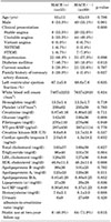
The baseline characteristics, coronary angiographic findings, and IVUS findings are summarized in Table 1. Patient's mean age was 62±10 years and about 59% of the patients had acute coronary syndrome. More than half of the patients had hypertension and one fourth of the patients had diabetes mellitus. Most of the target vessel was from the left anterior descending artery and about 80% of the lesion was located at the middle portion of the target vessel. All patients had TIMI grade 3 flow and angiographic calcium was observed in one third of the patients, and diameter stenosis was 57.1±10.8%. Lumen CSA was 3.4±0.4 mm2 and plaque burden was 64.1±7.0% at the MLA site.
During the two-year follow-up, 15 MACEs (14.4%) (including 1 death, 2 myocardial infarctions, 9 target lesion revascularizations, 10 target vessel revascularizations, and 2 cerebrovascular accident) occurred.
Baseline characteristics according to the presence or absence of MACEs within two years are summarized in Table 2. The prevalence of diabetes mellitus and family history of coronary artery disease were significantly higher, and the prevalence of hypertension also tended to be higher in patients with MACEs compared with those without MACEs. All laboratory findings were similar between both groups. At the two-year follow up, statins were used less frequently in patients with MACE compared with those without MACE.
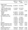
Coronary angiographic findings according to the presence or absence of two-year MACEs are summarized in Table 3. There were no significant differences in target vessels, lesion locations, ACC/AHA types, angiographic calcium, reference diameter, minimal lumen diameter, diameter stenosis, and angiographic lesion length between patients with MACE and those without MACE.
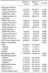
IVUS findings according to the presence or absence of two-year MACE are summarized in Table 4. At the lesion site, EEM CSA and plaque burden were significantly greater, and P&M CSA tended to be greater in patients with MACE compared with those without MACE. IVUS lesions were significantly longer and the remodeling index was significantly greater in patients with MACE compared with those without MACE.
Multivariate analysis was performed to identify independent predictors of two-year MACEs. The following variables were tested (all with p<0.1 in univariate analysis): hypertension, diabetes mellitus, family history of coronary artery disease, no statin use at two-year follow-up, plaque burden at the MLA site, IVUS lesion length, and remodeling index. Independent predictors of two-year MACEs included diabetes mellitus (odds ratio [OR]=3.41; 95% CI=1.43–8.39, p=0.020) and non-statin therapy (OR=3.11; 95% CI=1.14–6.50, p=0.027).
The present study demonstrated that 1) MACEs occurred in only 14.4% of the patients during two-year follow-up, 2) the prevalence of diabetes mellitus was significantly higher in patients with two-year MACEs compared with those without MACEs, 3) statins were used less frequently in patients with two-year MACE compared with those without MACE, and 4) diabetes mellitus and non-statin therapy were the independent predictors of MACE in patients with angiographically intermediate lesions with IVUS MLA <4 mm2 in non-intervened non-proximal epicardial coronary artery during two-year follow-up.
Because of the limitations of coronary angiography, intermediate coronary lesions remain an unresolved problem for interventional cardiologists treating patients with suspected coronary artery disease. If the lesion severity of intermediate lesions has been overestimated, patients will undergo unnecessary revascularization procedures. In contrast, if the lesion severity of intermediate lesions has been underestimated, patients will not undergo necessary revascularization procedures and critical arterial stenosis will be untreated.
Previous studies have suggested that the MLA correlated with coronary physiology, and a MLA of <4 mm2 was found to be the threshold for flow-limiting stenosis in most studies.123 Abizaid et al.1 reported that the major determinants of the coronary flow reserve in patients with coronary artery disease are lumen compromise (which is best assessed by the IVUS measurement of the MLA) and lesion length and that a MLA ≥4.0 mm2 has a high diagnostic accuracy in predicting a coronary flow reserve ≥2.0, especially before intervention. Nishioka et al.2 reported that the lesion MLA 4 mm2 is a simple and highly accurate criterion for significant coronary narrowing when compared with the results of stress myocardial perfusion imaging. Briguori et al.3 reported that IVUS area stenosis >70%, minimal lumen diameter <1.8 mm, MLA <4.0 mm2, and lesion length >10 mm reliably identified functionally critical intermediate coronary stenosis in 53 intermediate coronary lesions found by angiography. However, this criterion is only applicable to lesions located at non-proximal coronary arteries with a reference segment diameter >3 mm, limiting the use of IVUS-derived anatomic criteria to define the functional significance of a subset of lesions.
In the present study, we evaluated two-year clinical outcomes in patients with angiographically intermediate lesions with IVUS MLA <4 mm2 in non-proximal epicardial coronary artery. During the two-year follow-up, 15 MACEs (14.4%) occurred. Although it is difficult to directly compare the MACE rates because of the differences in the patient population, the events rates are relatively low in patients with intermediate lesions. Stone et al.10 reported that the 3-year cumulative rates of MACEs were 20.4% in patients with acute coronary syndrome who underwent percutaneous intervention. Furthermore, Bech et al.11 reported that the 2-year outcome after medical treatment and FFR guided the PCI of intermediate coronary stenosis without myocardial infarction or unstable angina in patients. It showed 20.3% MACE rates. Diabetes mellitus was more prevalent and statins were used less frequently in patients with MACEs compared with those without MACEs. Independent predictors of two-year MACEs included diabetes mellitus and non-statin therapy. Impaired glycemic homeostasis had a direct influence on the propagation of atherosclerotic plaque.12 There are several plausible explanations for the more rapid progression of disease in diabetic patients. Diabetic patients have atherogenic dyslipidemia including hypertriglyceridemia, low high-density lipoprotein cholesterol, the presence of small, dense low-density lipoprotein particles. Hyperglycemia and the potential generation of advanced glycation end products also seem to play an important role.13 Nicholls et al.14 reported that diabetes is accompanied by more extensive atherosclerosis and inadequate compensatory remodeling, Statin therapy could induce regression of coronary atherosclerosis and there was a strong linear relationship between achieved low-density lipoprotein cholesterol (LDL-C) levels and the course of atherosclerosis.151617 Development of new anti-atherosclerotic strategies is needed and intensive statin therapy is essential, especially in diabetic patients.
Recently, lower cut-off values of MLA in order to predict functionally significant coronary stenosis has been suggested. Kang et al.18 reported that the best cut-off value for MLA to predict fractional flow reserve <0.80 was <2.4 mm2 with a 90% sensitivity and 60% specificity. Nam et al.19 reported that both fractional flow reserve- and IVUS-guided percutaneous coronary intervention strategies for intermediate coronary artery disease were associated with favorable outcomes and the fractional flow reserve-guided percutaneous coronary intervention reduced the need for revascularization of many of these lesions. In the present study, event rates were relatively low with only medical therapy without any intervention. Therefore, the cut-off of IVUS MLA 4 mm2 might be too large to be used in patients with angiographically intermediate lesions in non-proximal epicardial coronary artery.
There are several limitations to be mentioned. First, this study is based on a small sample, thus raising the possibility of selection bias. In particular, only 7 diabetes patients and 6 other patients took statins in the MACE group which might be too small of a sample from which to make conclusions. Second, this was a retrospective single-center study. The results of this study should be verified by further prospective investigation. Furthermore, access to some detailed data was not available, such as the intensity of statin use, the type of statins used, and non-culprit lesion outcomes. Third, we did not evaluate the functional severity of stenosis. The heterogeneity of the study population, which included stable angina and acute coronary syndrome patients made it hard to identify differences only with anatomical evaluation. Additionally, we included CVA patients in the MACE group, but CVA risk might be not related moderate coronary lesion.
In conclusion, in patients with angiographically intermediate lesions in the non-proximal epicardial coronary artery, the event rates were relatively low with only medical therapy without any intervention, therefore the cut-off of IVUS MLA 4 mm2 may not be applied for predicting future cardiovascular events and the predictors of long-term MACE were diabetes mellitus and non-statin therapy.
Figures and Tables
 | FIG. 1Coronary angiographic (A) and intravascular ultrasound images (B) of angiographically intermediate lesions with intravascular ultrasound minimal lumen area (MLA) <4 mm2 in middle left anterior descending artery. |
TABLE 1
Baseline characteristics, coronary angiographic and intravascular ultrasound findings (n=104)

Values are n (%), mean±SD. NSTEMI: non-ST segment elevation myocardial infarction, STEMI: ST segment elevation myocardial infarction, NT-pro-BNP: N-terminal pro-B type natriuretic peptide, LDL: low-density lipoprotein, HDL: high-density lipoprotein, hs-CRP: high-sensitivity C-reactive protein, TIMI: Thrombolysis In Myocardial Infarction, ACC/AHA: American College of Cardiology/American Heart Association, EEM: external elastic membrane, CSA: cross-sectional area, P&M: plaque plus media, IVUS: intravascular ultrasound.
TABLE 2
Baseline characteristics according to the presence or absence of two-year major adverse cardiac events

ACKNOWLEDGEMENTS
This study was supported by a grant of the Korean Health Technology R&D Project, Ministry of Health and Welfare, Republic of Korea (HI13C0163), by a grant of the Korean Health Technology R&D Project, Ministry of Health and Welfare, Republic of Korea (HI17C2150), by a grant of the Korean Health Technology R&D Project, Ministry of Health and Welfare, Republic of Korea (HI18C0173), by a grant of Bio & Medical Technology Development Program of the National Research Foundation (NRF) & funded by the Korean government (MSIT) (2018M3A9E2024584), by a grant of the Korean Health Technology R&D Project, Ministry of Health and Welfare, Republic of Korea (HI14C2069), and by a grant of the Korean Health Technology R&D Project, Ministry of Health and Welfare, Republic of Korea (HI13C1527).
References
1. Abizaid A, Mintz GS, Pichard AD, Kent KM, Satler LF, Walsh CL, et al. Clinical, intravascular ultrasound, and quantitative angiographic determinants of the coronary flow reserve before and after percutaneous transluminal coronary angioplasty. Am J Cardiol. 1998; 82:423–428.

2. Nishioka T, Amanullah AM, Luo H, Berglund H, Kim CJ, Nagai T, et al. Clinical validation of intravascular ultrasound imaging for assessment of coronary stenosis severity: comparison with stress myocardial perfusion imaging. J Am Coll Cardiol. 1999; 33:1870–1878.

3. Briguori C, Anzuini A, Airoldi F, Gimelli G, Nishida T, Adamian M, et al. Intravascular ultrasound criteria for the assessment of the functional significance of intermediate coronary artery stenoses and comparison with fractional flow reserve. Am J Cardiol. 2001; 87:136–141.

4. Dhawan J, Bray CL. Are Asian coronary arteries smaller than caucasian? A study on angiographic coronary artery size estimation during life. Int J Cardiol. 1995; 49:267–269.

5. Lip GY, Rathore VS, Katira R, Watson RD, Singh SP. Do Indo-Asians have smaller coronary arteries. Postgrad Med J. 1999; 75:463–466.

6. Roberts WL, Moulton L, Law TC, Farrow G, Cooper-Anderson M, Savory J, et al. Evaluation of nine automated high-sensitivity C-reactive protein methods: implications for clinical and epidemiological applications. Part 2. Clin Chem. 2001; 47:418–425.

7. TIMI IIIB Investigators. Effects of tissue plasminogen activator and a comparison of early invasive and conservative strategies in unstable angina and non-Q-wave myocardial infarction. Results of the TIMI IIIB trial. Thrombolysis in myocardial ischemia. Circulation. 1994; 89:1545–1556.
8. Mintz GS, Nissen SE, Anderson WD, Bailey SR, Erbel R, Fitzgerald PJ, et al. American college of cardiology clinical expert consensus document on standards for acquisition, measurement and reporting of intravascular ultrasound studies (IVUS): a report of the American college of cardiology task force on clinical expert consensus documents. J Am Coll Cardiol. 2001; 37:1478–1492.

9. Nakamura M, Nishikawa H, Mukai S, Setsuda M, Nakajima K, Tamada H, et al. Impact of coronary artery remodeling on clinical presentation of coronary artery disease: an intravascular ultrasound study. J Am Coll Cardiol. 2001; 37:63–69.

10. Stone GW, Maehara A, Lansky AJ, de Bruyne B, Cristea E, Mintz GS, et al. A prospective natural-history study of coronary atherosclerosis. N Engl J Med. 2011; 364:226–235.

11. Bech GJ, de Bruyne B, Pijls NH, de Muinck ED, Hoorntje JC, Escaned J, et al. Fractional flow reserve to determine the appropriateness of angioplasty in moderate coronary stenosis: a randomized trial. Circulation. 2001; 103:2928–2934.

12. Boyle PJ. Diabetes mellitus and macrovascular disease: mechanisms and mediators. Am J Med. 2007; 120:9 Suppl 2. S12–S17.

13. Stancoven A, McGuire DK. Preventing macrovascular complications in type 2 diabetes mellitus: glucose control and beyond. Am J Cardiol. 2007; 99:5H–11H.

14. Nicholls SJ, Tuzcu EM, Kalidindi S, Wolski K, Moon KW, Sipahi I, et al. Effect of diabetes on progression of coronary atherosclerosis and arterial remodeling: a pooled analysis of 5 intravascular ultrasound trials. J Am Coll Cardiol. 2008; 52:255–262.

15. Nissen SE, Tuzcu EM, Schoenhagen P, Crowe T, Sasiela WJ, Tsai J, et al. Reversal of Atherosclerosis with Aggressive Lipid Lowering (REVERSAL) Investigators. Statin therapy, LDL cholesterol, C-reactive protein, and coronary artery disease. N Engl J Med. 2005; 352:29–38.

16. Nissen SE, Nicholls SJ, Sipahi I, Libby P, Raichlen JS, Ballantyne CM, et al. Effect of very high-intensity statin therapy on regression of coronary atherosclerosis: the ASTEROID trial. JAMA. 2006; 295:1556–1565.

17. Nicholls SJ, Tuzcu EM, Sipahi I, Grasso AW, Schoenhagen P, Hu T, et al. Statins, high-density lipoprotein cholesterol, and regression of coronary atherosclerosis. JAMA. 2007; 297:499–508.


 PDF
PDF ePub
ePub Citation
 Print
Print


 XML Download