

 PDF
PDF ePub
ePub Citation
 Print
Print


INTRODUCTION
Advanced gastric cancer (AGC) is one of the most common malignancies resulting in cancer-related deaths in Asian populations [123]. Radical surgery is the standard treatment option for patients with resectable disease; however, the prognosis remains poor with a 5-year survival of approximately 30% [3]. Recurrence after curative surgery often occurs in 12%−54% of cases and is the main cause of cancer-related death [4]. Hence, stratifying the patient's risk and prediction of prognosis are key issues in the postoperative management of AGC patients. The tumor, node, metastasis (TNM) classification system by the American Joint Committee on Cancer (AJCC) has been used to predict patients' prognoses. However, the conventional clinicopathological approach does not adequately represent the complexity of gastric cancer biology [56].
Current guidelines for staging AGC include gastrofiberscopy; contrast-enhanced computed tomography (CECT) of the thorax, abdomen, and pelvis; and laparoscopy. 18F-fluorodeoxyglucose (18F-FDG) positron emission tomography/computed tomography (PET/CT) is preferred to evaluate metastatic disease [78]. 18F-FDG PET/CT has been shown to be useful in AGC patients in many aspects. 18F-FDG PET/CT can determine the operability of locally advanced gastric cancer [9] and facilitate the detection of recurrence during follow-up after surgery [1011]. Several studies have shown the usefulness of 18F-FDG PET/CT for the prognosis of AGC patients undergoing chemotherapy [1213]. However, the independent role of preoperative 18F-FDG PET/CT in AGC still needs to be investigated.
Lymph node (LN) metastasis is a substantial factor in the staging and prognosis of patients with AGC [14]. Although 18F-FDG PET/CT has low diagnostic sensitivity for LN metastasis, evaluation of LN metabolism can be useful to predict prognosis [15]. The advantage of 18F-FDG PET/CT is the simultaneous assessment and comparison of metabolism in primary tumors and metastatic LNs, which provide prognostic information [1617]. A comprehensive evaluation of the metabolic aggressiveness of metastatic LNs in the preoperative setting can be used as a potential biomarker in AGC.
In this study, we evaluated the metabolic characteristics of primary tumors and/or metastatic LNs and compared them with survival data after surgery in patients with AGC to determine the clinical impact of preoperative 18F-FDG PET/CT.
MATERIALS AND METHODS
Patients
We analyzed the medical records of consecutive AGC patients in our hospital between July 2010 and December 2014. Among them, 168 patients with AGC who underwent curative surgical resection were enrolled in this study. The inclusion criteria were: 1) initial diagnosis of gastric cancer, 2) preoperative staging with 18F-FDG PET/CT in our hospital, 3) pathologic staging of T2–T4, and 4) a histological type of common epithelial tumor according to the Japanese Gastric Cancer Association system [18]. The exclusion criteria were: 1) early gastric cancers, 2) double primary malignancies, 3) distant organ metastasis, 4) loss to follow-up within 3 months after surgery, and 5) treatment with palliative/non-curative surgery. Patients' clinical and histopathologic characteristics analyzed in this study were acquired prospectively at the time of surgery. The Institutional Review Board of our hospital approved this retrospective clinical study (AN17196-001), and the requirement for informed consent was waived. All procedures performed in this study involving human participants were in accordance with the 1964 Helsinki declaration and its later amendments or comparable ethical standards.
All patients underwent total or subtotal gastrectomy according to the anatomic location of the primary tumor, accompanied by lymphadenectomy according to the General Rules for Gastric Cancer [18]. Curative resection was defined by the complete absence of visible tumor tissue with negative margins. In cases of subtotal gastrectomy, tumor-free margins of >3 cm and 5 cm were obtained in patients with well-marginated and infiltrative primary tumors, respectively. Histopathologic classification of the primary tumors included subtype (signet ring cell vs. non-signet ring cell, which included adenocarcinoma with papillary, tubular, poorly differentiated, and mucinous subtypes), Lauren classification (intestinal vs. non-intestinal, which included diffuse and mixed types), and Bormann classification. After surgery, patients underwent adjuvant chemotherapy based on their histopathologic results and performance status. Subsequently, patients were regularly followed using serum tumor markers, gastrofiberscopy, and/or CECT.
18F-FDG PET/CT
18F-FDG PET/CT was performed using a dedicated scanner (Gemini TF 16, Philips Medical Systems, Andover, MA, USA). Patients fasted for 6 hours, and blood glucose level was <200 mg/dL at the time of 18F-FDG injection. PET/CT image acquisition was performed 60 minutes after the administration of 18F-FDG (approximately 5.18 MBq/kg). A low-dose CT scan was acquired for attenuation correction (50 mA, 120 kVp, slice thickness 4 mm, matrix size 512×512). Emission PET images were acquired for 1 minute in each bed position. PET images were reconstructed using a 3-dimensional iterative algorithm (row action maximum likelihood algorithm) with TOF function (3 iterations, 33 subsets, matrix size 144×144).
Image analysis
All 18F-FDG PET/CT images were reviewed by 2 experienced nuclear medicine physicians in consensus. 18F-FDG avidity for both primary tumors and LNs were determined using quantitative and qualitative methods. Quantitative parameters included the maximum standardized uptake value (SUVmax), metabolic tumor volume (MTV), total lesion glycolysis (TLG), and the LN-to-tumor ratio (LTR). The SUVmax of the lesion was measured in volume of interest (VOI). VOI of the primary tumor was determined from the hottest point in the lesion in transaxial PET/CT images. In the case of a non-discernible primary lesion on PET/CT, VOI was drawn on the stomach wall according to the tumor location based on CECT and gastrofiberscopy. SUV was calculated as (radioactivity [kBq] per unit volume [mL]/injected 18F-FDG activity [kBq] per body mass [g]). The MTV was defined as the volume (cm3) within the VOI. The TLG was calculated as the product of the MTV and mean SUV within the VOI. The ratio of the SUVmax of the LTR was calculated. The LTR in non-discernible LNs was calculated by coding the SUVmax of the LNs as 1. All image analyses were performed using a dedicated workstation (Extended Brilliance Workspace 4.0, Philips Healthcare, Amsterdam, The Netherlands).
Criteria for determining 18F-FDG avidity for both primary tumors and LNs were defined as 1) focally increased 18F-FDG uptake by visual analysis and 2) a reasonable isocontouring result of MTV with a cutoff value of 50% of the SUVmax (Fig. 1).
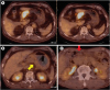 | Fig. 1Representative cases of 18F-FDG-avid primary tumor (A, left) and LNs (A, right), non-avid primary tumor (B), and non-avid LN (C). The metabolic tumor volume of the primary tumor and LNs in the patient (A) was 18.8 cm3 and 0.2 cm3, respectively (yellow arrow: primary tumor, red arrow: LN).
18F-FDG = 18F-fluorodeoxyglucose; LN = lymph node.
|
Clinicopathologic and survival data
Clinical data obtained from medical records included age at surgery, sex, preoperative serum carcinoembryonic antigen (CEA), histopathologic results, and pathologic T (pT) and N (pN) staging. Pathologic staging was based on the 7th edition of the AJCC System. Recurrence-free survival (RFS) was defined as the interval from the date of surgery to the date of cross-sectional imaging when recurrence (by pathologic confirmation or clinical decision) was first noted. Overall survival (OS) was defined as the interval from the date of surgery to the date of the last hospital visit.
Statistical analyses
Numeric data were represented as the mean±standard deviation. Differences among variables between groups were compared using Student's t-test, the Kruskal-Wallis test, and the χ2 test. Diagnostic performance of 18F-FDG avidity for detecting primary tumors and metastatic LNs was evaluated by calculating the sensitivity, specificity, positive predictive value (PPV), and negative predictive value (NPV). Survival time was estimated using the log-rank test. Univariate Cox regression analysis was performed for evaluating the statistical significance of clinicopathologic parameters. Continuous variables were analyzed directly or dichotomized using the optimal cutoff value by receiver operating characteristics curve analysis. Multivariate Cox regression analysis was performed to evaluate the effect of LN-related parameters on other significant parameters. Statistical analyses were performed using SPSS statistics software (version 18.0, SPSS Inc., Chicago, IL, USA), and a P value of less than 0.05 was considered statistically significant.
RESULTS
Patient characteristics
Of the 168 enrolled patients, 51 (30.4%) experienced recurrence during the follow-up period (median follow-up duration, 35 months; range, 3–81 months), and 32 (19.0%) were documented deaths. Age, sex, and operation type were not significantly different between patients with and without recurrence (Table 1). The history of adjuvant chemotherapy, pT stage, pN stage, TNM staging, tumor size, and the presence of lymphovascular invasion (LVI) were significantly different according to recurrence (P<0.05). For histopathologic results, the frequency of signet ring cell-type was not significantly different according to recurrence (P=0.288). The frequency of intestinal-type was higher in the non-recurring group (P=0.003). However, the Bormann classification results did not vary significantly in terms of recurrence (P=0.062).
Table 1
Patient characteristics
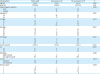
Tx = therapy; pT = pathologic tumor; pN = pathologic node; TNM = tumor, node, metastasis; LVI = lymphovascular invasion.
*Three, †5 unclassified.
![]()
18F-FDG avidity of tumor lesions: diagnostic evaluation
We detected 18F-FDG-avid primary tumors in 119 (70.8%) patients. However, the presence of 18F-FDG-avid primary tumors was not significantly different between patients with (n=37, 72.5%) and without (n=82, 70.1%) recurrence (P=0.747). The SUVmax, MTV, and TLG values of 18F-FDG-avid primary tumors showed no significant difference according to recurrence (P=0.561, 0.551, and 0.800, respectively). The SUVmax of all primary tumors (including non-avid tumors) also showed no difference between patients with and without recurrence (5.73±4.26 vs. 5.75±5.08; P=0.979). We found that 33 (19.6%) patients showed more than one 18F-FDG-avid LN on 18F-FDG PET/CT. 18F-FDG-avid LNs tended to be more frequent in recurring cases (n=14, 27.5%) than in non-recurring cases (n=19, 16.2%; P=0.093). Similar to primary tumors, the SUVmax, MTV, and TLG values of 18F-FDG-avid LNs were not different between patients with and without recurrence (P=0.854, 0.864, and 0.661, respectively). The calculated LTR was also not significantly different between patients with and without recurrence (0.38±0.26 vs. 0.36±0.29; P=0.749).
The diagnostic evaluation of 18F-FDG PET/CT for high T stage (pT 3–4) primary tumors showed a sensitivity of 73.8%, a specificity of 38.1%, a PPV of 78.2%, and an NPV of 32.7% (Table 2). For the detection of high N stage (pN 2–3), the sensitivity, specificity, PPV, and NPV of 18F-FDG PET/CT were 29.7%, 92.2%, 81.8%, and 52.6%, respectively. For the detection of metastatic LNs (pN 1–3), the sensitivity, specificity, PPV, and NPV of 18F-FDG PET/CT were 26.2%, 97.8%, 97.0%, and 33.3%, respectively (Table 3).


Prognostic factors for RFS and OS
The median RFS and OS were 34.9 and 36.5 months, respectively. The significance of 18F-FDG avidity for recurrence was evaluated (Fig. 2). The 18F-FDG avidity of primary tumors was not significantly correlated with RFS or OS (P=0.532 and 0.658, respectively). However, the 18F-FDG avidity of LNs showed significant correlation with a shorter RFS (P=0.012). The mean RFS of patients with 18F-FDG-avid LNs was 36.5±4.5 months, whereas that of patients with non-avid LNs was 60.4±2.7 months. The relationship between 18F-FDG avidity of LNs and OS did not show statistical significance (P=0.181).
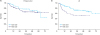 | Fig. 2Cumulative RFS curve according to the 18F-FDG avidity of (A) primary tumors and (B) LNs in enrolled patients (red: non-avid group, blue: 18F-FDG-avid group).RFS = recurrence-free survival; LN = lymph node.
|
Univariate analysis was performed to evaluate the prognostic significance of the clinicopathologic parameters (Table 4). Age and sex were not correlated with patients' survival. The size of the primary tumor and the number of metastatic LNs proven histopathologically were significantly correlated with both RFS and OS (P≤0.001). High pT (≥3) and pN (≥2) stages were also significantly correlated with patients' survival. The histopathologic subtype of signet ring cell cancer was a prognostic factor for OS (P=0.044), but not for RFS (P=0.377). Non-intestinal subtype and LVI were significant prognostic factors for both RFS and OS. Preoperative CEA level was not related to RFS (P=0.536) and OS (P=0.351). 18F-FDG avidity of primary tumors was not correlated with RFS and OS, but 18F-FDG avidity of LNs was a significant prognostic factor for RFS (P=0.015). The correlation between 18F-FDG avidity of LNs and OS was not significant (P=0.187). Other 18F-FDG-PET/CT-driven parameters, including the SUVmax of primary tumors and the LTR, were not significantly correlated with patients' survival.
Table 4
Univariate Cox regression analysis for prognostic factors related to survival

RFS = recurrence-free survival; OS = overall survival; HR = hazard ratio; CI = confidence interval; LN = lymph node; pT = pathologic tumor; pN = pathologic node; LVI = lymphovascular invasion; CEA = carcinoembryonic antigen; 18F-FDG = 18F-fluorodeoxyglucose; SUVmax = maximum standardized uptake value; LTR = lymph node-to-tumor ratio.
![]()
Construction of the multivariate survival models was based on significant parameters driven from univariate analysis, and metastatic LN-related parameters were combined to compare the effect in each model: 1) Model with 18F-FDG avidity of LNs, 2) Model with number of metastatic LNs, and 3) Model with pN stage (≥2) (Tables 5 and 6). For RFS, the size of the primary tumor was an independent prognostic factor in all models (Table 5). Non-intestinal subtype and LVI were independent factors in models 1 and 2, but significance was not observed in model 3. 18F-FDG avidity of LNs was an independent prognostic factor for predicting RFS. For OS, the size of the primary tumor was an independent prognostic factor in all models (Table 6). Other histopathologic factors did not show statistical significance. Among the LN-related parameters, only pN stage was an independent factor for predicting OS.
Table 5
Multivariate Cox regression models for RFS

RFS = recurrence-free survival; LN = lymph node; pN = pathologic node; HR = hazard ratio; CI = confidence interval; LVI = lymphovascular invasion; 18F-FDG = 18F-fluorodeoxyglucose; N/A = not assessed.
![]()
Table 6
Multivariate Cox regression models for OS

OS = overall survival; LN = lymph node; pN = pathologic node; HR = hazard ratio; CI = confidence interval; LVI = lymphovascular invasion; 18F-FDG = 18F-fluorodeoxyglucose; N/A = not assessed.
![]()
Subgroup analysis was performed for patients with histopathologically proven adenocarcinoma (n=154), and histologic grade of the primary tumor was added in the survival analysis. As in the total patient population, tumor size, number of metastatic LNs, pathologic staging, and LVI showed significance for predicting RFS and OS (Supplementary Table 1). Poorly differentiated subtype was a significant prognostic factor for RFS (P=0.005) but not for OS (P=0.175). Lauren nonintestinal subtype was also a significant factor for RFS (P=0.001) but not for OS (P=0.137). The 18F-FDG avidity of LNs was a significant factor for RFS in the univariate analysis (P=0.031) but not in the multivariate analysis (P=0.120, Supplementary Table 2).
DISCUSSION
This study evaluated the diagnostic and prognostic value of preoperative 18F-FDG PET/CT in patients with resectable AGC. Current evidence suggests that primary tumor resection and standardized D2 lymphadenectomy under best post-operative care lower recurrence, death rate, and morbidity/mortality [19]. Standardized preoperative workup is recommended to evaluate the disease status based on patient examination, laboratory profile, endoscopy procedure, and CECT [7]. However, the 5-year survival rate after curative resection remains disappointing. Randomized clinical trials have reported varying results for adjuvant chemotherapy according to regional differences [2021]. These findings suggest the heterogeneous biology of gastric cancer and the need for effective prognostic biomarkers for AGC patients [22]. Evaluation of tumor metabolism by 18F-FDG PET/CT may play a potential role as a surrogate marker of tumor aggressiveness, which is correlated with patients' prognoses.
The diagnosis of LN metastasis is one of the most important prognostic factors in AGC patients after curative surgery [23]. LN metastasis predicts accurate and stage-specific survival in the subclassification of patients with a similar pT stage [24]. The number of metastatic LNs is directly correlated with survival time [25]. Hence, a comprehensive analysis of LN metastasis is needed to evaluate AGC patients, especially those contemplating curative surgery. Endoscopic ultrasonography showed a high predictive value (approximately 92.6%) for node-negative, early gastric cancer but played a limited role in predicting extra-perigastric LN metastasis [2627]. CECT has been widely used for the evaluation of the degree of LN metastasis; however, the size criteria for determining LN metastasis often have limited sensitivity and yield false-negative results [28]. The diagnostic accuracy of preoperative 18F-FDG PET/CT for LN metastasis in patients with AGC has long been discussed. The diagnostic sensitivity for metastasis of N1 LNs was below 50%, which was lower than for CECT. However, 18F-FDG PET/CT outperformed CECT for the specificity of N staging. Especially in advanced N stage, the specificity by 18F-FDG PET/CT was 96% and 99% in N2 and N3 lymph nodes, respectively [293031]. In view of resectability, accurate staging of advanced disease is essential for planning the surgical approach and extent of en bloc resection. This study also showed comparable accuracy with high specificity (92.2%) for the diagnosis of advanced N stage, although 18F-FDG avidity of LNs was determined by combining qualitative and quantitative criteria. We suggest that the diagnostic value of 18F-FDG PET/CT might be related directly with prognostic information to determine patients' survival.
Until now, a few studies have reported the correlation between 18F-FDG uptake by LNs and the prognoses of patients with AGC. A recent study revealed that a high SUVmax of LNs is an independent factor for patients' RFS and OS [15]. Metabolic information for LNs was correlated with patients' OS, whereas the size of lymphadenopathies did not show a significant relationship. Additionally, 18F-FDG PET/CT revealed metastatic disease that was not detected on CT [32]. In this study, the 18F-FDG avidity of LNs in patients with AGC was significantly correlated with disease recurrence as well as other histopathologic parameters for determining metastatic LNs. Patients with non-avid LNs had a 2-year RFS rate of 82.2% (111/135), whereas patients with 18F-FDG-avid LNs showed a 2-year RFS rate of 60.6% (20/33). The mean difference in RFS according to the 18F-FDG avidity of LNs was approximately 24 months. The results suggest the need for revisiting the role of preoperative 18F-FDG PET/CT for accurate prediction of recurrence in patients with AGC.
Studies for evaluating the utility of 18F-FDG PET/CT in AGC focused on 18F-FDG uptake by primary tumors. High 18F-FDG uptake by primary tumors was associated with 2-year RFS after curative resection [3334]. In this study, the 18F-FDG avidity of primary tumors was not significantly correlated with recurrence. 18F-FDG uptake by primary tumors is partly affected by background gastric wall activity and histopathologic subtype [35]. Furthermore, several parameters for measuring 18F-FDG uptake by primary tumors have been proposed, including SUVmax, MTV, and tumor-to-liver ratio [1136]. Further studies for optimizing 18F-FDG PET/CT-driven parameters are needed to investigate the correlation between glucose metabolism in gastric cancer and patients’ prognoses. In the present study, the 18F-FDG avidity of LNs was determined dichotomously instead of using continuous SUVmax values. The SUVmax of small perigastric LNs is often hard to standardize because of the partial-volume effect and spill-over effect [37]. 18F-FDG avidity of LNs used in this study had high specificity for determining nodal metastases and represents an independent prognostic factor for RFS.
This study has several limitations. First, the 18F-FDG avidity of LNs was correlated with patients' RFS but not with OS, probably due to the relatively small sample and event size. Second, the prognosis-predicting model in this study was not entirely based on pre-operative clinical parameters. Further studies are needed to evaluate the significance of preoperative 18F-FDG PET/CT in terms of clinical staging. Finally, this retrospective single-center study might be inevitably associated with a patient selection bias.
In conclusion, this study demonstrates that 18F-FDG avidity of LNs evaluated by PET/CT scan is an independent factor contributing to RFS following curative resection in patients with AGC. We suggest that preoperative 18F-FDG PET/CT should be used for predicting prognosis aimed at appropriate risk monitoring of AGC patients during follow-up after surgery.
 XML Download