

 PDF
PDF ePub
ePub Citation
 Print
Print


INTRODUCTION
In recent years, the incidence of adenocarcinoma located in the gastroesophageal junction (GEJ) has increased rapidly in Western countries, while that of squamous cell carcinoma has gradually decreased [1234]. A similar trend has been observed in Asia, possibly due to the current availability of Helicobacter pylori eradication therapy, a high prevalence of gastroesophageal reflux disease and obesity, and dietary factors [567]. The TNM Classification of Malignant Tumors, 8th Edition, defines GEJ adenocarcinoma as adenocarcinoma with an epicenter within 5 cm of the GEJ and extending into the esophagus [8]. GEJ adenocarcinoma is usually classified into three subtypes according to the Siewert classification, which is based on the location of the tumor epicenter [9]. Siewert type I cancers are located between 1 and 5 cm above the GEJ and are generally considered to arise against the background of Barrett's esophagus in the lower esophagus; Siewert type II cancers are located between 1 cm above and 2 cm below the GEJ and are considered to be true gastric cardia tumors; Siewert type III cancers are located between 2 and 5 cm below the GEJ, with invasion of the esophagus, and are considered to be subcardial gastric cancers. In Japan, Nishi's classification system defines GEJ cancer as a tumor with an epicenter between 2 cm above and 2 cm below the GEJ, regardless of its histological subtype [101112].
A unique characteristic of GEJ cancer is that its lymphatic drainage pathways include both the mediastinal and abdominal fields [1314]. To determine the optimal treatment for GEJ cancers, identifying the exact tumor location is of great importance because this strongly influences the pattern of lymph node (LN) metastasis. Other factors that should be considered are the extent of LN dissection, the type of esophagectomy and gastrectomy, and whether paraaortic lymph node dissection (PAND) should be performed or not.
In this review, the focus was on adenocarcinoma of the GEJ using the Siewert classification as recommended by international consensus. This study aimed to provide an update on the surgical treatment of GEJ adenocarcinoma by reviewing previous reports and propose recommended surgical approaches. In addition, we introduced a currently ongoing Japanese nationwide prospective trial that is being conducted through the collaboration of the Japanese Gastric Cancer Association (JGCA) and the Japan Esophageal Society (JES) to determine the optimal extent of LN dissection in GEJ cancers.
MEDIASTINAL LN METASTASES FROM GEJ CANCERS
The incidence of LN metastasis in patients with GEJ cancer is considered to be high, and lymphatic drainage occurs into both the mediastinal and abdominal fields. To investigate the incidence of mediastinal LN metastasis in patients with GEJ cancer, a multicenter retrospective study was conducted in Japan [15]. In 315 patients with pT2-4 Siewert type II cancers, the overall incidence rates of metastasis or recurrence were 3.8% (12/315) and 7.0% (22/315) in the upper and middle mediastinal LNs, respectively. The 5-year overall survival (OS) rate of patients with metastasis in the upper or middle mediastinal LNs was 16.7%. This study also demonstrated that the rate of GEJ adenocarcinoma metastasis or recurrence in the upper or middle mediastinal LNs was significantly higher when the length of esophageal invasion was >3 cm, while in the lower mediastinal LNs, this rate was higher when the esophageal invasion length was >2 cm. On the basis of these results, GEJ adenocarcinoma with a length of esophageal invasion of >3 cm has been suggested to be resected together with the upper and middle mediastinal LNs. However, no prospective studies have examined the incidence of mediastinal LN metastasis; therefore, considering the possibility of selection bias when basing treatment on the results of retrospective studies is important [1416171819]. Randomized controlled trials are warranted to evaluate the efficacy of mediastinal LN dissection.
SURGICAL APPROACHES FOR MEDIASTINAL LN DISSECTION
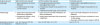
Three principal approaches are used to remove the mediastinal LNs: the right transthoracic (RT) approach with right thoracotomy and laparotomy; the left transthoracic (LT) approach with left thoracotomy and laparotomy, which is less commonly used; and the transhiatal (TH) approach without thoracotomy. Each technique has potential advantages and disadvantages (Table 1).
Table 1
Advantages and disadvantages of 3 approaches for dissecting mediastinal LNs
![]()
The RT approach is almost the same as the approach used for esophageal squamous cell carcinoma, and it is possible to ensure a sufficient proximal margin even in advanced GEJ cancers with long esophageal invasion. It is also possible to remove the upper mediastinal LNs, including those around the left and right recurrent nerves. However, the surgical stress associated with thoracotomy is significant, and careful attention should be paid to avoid recurrent nerve paralysis and postoperative pneumonia. Furthermore, an intraoperative change in body position is required when transitioning from thoracotomy to laparotomy. Regarding the extent of LN dissection, the Ivor Lewis procedure without upper mediastinal LN dissection via right thoracotomy is frequently performed in western countries.
The LT approach is classified into two types: the left thoracoabdominal (LTA) approach, with an oblique incision from the left thorax to the abdomen, and left thoracophrenolaparotomy, which includes laparotomy and transdiaphragmatic thoracotomy. Unlike the RT approach, neither of these techniques requires an intraoperative change in body position. Surgical procedures around the esophageal hiatus can be performed easily under direct visual control; however, the upper mediastinal LNs, as well as some of the middle nodes, cannot be dissected by this approach. In addition, surgical stress due to thoracotomy is pronounced, and care should be taken to prevent postoperative complications.
The final technique, the TH approach, consists of TH surgery on the abdomen and lower mediastinum and does not require thoracotomy. The procedures in the lower mediastinum include lower esophagectomy and only en bloc peri-esophageal LN dissection. Surgical stress, particularly respiratory damage, appears to be less significant than with the other approaches. Although en bloc resection of the lower mediastinal LNs is possible, the surgical view of the mediastinum of this approach in open surgery is worse compared with the other approaches; therefore, the TH approach is not suitable for thorough LN dissection. Instead, laparoscopic surgery allows for an improved surgical view of the mediastinum. However, great attention to detail is required regarding the anastomotic procedure. Furthermore, ensuring the proximal margin is difficult, and this approach is therefore not suitable for advanced tumors with a length of esophageal invasion of >3 cm.
RANDOMIZED CONTROLLED TRIALS COMPARING SURGICAL APPROACHES
In the 1990s, two randomized controlled trials comparing approaches for mediastinal LN dissection were conducted in the Netherlands and in Japan (Table 2). The Dutch study was a phase III trial that compared the RT approach with the TH approach [20]. A total of 205 patients, including those with Siewert type I (n=90) and type II (n=105) cancer, were enrolled, and 110 patients underwent esophagectomy via the RT approach, while 95 patients underwent esophagectomy via the TH approach. In terms of short-term postoperative outcomes, the incidence of pulmonary complications was higher in the RT approach group than in the TH approach group (57% vs. 27%, respectively; P<0.001), and the overall in-hospital mortality rates were 4% and 2% in the RT approach and TH approach groups (P=0.45), respectively. As for the long-term postoperative outcomes, the 5-year OS rates were 34% and 36% in the RT approach and TH approach groups (P=0.71, per protocol analysis), respectively. In a subgroup analysis, no survival difference between the RT approach and TH approach groups was seen in patients with Siewert type II cancer (P=0.81), while a substantial difference in survival was seen in patients with Siewert type I cancer (51% in the RT approach group and 37% in the TH approach group; P=0.33) [21]. Based on these results, RT approach has been recommended for Siewert type I cancer and TH approach for Siewert type II cancer.
Table 2
Phase III trials comparing surgical approaches for mediastinal LN dissection
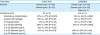
LN = lymph node; RT = right transthoracic; TH = transhiatal; LTA = left thoracoabdominal; OS = overall survival.
*Per protocol analysis.
![]()
The JCOG9502 trial was a Japanese phase III trial that compared extended dissection of the lower mediastinal nodes via the LTA approach and limited dissection via the TH approach in patients with gastric or GEJ adenocarcinoma with esophageal invasion of 3 cm or less [22]. The study participants included 167 patients, including those with Siewert type II (n=95) and type III (n=63) cancer, and they were randomly assigned to the LTA approach (n=85) or the TH approach (n=82). The incidence of pneumonia was significantly higher in the LTA approach group than in the TH approach group (13% vs. 4%, respectively; P=0.048), and the 5-year OS rates were 37.9% and 52.3% in the LTA approach and TH approach groups, respectively. After the first interim analysis, the predicted probability of the LTA approach group having a significantly better OS than the TH approach group at the final analysis was only 3.65%, and the trial was immediately terminated. Body weight loss, vital capacity, and postoperative quality of life were also significantly poorer in the LTA approach group than in the TH approach group [23]. Even in the final analysis after 10 years of follow-up, the 5-year OS in the LTA approach group was worse than that in the TH approach group (37% vs. 51%, respectively; 2-sided P=0.060) [24]. In a subgroup analysis, the 5-year OS rate was lower in the LTA approach group than in the TH approach group in Siewert type II cancer (42% vs. 50%, respectively; P=0.496) compared with Siewert type III cancer (36% vs. 59%, respectively; P=0.102), although the differences were not statistically significant. Based on these results, the TH approach with en bloc lower mediastinal dissection has been recommended for patients with Siewert type II or III cancer with esophageal invasion of 3 cm or less.
ABDOMINAL LN METASTASIS IN GEJ CANCER
LN metastasis in GEJ cancer is most frequently observed in the abdominal LNs around the stomach. Several retrospective studies reported the incidence of abdominal LN metastasis in GEJ cancer [141617252627]. A multicenter retrospective study in Japan examined the incidence of abdominal LN metastasis and the 5-year OS rate of patients with metastasis to each LN station in Siewert type II cancer [16]. To evaluate the theoretical therapeutic impact of dissecting each LN station, the study adopted a method using the therapeutic value index, which was introduced in a previous study [28]. The index was calculated by multiplying the incidence of metastasis to each station by the corresponding 5-year survival rate. The LN stations with a metastatic incidence >10% were the lesser curvature nodes (No. 3), the right paracardial nodes (No. 1), the left paracardial nodes (No. 2), the nodes at the root of the left gastric artery (No. 7), the nodes at the celiac artery (No. 9), and the nodes along the proximal splenic artery (No. 11p). By contrast, the nodes along the greater curvature (Nos. 4sa, 4sb, 4d, and 6), the suprapyloric nodes (No. 5), and the nodes along the proper hepatic artery (No. 12a) had a metastatic incidence below 5%. Furthermore, a nationwide retrospective study in Japan of GEJ cancers <4 cm in diameter whose epicenters were located within 2 cm proximal or distal to the GEJ reported that LN metastases frequently involved the LNs in the abdominal field, especially those at the right and left cardia, lesser curvature, and along the left gastric artery [17]. Therefore, the nodes along the distal portion of the stomach were much less likely to develop metastases, and their dissection seemed unlikely to be beneficial. Therefore, it was suggested that total gastrectomy for dissecting the LNs along the greater curvature (Nos. 4sa, 4sb, 4d, and 6) and the suprapyloric nodes (No. 5) was considered unnecessary for Siewert type II cancer. However, when the distance from the GEJ to the anal edge of the tumor exceeded 5 cm, the incidence of metastasis to these LNs was high, and total gastrectomy was considered necessary [29].
As for Siewert type I and type III cancers, another retrospective study reported that the incidence of metastasis in LN stations other than Nos. 1, 2, 3, and 7 was extremely rare in Siewert type I cancer, while LN stations including Nos. 4sa, 4sb, 4d, 8a, 9, and 11p showed a high incidence of metastasis in Siewert type III cancer [30]. These results were consistent with previous studies of GEJ cancer [1431]. Based on these results, total gastrectomy to remove the LNs along the distal portion of the stomach is considered unnecessary in Siewert type I cancer. Meanwhile, Siewert type III cancer has a high incidence of LN metastasis in No. 4d, and total gastrectomy is considered necessary. In terms of LN stations like No. 19 (infradiaphragmatic nodes) and No. 20 (paraesophageal nodes at the esophageal hiatus of the diaphragm), these are anatomically very close to the GEJ and should be dissected regardless of Siewert classification.
On the basis of the results described above, we propose tentative recommended surgical approaches for cT2-4 GEJ adenocarcinoma according to the tumor location (Table 3).
Table 3
Tentative recommended surgeries for cT2-4 GEJ adenocarcinoma according to tumor location
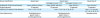
GEJ = gastroesophageal junction; LN = lymph node; RT = right transthoracic; TH = transhiatal; Nos. = numbers.
![]()
PAND
The PANDs are considered to be extraregional in the TNM classification, and PAND is not a standard treatment in GEJ cancer [8]. However, previous studies reported that lymphatic pathways from the GEJ lead directly into the paraaortic area, and a particularly high incidence of metastasis was observed in LN station No. 16a2 (paraaortic LNs between the upper margin of the origin of the celiac artery and the lower border of the left renal vein) [323334]. Another previous study reported that patients with Siewert type II cancer developed metastasis in paraaortic LN station No. 16a2 in 14.4% of cases, with a 5-year OS rate of 16.7%. Therefore, the paraaortic LNs may be a target of nodal dissection in patients with GEJ cancer [16]. In addition, a retrospective study at a single institution reported that PAND was an independent prognostic factor in patients with Siewert type II cancer [35].
Meanwhile, prophylactic PAND did not improve the survival rate in patients with cT3-4 gastric cancer in a phase III trial (JCOG9501) [36]. However, this trial did not include GEJ cancer, and a subgroup analysis showed that the therapeutic value of dissecting the paraaortic LNs was higher for cancers located in the upper region than in other regions. These results suggest that further data is needed to determine whether paraaortic LNs should be dissected in patients with GEJ cancer.
JAPANESE NATIONWIDE PROSPECTIVE TRIAL OF GEJ CANCER
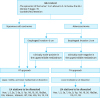
Although we have discussed surgical treatment for GEJ cancer from the perspective of numerous retrospective studies, a few prospective studies have been conducted. For this reason, JGCA and JES are jointly conducting a nationwide prospective trial evaluating the incidence of LN metastasis, including the mediastinal, abdominal, and paraaortic LNs, to determine the optimal extent of LN dissection in GEJ cancers. The trial includes patients with cT2-4 GEJ adenocarcinoma and squamous cell carcinoma with an epicenter between 2 cm above and below the GEJ. Patients with GEJ cancer with a length of esophageal invasion of 3 cm or less and clinically node-negative upper and middle mediastinal fields undergo lower esophagectomy with lower mediastinal LN dissection via the TH approach, while other patients undergo subtotal esophagectomy with upper, middle, and lower mediastinal LN dissection via the RT approach (Fig. 1). All patients undergo PAND (No. 16a2). The primary endpoint is the incidence of metastasis in each LN, and the secondary endpoints are recurrence-free survival, OS, the therapeutic value index of each LN, R0 resection rate, postoperative complications, and recurrence sites. Patient enrollment was completed with 371 patients in September 2017. Although the results have not been reported yet, this trial is expected to be valuable in determining the optimal extent of LN dissection and should help establish standard treatment approaches for GEJ cancer.
CONCLUSIONS
Tumor location is considered essential when determining the treatment strategy for GEJ cancer. The Siewert classification is used worldwide and is beneficial for choosing the optimal surgical procedure: partial gastrectomy and subtotal esophagectomy with thorough mediastinal LN dissection via RT approach for Siewert type I and total gastrectomy with lower mediastinal LN dissection via TH approach for Siewert type III. However, no consensus has been reached concerning the treatment of Siewert type II, because few randomized trials have been conducted for GEJ cancer as a separate entity. In addition, there is room for debate in terms of PAND in patients with GEJ cancer. A currently ongoing Japanese nationwide prospective trial evaluating the optimal extent of LN dissection in GEJ cancer is expected to lead to the standardization of surgical approaches for GEJ cancer in the future.
 XML Download