

 PDF
PDF ePub
ePub Citation
 Print
Print


INTRODUCTION
Papillary thyroid carcinoma (PTC) is the most common type of thyroid cancers. Its incidence rate is recently growing worldwide because of rapid development of imaging instruments and diagnostic techniques such as fine-needle aspiration biopsy (FNAB) (123). In addition, most PTCs are known as an indolent mild cancer with favorable prognosis, having a survival rate of 30 years in >90% of cases (4).
The first-line treatment of PTC is thyroidectomy (5). When only unilateral lobectomy was performed, thyroid cancer in the contralateral lobe should be excluded before surgery. However, occult malignant foci (OMF) in the contralateral lobe not found before surgery (6). Preoperatively undiagnosed OMF in contralateral thyroid lobes can cause postoperative remnant disease not only may lead to an additional surgery, but also can be associated with regional and distant metastasis (7).
The primary objective of this study was to assess the presence of contralateral OMF in patients with unilateral confined PTC.
METHODS
We retrospectively evaluated 714 patients who received total thyroidectomy with central lymph node (LN) dissection after being diagnosed as having unilaterally confined PTC, between January 2010 and December 2012. Preoperative ultrasonography (US) and US-guided FNAB was performed for all the patients. Neck computed tomography (CT) was further performed if an extrathyroidal extension (ETE) is suspected from US findings, or central or lateral LN metastasis was present.
The patient group of 2010 to 2012, according to 2009 American Thyroid Association (ATA) Guideline (5) or 2010 Korean Thyroid Association (KTA) Management Guidelines (89) went through total thyroidectomy for thyroid cancer larger than 1 cm or <1 cm with multifocality, ETE (included gross ETE and micro/minimal/minor ETE by American Joint Committee on Cancer (AJCC) Cancer Staging Manual, 7th edition (10), central LN metastasis (only level 6, AJCC 7th edition), a personal history of radiation therapy to the head and neck, or familial thyroid carcinoma (5). Additionally, when gross finding and operator's empirical decision at the operation field favored it, total thyroidectomy was done.
The contralateral OMF was defined as foci found in the pathological examination after surgery, but not diagnosed based on preoperative radiological evaluation and FNAB results. By dividing the patients into 2 groups according to the presence or absence of contralateral OMF, the relationship between OMF and clinicopathological factors such as age, sex, tumor size, multifocality (more than 2 malignant nodules in unilateral lobe), chronic lymphocytic/Hashimoto's thyroiditis, contralateral lobe benign nodule, ETE and central LN metastasis were analyzed by using SPSS 18.0 (SPSS Inc., Chicago, IL, USA). The relationship between the predictive factors and OMF was analyzed by using χ2 test. A multivariate analysis was performed by using multivariate logistic regression analysis. Data were considered statistically significant when the significance level was <0.05
RESULTS
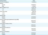
Among the 714 patients included in the present study, 637 were women and 77 were men. The mean age was 47.5 years (range, 8–83 years) at the time of surgery.
After performing total thyroidectomy, contralateral OMF was found in 61 patients (8.5%) on pathological examination. The mean size of the tumors in 61 patients with contralateral malignant nodules was 0.2 cm (range, 0.1–0.3 cm). Furthermore, all of the OMF were papillary thyroid microcarcinoma (PTMC).
The mean size of the primary malignant tumors was 1.25 cm (range, 0.1–7 cm). The mean size of the primary malignant tumors with OMF was 1.43 cm (range, 0.4–4 cm). Of the patients, 280 (39.2%) had multifocal PTCs with ≥ malignant 2 nodules in one lobe, with a maximum of 4 nodules found. ETE of tissues surrounding the thyroid, and metastasis to the central neck LN occurred in 321 (45%) and 370 (51.8%), respectively. Chronic lymphocytic/Hashimoto's thyroiditis was found in 148 patients (20.7%; Table 1).
Table 1
Demographic characteristics of 714 patients with papillary thyroid carcinoma
![]()
When the patients were divided into 2 groups depending on the presence or absence of OMF, the mean (standard deviation) ages of the patients in the 2 groups were 48.05±11.30 and 47.49±11.36, respectively. OMF were more frequently found in the group aged >45 years, although the difference was not statistically significant (P=0.753). The sex distribution was 58 women and 3 men (P=0.122). When the patients were subdivided based on primary tumor size, OMF in the contralateral lobe were more frequently found as the tumor sizes increased, with statistical significance (P=0.012). In the patients with multifocal thyroid cancer in the lobe that had primary tumor, OMF was observed in the other lobe regardless of the number of primary malignant nodules, and the difference was statistically significant (P=0.001). However, accompanying chronic lymphocytic/Hashimoto's thyroiditis, contralateral lobe benign nodule, ETE or central LN metastasis had no significant relationship with the presence or absence of OMF (Table 2).
Table 2
Clinicopathologic factors in relation to OMF of contralateral lobe in 714 patients with papillary thyroid carcinoma
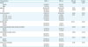
Values are shown as number (%) or mean±standard deviation.
OMF = occult malignant foci; m = mass; ETE = extrathyroidal extension; LN = lymph node.
![]()
Multivariate logistic regression analysis was performed for primary tumor size and multifocality, which were statistically significant on univariable analysis. OMF in the other lobe were more frequently found as the primary tumor sizes (P=0.027) and multifocality (P<0.001) increased (Table 3).
Table 3
Multivariate analysis of clinicopathologic factors associated with OMF in patients with unilateral papillary thyroid carcinoma

| Characteristics | B | SE | Sig. | Exp (B) | 95% CI | |
|---|---|---|---|---|---|---|
| Lower | Upper | |||||
| Tumor size | 0.317 | 0.144 | 0.027 | 1.374 | 1.036 | 1.821 |
| Multifocality | 0.852 | 0.116 | <0.001 | 2.345 | 1.868 | 2.943 |
| Constant | −4.434 | 0.370 | 0.000 | 0.012 | ||
OMF = occult malignant foci; B = coefficient of regression; SE = standard error; Sig. = significance probability; Exp (B) = odds ratio; CI = confidence interval.
![]()
DISCUSSION
Decisions regarding the extent of thyroid surgery are influenced by several factors (11).
Older age (>45 years), contralateral thyroid nodules may be criteria for recommending a bilateral procedure either because of plans for radioactive iodine (RAI) therapy, to facilitate follow-up strategies, or to address suspicions of bilateral disease (12131415). However, in this study, age (<45 years, ≥45 years), contralateral lobe benign nodules had no significant (P=0.248) relationship with the presence or absence of OMF (Table 2). The 2017 AJCC Cancer Staging Manual, 8th edition (16) suggest the age at diagnosis cutoff used for staging was increased from 45 years to 55 years. The significant clinical benefit of preventing upstaging based only on age of diagnosis between 45 and 55 years in patients who otherwise would be considered low-risk (stage I or II). Other investigators have endorsed using an age cutoff of 55 years as the optimal single time point for papillary thyroid cancer prognostic models (1718192021).
Kasai and Sakamoto (22) reported that the invasiveness differed depending on the primary tumor size. 2015 ATA Guidelines (23) suggest that if the primary thyroid carcinoma is >4 cm, near-total or total thyroidectomy is necessary if the overall strategy is to include RAI therapy post-operatively (2015 ATA). for tumors that are between 1 and 4 cm in size, either a bilateral thyroidectomy (total or near-total) or a unilateral procedure (thyroid lobectomy) may be suitable as treatment plan (2015 ATA). Based on our study, the mean size of the primary malignant nodules with OMF was 1.43 cm (range, 0.4–4 cm). When the patients were subdivided based on primary tumor size, OMF in the contralateral lobe were more frequently found as the tumor sizes increased, with statistical significance (P=0.012).
2016 KTA Management Guidelines (24) and 2017 The Korean Association of Thyroid and Endocrine Surgeons (KATES) Guidelines (25) suggest that the initial surgical procedure of gross extrathyroidal extension should include a near-total or total thyroidectomy (2425). In this study, ETE had no significant (P=0.909) relationship with the presence or absence of OMF (Table 2). The limitation of this study is our data included grossETE and micro/minimal/minor ETE. Pathologically, the thyroid has an incomplete capsule. Because, the thyroid gland may contain adipose tissue and skeletal muscle under normal circumstances. According to the College of American Pathologists, defining (minimal) ETE may be problematic and subjective (26). Microscopic ETE is not an independent prognostic factor for persistent/recurrent disease. The disease-free survival is equivalent in patients with microscopic ETE and those with completely intrathyroidal tumors (2627282930). The 2017 AJCC Cancer Staging Manual 8th edition suggested minor ETE was removed from the definition of T3 disease. As a result, minor ETE does not affect either T category or overall stage.
In the present study, the primary tumor multifocality is a risk factor that can predict the presence of contralateral lobe occult cancers. Kim et al. (31) published that multifocality is a unique factor that can predict the incidence of contralateral cancer after unilateral surgery of thyroid cancer. In addition, the presence of PTMC in the contralateral lobe is related with the multifocality of primary cancer (3233). Park et al. (34) and Giannini et al. (35) reported that each multifocal cancer occurs independently by studying BRAF mutation rate. Sugg et al. (36) thought that individual tumors are associated with multifocality via a distinct pattern of RET/PTC rearrangement. According to Wang et al. (37), if bilateral PTCs are observed, the disease would have already progressed, disease-free survival duration would be shorter, different biological characteristics would be observed, and the stage would be more advanced than that of unilateral PTC. Patients with indeterminate nodules who have bilateral nodular disease, those with significant medical comorbidities, or those who prefer to undergo bilateral thyroidectomy to avoid the possibility of requiring a future surgery on the contralateral lobe, may undergo total or near-total thyroidectomy, assuming completion thyroidectomy would be recommended if the indeterminate nodule proved malignant following lobectomy. including the estimated pre-surgical likelihood of malignancy based upon clinical risk factors (>4 cm, family history, and/or radiation history) (38394041). However, not only the growing rate and prognosis of OMF after thyroid lobectomy, but also the usefulness of routine total thyroidectomy for detection of thyroid cancer of <0.5 cm in size are controversial. In PTMC patients, delayed surgery was not associated with higher risk of structural recurrent/persistent disease compared to immediate surgery. These findings support the notion that surgical treatment can be safely delayed in patients with PTMC under close monitoring (42). Ito et al. (43) proposed active surveillance is the optimal first line of management for all adult patients with low-risk papillary microcarcinomas.
In this retrospective study, the incidence of contralateral OMF among 714 patients who received total thyroidectomy after being diagnosed as having unilateral PTC was 61 patients (8.5%), which is lower than that reported in a previous study (15.6%–27%) (4445). In the our study, the mean size of the contralateral OMF was 0.2 cm (range, 0.1–0.3 cm) and all of the OMF were PTMCs. As small PTMC grows slowly, has favorable prognosis, and shows a pattern like that of benign lesion (46), active monitoring and observation are necessary when unilateral lobectomy is performed by considering primary tumor size and multifocality.
Among the clinicopathological factors related to the presence of contralateral OMF before surgery, age, sex, chronic lymphocytic/Hashimoto's thyroiditis, benign nodule, ETE, and central LN metastasis were not significant predictive factors, but the primary tumor size and multifocality had a significant relationship with the incidence of contralateral OMF (Table 3). However, in this study, the mean size of the contralateral OMF was very small 0.2 cm (range, 0.1–0.3 cm) and all of the OMF were PTMCs. So if the lobectomy was performed already, meticulous follow-up is necessary about remnant lobe than completion thyroidectomy.
 XML Download