Abstract
Background/Aims
Ligation-assisted endoscopic submucosal resection (ESMR-L) is preferred for the treatment of rectal neuroendocrine tumors because its results are better than those for endoscopic mucosal resection (EMR), and the procedure time is shorter and the incidence of complications is lower than endoscopic submucosal dissection. The aim of this study was to evaluate the clinical usefulness of ESMR-L compared with EMR for rectal neuroendocrine tumors.
Methods
From March 2007 to May 2017, 148 patients diagnosed with rectal neuroendocrine tumors were divided into ESMR-L and EMR groups and analyzed retrospectively.
Go to : 
References
1. Cho MY, Kim JM, Sohn JH, et al. Current trends of the incidence and pathological diagnosis of Gastroenteropancreatic Neuroendocrine Tumors (GEP-NETs) in Korea 2000–2009: multicenter study. Cancer Res Treat. 2012; 44:157–165.

2. Scherübl H. Rectal carcinoids are on the rise: early detection by screening endoscopy. Endoscopy. 2009; 41:162–165.

3. Modlin IM, Oberg K, Chung DC, et al. Gastroenteropancreatic neuroendocrine tumours. Lancet Oncol. 2008; 9:61–72.

4. Mashimo Y, Matsuda T, Uraoka T, et al. Endoscopic submucosal resection with a ligation device is an effective and safe treatment for carcinoid tumors in the lower rectum. J Gastroenterol Hepatol. 2008; 23:218–221.

5. Kim HH, Park SJ, Lee SH, et al. Efficacy of endoscopic submucosal resection with a ligation device for removing small rectal carcinoid tumor compared with endoscopic mucosal resection: analysis of 100 cases. Dig Endosc. 2012; 24:159–163.

6. Kim YJ, Lee SK, Cheon JH, et al. Efficacy of endoscopic resection for small rectal carcinoid: a retrospective study. Korean J Gastroenterol. 2008; 51:174–180.
7. Kim KM, Eo SJ, Shim SG, et al. Treatment outcomes according to endoscopic treatment modalities for rectal carcinoid tumors. Clin Res Hepatol Gastroenterol. 2013; 37:275–282.

8. Abe T, Kakemura T, Fujinuma S, Maetani I. Successful outcomes of EMR-L with 3D-EUS for rectal carcinoids compared with historical controls. World J Gastroenterol. 2008; 14:4054–4058.

9. Saha S, Hoda S, Godfrey R, Sutherland C, Raybon K. Carcinoid tumors of the gastrointestinal tract: a 44-year experience. South Med J. 1989; 82:1501–1505.
10. Higaki S, Nishiaki M, Mitani N, Yanai H, Tada M, Okita K. Effectiveness of local endoscopic resection of rectal carcinoid tumors. Endoscopy. 1997; 29:171–175.

11. Soga J. Early-stage carcinoids of the gastrointestinal tract: an analysis of 1914 reported cases. Cancer. 2005; 103:1587–1595.
12. Shirouzu K, Isomoto H, Kakegawa T, Morimatsu M. Treatment of rectal carcinoid tumors. Am J Surg. 1990; 160:262–265.

13. Matsui K, Iwase T, Kitagawa M. Small, polypoid-appearing carcinoid tumors of the rectum: clinicopathologic study of 16 cases and effectiveness of endoscopic treatment. Am J Gastroenterol. 1993; 88:1949–1953.
14. Schindl M, Niederle B, Häfner M, et al. Stage-dependent therapy of rectal carcinoid tumors. World J Surg. 1998; 22:628–633. discussion 634.

15. Kobayashi K, Katsumata T, Yoshizawa S, et al. Indications of endoscopic polypectomy for rectal carcinoid tumors and clinical usefulness of endoscopic ultrasonography. Dis Colon Rectum. 2005; 48:285–291.

16. Onozato Y, Kakizaki S, Ishihara H, et al. Endoscopic submucosal dissection for rectal tumors. Endoscopy. 2007; 39:423–427.

Go to : 
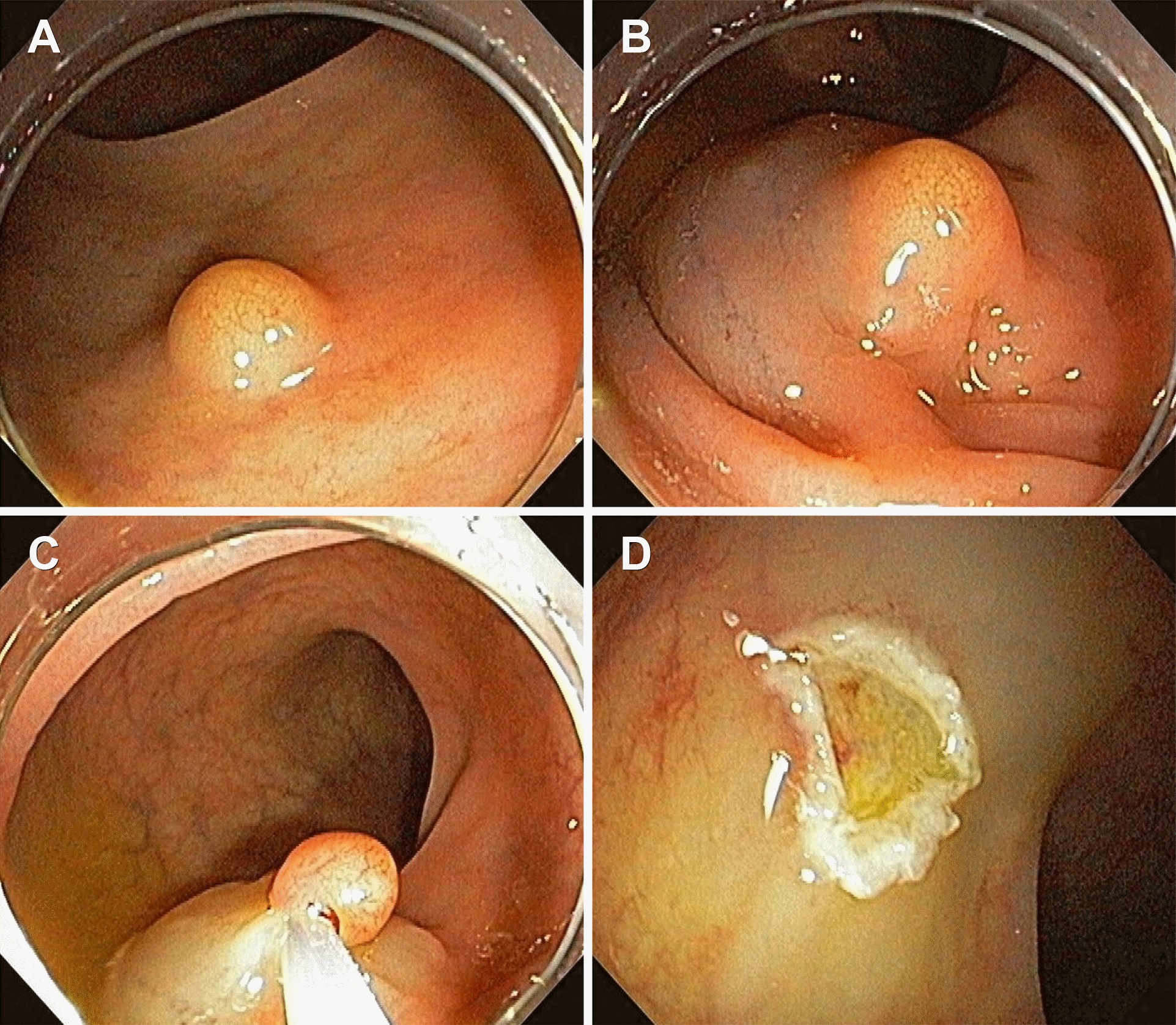
 | Fig. 1.Endoscopic mucosal resection. (A) Submucosal tumor is observed. (B) Submucosal saline mixed with epinephrine and indigo carmine solution is injected beneath the neuroendocrine tumor. (C) Elevated tumor is snared. (D) Tumor is resected using high-frequency currents. |
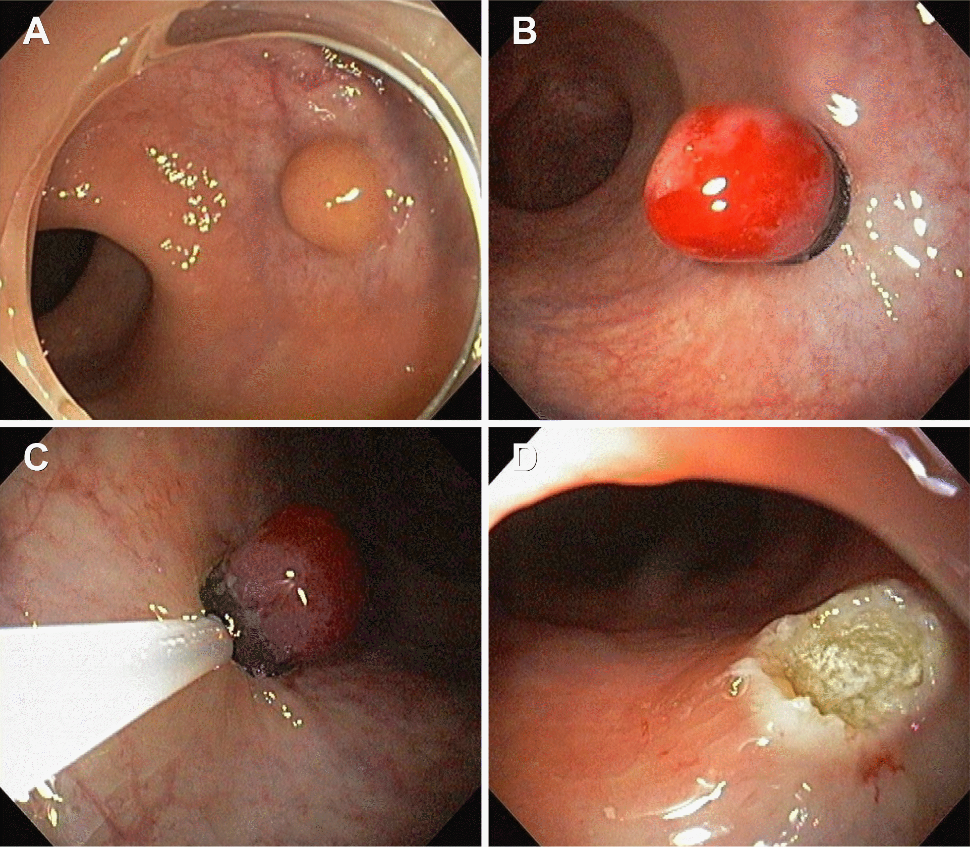
 | Fig. 2.Endoscopic submucosal resection with a ligation device. (A) Submucosal saline mixed with epinephrine and indigo carmine solution is injected beneath the neuroendocrine tumor. (B) The lesion is aspirated into the ligation device and elastic band is deployed. (C) Snaring below the elastic band. (D) After resection, clear ulcer surface is observed. |
Table 1.
Baseline Characteristics of the Study Groups
Table 2.
Characteristics of Tumor
Table 3.
Clinical Outcomes according to Endoscopic Treatment Modality
| ESMR-L (n=120) | EMR (n=28) | p-value | |
|---|---|---|---|
| Procedure time (min) | 7.4±2.1 | 6.6±4.6 | 0.336 |
| Complete resection | 112 (93.3) | 21 (75.0) | 0.009 |
| Curative resection | 111 (92.5) | 20 (71.4) | 0.003 |
| Noncurative resection | |||
| Resection margin involvement | 8 (6.7) | 7 (25.0) | 0.009 |
| Base margin involvement | 3 (2.5) | 6 (21.4) | 0.002 |
| Lateral margin involvement | 0 (0.0) | 0 (0.0) | |
| Both margin involvement | 5 (4.0) | 1 (3.6) | 1.000 |
| Lymphatic invasion | 1 (0.8) | 1 (3.6) | 0.344 |
| Vascular invasion | 1 (0.8) | 0 (0.0) | 1.000 |
| Post-procedure complications a | 7 (5.8) | 1 (3.6) | 1.000 |
| Follow up period, month | 41.8±27.4 | 51.1±24.8 | 0.111 |
| Follow up after non-curative resection | |||
| Surgery | 3 (2.5) | 0 (0.0) | |
| Endoscopic follow up | 6 (5.0) | 5 (17.9) | |
| Follow up loss | 0 (0.0) | 3 (10.7) | |
| Recurrence | 3 (3.2) | 2 (8.0) | 0.278 |
| Follow up loss | 25 (20.8) | 3 (10.7) |
Table 4.
Clinical Features of Patients with Recurrence after Resection of Rectal Neuroendocrine Tumor
|
ESMR-L |
EMR |
||||
|---|---|---|---|---|---|
| 1 | 2 | 3 | 4 | 5 | |
| Recurrent interval (month) | 22 | 13 | 20 | 33 | 35 |
| Recurrent location, from anal verge | 10 cm | 5 cm | 5 cm | 1 cm | 20 cm |
| Previous resection site recurrence | Yes | Yes | Yes | Yes | No |
| Previous curative resection | No a | Yes | Yes | Yes | Yes |
| Previous tumor size (mm) | 5×5 | 5×5 | 5×5 | 5×5 | 6×6 |
| Tumor with central depression or ulceration | No | No | No | Yes | No |


 PDF
PDF ePub
ePub Citation
 Print
Print


 XML Download