Journal List > Korean Circ J > v.48(10) > 1101173

Kaneko, Kobayashi, Hachinohe, Sumino, Furugen, Kawamura, Doi, and Fujita: Successful Direct Iliac Transcatheter Aortic Valve Implantation to Overcome Significant Tortuosity of the Thoracic Aorta
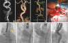
An 87-year-old woman with symptomatic severe aortic stenosis underwent transcatheter aortic valve implantation (TAVI). Multi-detector computed tomography showed a significantly tortuous descending thoracic aorta (Figure 1A and B). Due to the high risk of coronary obstruction and a horizontal aorta, a SAPIEN 3 (Edwards LifeSciences, Irvine, CA, USA) prosthesis via transfemoral access was considered. However, the total length of the Edwards expandable introducer sheath (eSheath, 360 mm) was not sufficient to reach the acute bend in the thoracic aorta, which measured 415 mm in length. Therefore, direct right common iliac access obtained by retroperitoneal approach was chosen to insert the eSheath beyond the acute bend of the thoracic aorta (Figure 1C and D). After insertion of a single stiff wire, the tip of the eSheath was successfully placed beyond the acute bend of the thoracic aorta with no resistance (Figure 1E). Using buddy wire technique, smooth delivery and implantation of a 26-mm SAPIEN 3 was successful without vascular complication (Figure 1F-H).
An extremely tortuous thoracic aorta can be a major hindrance to transfemoral access and is associated with fatal complications, such as aortic dissection or rupture.1)2) Compared with advancement of bulky TAVI device, insertion of the eSheath beyond the acute bend via a direct iliac access can be easier and less traumatic due to its tapered tip and hydrophilic coating, finally yielding an excellent result. A severely calcified aorta may not be straightened even after insertion of double stiff wires. Therefore, the calcium extent and distribution of aorta will be important factors for successful sheath insertion.

Figures and Tables

Figure 1

(A-C) Pre-procedural MDCT (A) Left anterior oblique view of maximum intensity projection showing a markedly tortuous thoracic aorta and no significant calcification around the acute bend (red arrows). (B) Posterior volume-rendering image of the aorta. (C) Volume-rendering image of the aorta showing the distance from the Rt.CFA (white bidirectional arrow) and the Rt.CIA (yellow bidirectional arrow) to the acute bend in the thoracic aorta. (D) Direct iliac approach. (E-H) Fluoroscopic images during the procedure. (E) Facilitating smooth insertion of the eSheath (yellow arrow) beyond the tortuous segment of the thoracic aorta (red arrows). (F) Successful implantation of the prosthetic valve. (G) Aortography showing trivial paravalvular leak. (H) Final aortography showing no vascular complication.

MDCT = multi-detector computed tomography; Rt.CFA = right common femoral artery; Rt.CIA = right common iliac artery.
kcj-48-949-g001

Notes

Conflict of Interest The authors have no financial conflicts of interest.

Author Contributions

  • Conceptualization: Kobayashi K, Hachinohe D, Furugen A, Sumino S.

  • Formal analysis: Kawamura T.

  • Supervision: Doi H, Fujita T.

  • Writing - original draft: Kaneko U.

References

1. Aminian A, Lalmand J, El Nakadi B. Perforation of the descending thoracic aorta during transcatheter aortic valve implantation (TAVI): an unexpected and dramatic procedural complication. Catheter Cardiovasc Interv. 2011; 77:1076–1078.
crossref
2. de Agustin JA, Jiménez-Quevedo P, Nombela-Franco L, et al. Descending aorta rupture during transcatheter aortic valve replacement. Circulation. 2016; 133:e14–e17.
crossref
TOOLS
ORCID iDs

Umihiko Kaneko
https://orcid.org/0000-0003-2392-5084

Daisuke Hachinohe
https://orcid.org/0000-0003-4828-1836

Similar articles