

 PDF
PDF ePub
ePub Citation
 Print
Print


Abstract
Chopart joint fracture and dislocation are rare injuries compared with other joint injuries with various clinical manifestations. Moreover, there is a lack of knowledge of the radiological findings of the joints, and thus, the extent of joint ligament damage may be underestimated, leading to improper treatment. This paper reports three cases of Chopart joint injury and seeks to reconsider the importance of Chopart joint evaluation and treatment.
References
1. Wolf JH. François Chopart (1743–1795): inventor of the partial foot amputation at the tarsometatarsal articulation. Orthop Traumatol. 2000; 8:314–7.
2. Rammelt S, Schepers T. Chopart injuries: when to fix and when to fuse? Foot Ankle Clin. 2017; 22:163–80.
3. Rammelt S, Grass R, Schikore H, Zwipp H. [Injuries of the Chopart joint]. Unfallchirurg. 2002; 105:371–83. ; quiz 384–5. German.
4. Schneiders W, Rammelt S. Joint-sparing corrections of malunited Chopart joint injuries. Foot Ankle Clin. 2016; 21:147–60.

5. Kutaish H, Stern R, Drittenbass L, Assal M. Injuries to the Chopart joint complex: a current review. Eur J Orthop Surg Traumatol. 2017; 27:425–31.
6. Astion DJ, Deland JT, Otis JC, Kenneally S. Motion of the hindfoot after simulated arthrodesis. J Bone Joint Surg Am. 1997; 79:241–6.
7. Zwipp H. Chirurgie des fusses. Wien: Springer-Verlag;1994.
8. Richter M, Thermann H, Huefner T, Schmidt U, Goesling T, Krettek C. Chopart joint fracture-dislocation: initial open reduction provides better outcome than closed reduction. Foot Ankle Int. 2004; 25:340–8.
9. Kotter A, Wieberneit J, Braun W, Rüter A. [The Chopart dislocation. A frequently underestimated injury and its sequelae. A clinical study]. Unfallchirurg. 1997; 100:737–41. German.
10. Rammelt S, Marti RK, Zwipp H. [Arthrodesis of the talonavicular joint]. Orthopade. 2006; 35:428–34. German.
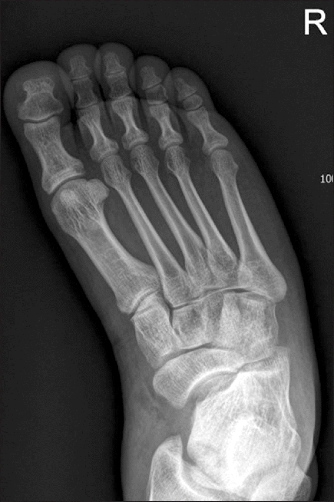
Figure 1.
Preoperative simple radiographs of the foot anteroposterior (A) and lateral (B) show navicular and cuboid bone fractures, combined with talonavicular and calcaneocuboid joint dislocations.
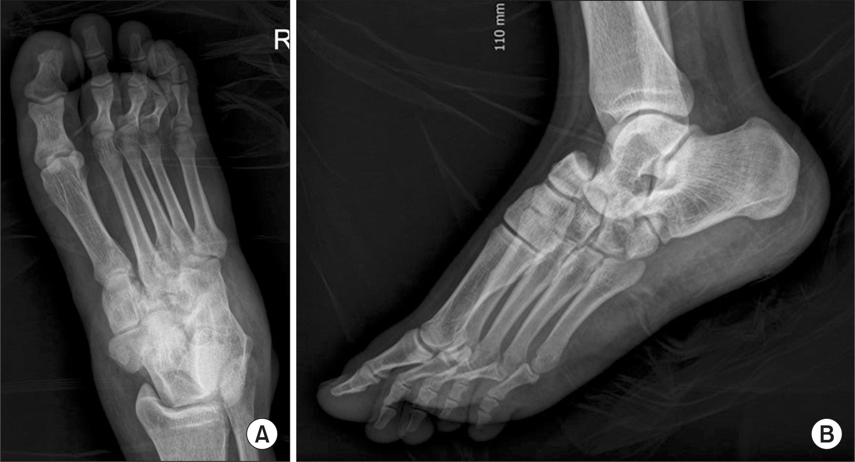
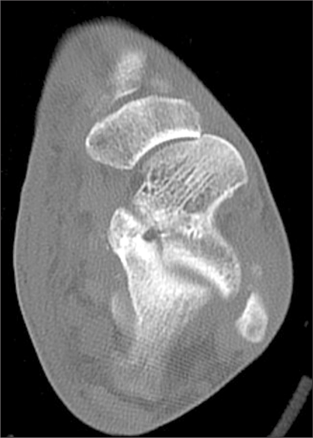
Figure 2.
Sagittal views of computed tomography show navicular bone fracture (A), dislocation of talonavicular joint (B), and fracture and dislocation of calcaneocuboid joint (C).
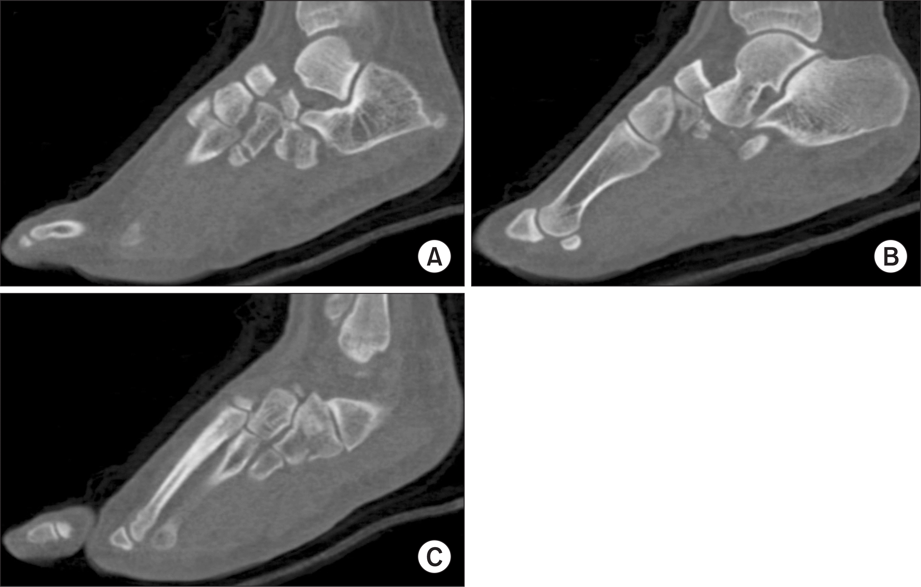
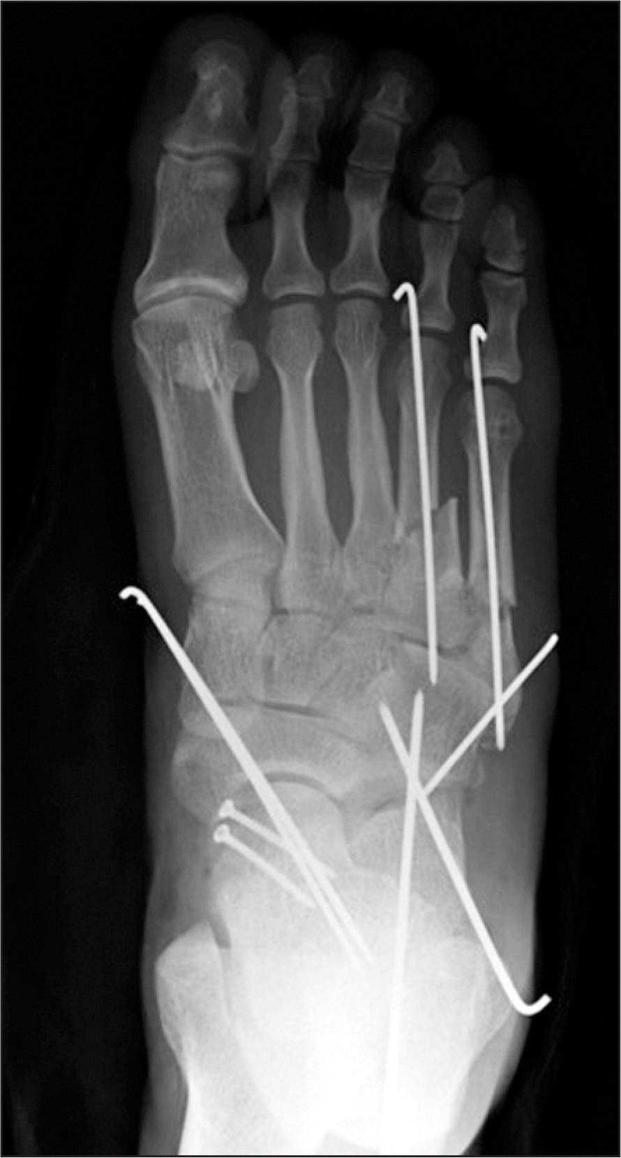
Figure 4.
Postoperative simple radiographs of the foot anteroposterior (A) and lateral (B) show the percutaneous K-wire fixation after open reduction for talonavicular injury, and the external fixation after closed reduction for calcaneocuboid injury.
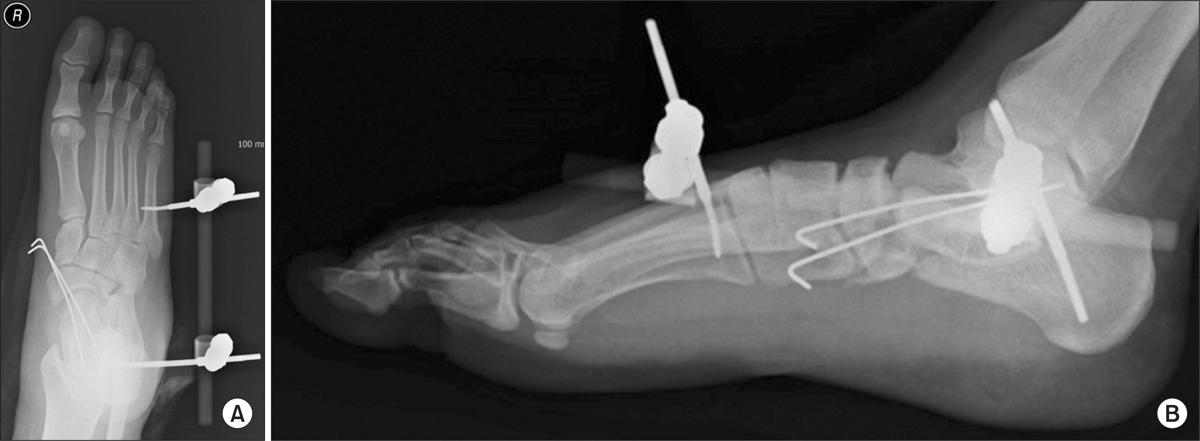
Figure 5.
Conversion operation was done. Postoperative simple radiograph of the foot anteroposterior shows that only one of the two percutaneous K-wire and external fixator were removed. K-wire, cortical screw fixation and bridging plate internal fixation were done respectively in navicular bone fracture and cuboid bone fracture.
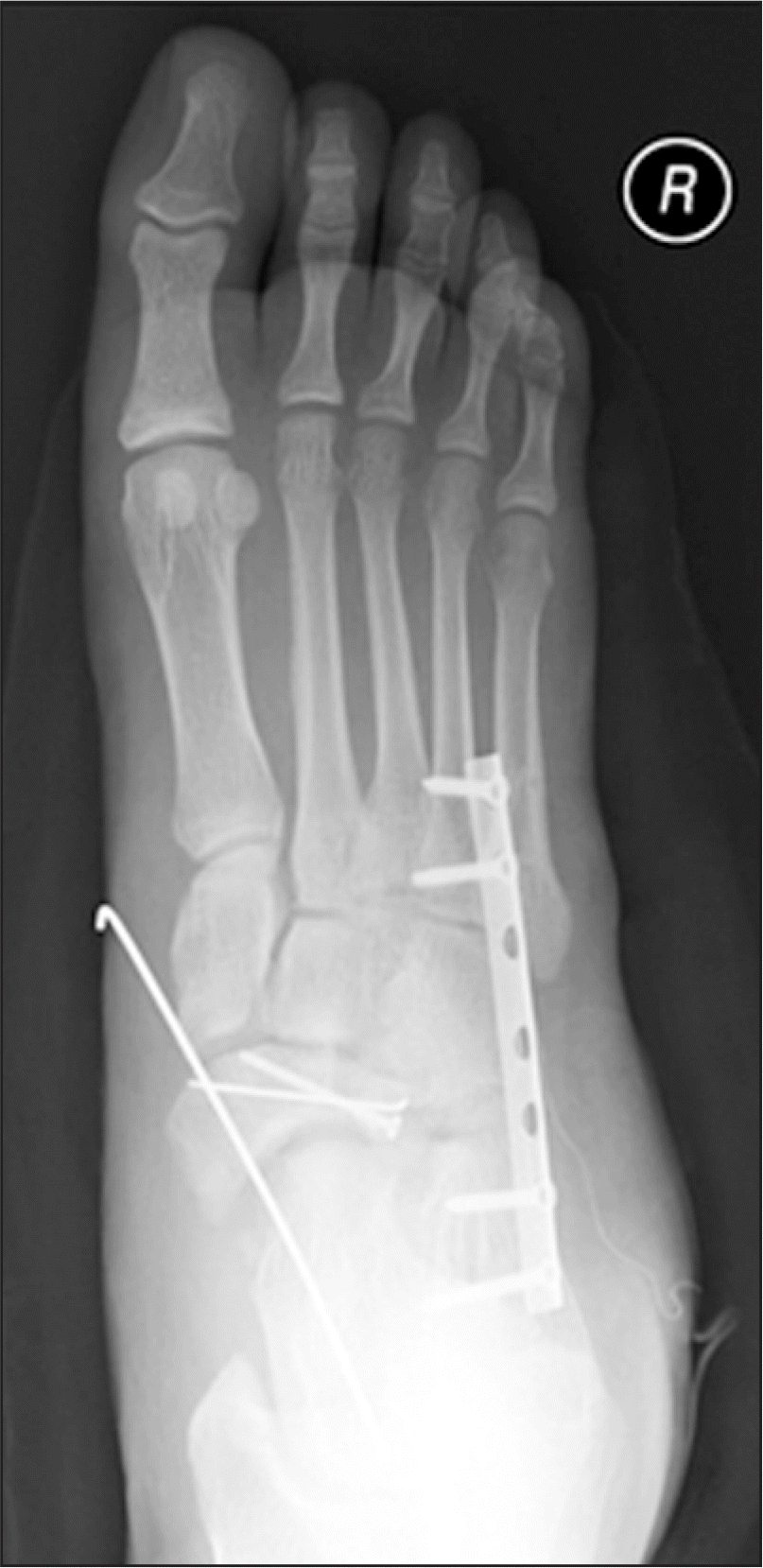
Figure 6.
After 6-month (A) and 7-month (B) following the conversion operation, bony union of navicular and cuboid bones were confirmed on standing lateral views of simple radiograph. Bridging plate was removed (B). Dislocation of talonavicular jointand flat foot deformity were observed (A, B).

Figure 7.
Postoperative simple radiographs of the foot anteroposterior (A) and lateral (B) show that arthrodesis with auto iliac bone graft was done in talonavicular joint.
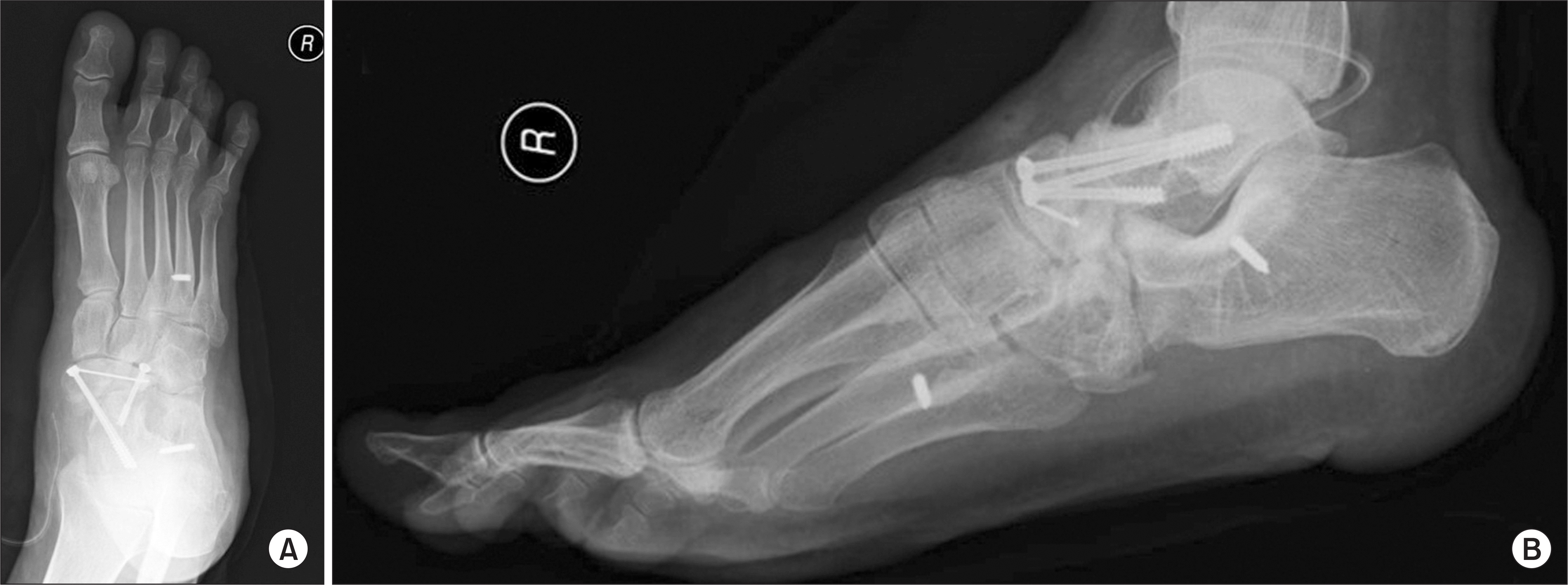
Figure 8.
Preoperative simple radiographs of the foot anteroposterior (A) and lateral (B) show that 3rd to 5th metatarsal bone fractures, talar head and cuboid bone fractures were observed and dislocations were observed in talonavicular joint, calcaneocuboid joint.
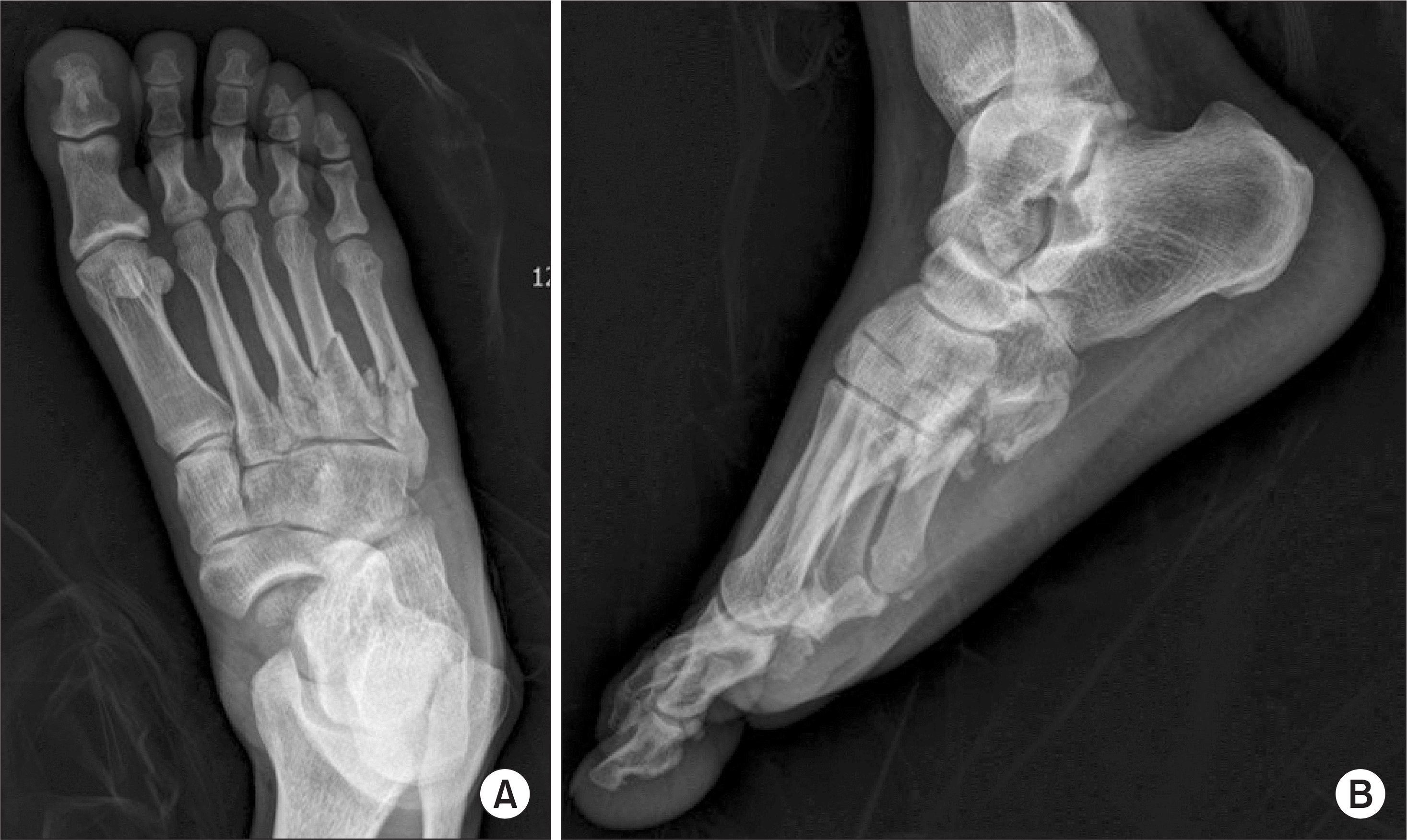
Figure 9.
Sagittal views of computed tomography show dislocation of talonavicular joint (A), talar head fracture (B), and fracture and dislocation of calcaneocuboid joint (C).
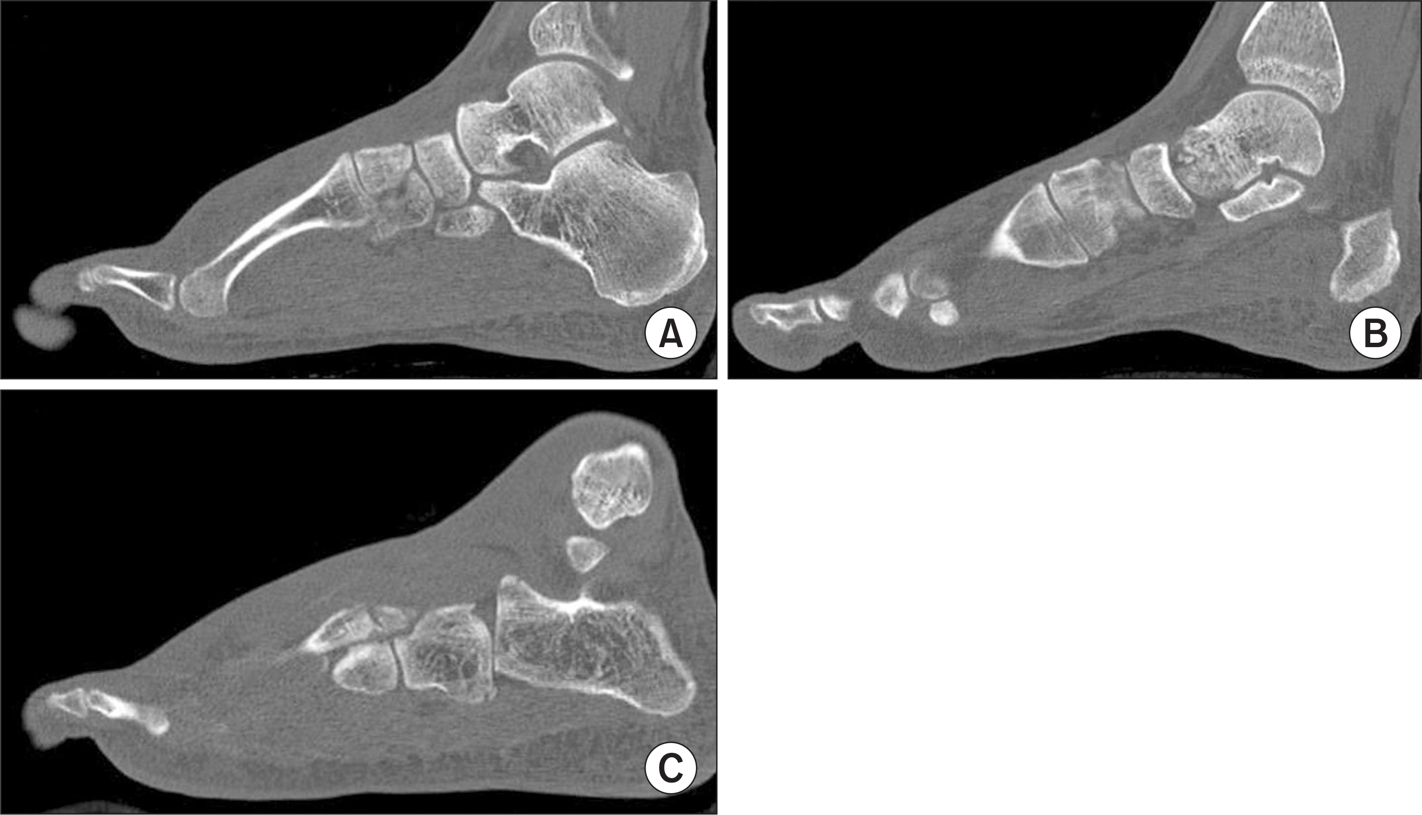
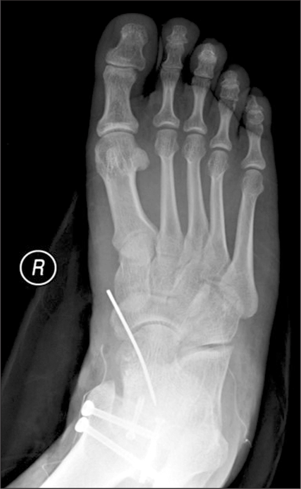
Figure 10.
Postoperative simple radiograph of the foot anteroposterior shows that percutaneous K-wire fixation, cortical screw fixation with open reduction in talonavicular lesion and percutaneous K-wire fixation with closed reduction in calcaneocuboid lesion and 4th to 5th metatarsal bone fracture were done.
 XML Download