

 PDF
PDF ePub
ePub Citation
 Print
Print


Introduction
Odontogenic lesions appear in the jaw, and various imaging modalities have been developed to assist dentists in detecting and diagnosing these lesions, as well as planning and performing follow-up procedures. Plain radiography is the first method used to diagnose intraosseous and extraosseous jaw lesions.12 Computed tomography (CT), cone-beam computed tomography (CBCT), and magnetic resonance imaging (MRI) are useful techniques for evaluating the margins, dimensions, and exact anatomical site of lesions.345 However, CT scans are associated with high radiation doses,6 and MRI equipment is very expensive and cannot be used in patients with cardiac pacemakers and certain types of surgical clips.7
The traditional method for assessing healing after surgery is a clinical examination coupled with recall radiographs. Previous studies showed that conventional radiographs were not an ideal tool for monitoring healing,89 because many errors can take place at any step during the developing process. Film processing consists of a series of steps that produce a visible permanent image on a dental radiograph. These steps are development, rinsing, fixing, washing, and drying. Moreover, the temperature and concentration of the developer and fixer strongly affect the time needed for the process to be optimal.
Additionally, radiography has several limitations, such as the superimposition of anatomical structures, inability to observe small changes in bone density, and interobserver variations in the interpretation of radiographs. Moreover, to monitor healing, multiple radiographs are required, exposing patients to unnecessary radiation, especially if tomographic images such as CBCT or CT are used instead of plain radiographs.8 In the healing phase, vascular changes are the most important feature, and conventional radiographs cannot reveal these changes in the bone.10 Ultrasonography (US) therefore plays an important role in evaluating the solid and cystic content of jaw lesions.1112 US imaging technology has been used in medicine for many years, but in the dental field, its use has been limited and mainly restricted to soft tissue applications.13 These applications are improved by their combination with color power Doppler to assess blood flow, which recently has been found to be useful in the diagnosis of jaw lesions and follow-up during the healing process. Rajendran and Sundaresan in 2007,8 Tikku et al. in 2010,14 and Maity et al. in 201115 demonstrated the value of ultrasound and color Doppler for monitoring the healing of periapical lesions.
The purpose of this study was to assess the reliability of US in monitoring the healing of jaw lesions by assessing its ability to show alterations in both lesion size and blood-flow velocity around the lesion.
Materials and Methods
After obtaining approval from the ethics committee of the Faculty of Dentistry of Damascus University, 21 patients aged between 11 and 50 with intraosseous odontogenic radiolucent lesions in the mandible or maxilla referred to the Department of Oral Surgery were selected for this study. All patients were informed about the study and provided consent before enrollment. An expert dental radiologist supervised the analysis and the calculation of the dimensions of the CBCT and US images. CBCT images were obtained for every lesion using a Pax-Uni3D device (Vatech, Hwaseong, Korea). The field of view and the voxel size were set at 10 cm × 8.5 cm and 0.2 mm, respectively. Ez3D2009 3D CD Viewer ver. 1.2.0.0 (Vatech, Hwaseong, Korea) was used to analyze the CBCT images. Like most viewers, it contains basic tools, including those for dimension measurements and multi-planar reconstruction. All patients underwent a US examination using an HS-4000 US device (Honda Electronics, Toyohashi, Japan) at the Faculty of Dentistry of Damascus University to detect the lesions and to calculate their dimensions. Another color-coded duplex scanner (ATL 4000 Ultrasound, Bothell, WA, USA) at Al-Assad University Hospital in Damascus was used to calculate the average blood-flow velocity around the lesion sites.
The work was divided into 3 stages: stage 1, before the surgical procedure; stage 2, at 1 week after the surgical procedure; and stage 3, at 6 months after the surgical procedure.
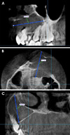
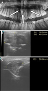
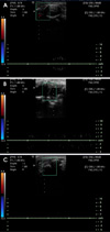
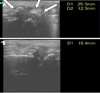
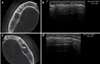
In stage 1, the anteroposterior, superoinferior, and mesiodistal dimensions of each lesion were measured on CBCT and US images. On the CBCT images, the anteroposterior dimensions were calculated based on the axial view of the lesion, the superoinferior dimensions based on the coronal view, and the mesiodistal dimensions based on the sagittal view (Fig. 1). All dimensions were measured in the slice where the lesion had the largest dimension. On the US images, anteroposterior and mesiodistal dimensions were measured on the longitudinal scan by placing the probe outside the mouth on the skin overlying the intraosseous lesion in a parallel direction to the inferior jaw border. The superoinferior dimension was measured on the transverse scan by placing the probe perpendicular to the inferior jaw border. Moreover, all dimensions were measured in the slice where the lesion had the largest dimension (Fig. 2). According to the US findings, the jaw lesions were classified as cystic, solid, or mixed. Cystic lesions showed posterior enhancement and an anechoic area with or without limited internal echoes. Solid lesions had a moderate echo pattern and no liquid component or posterior enhancement. Mixed lesions had cystic and solid areas combined within the same lesion. Color Doppler ultrasound examinations were then performed using a linear-array transducer to measure the average blood-flow velocity around the lesion site (Fig. 3A). After the US examination, surgery was performed and the lesions were sent for a histological examination.
In stage 2 (at 1 week after the surgical procedure), blood-flow velocity was measured around the surgical site in several places using color Doppler, then the average velocity value was calculated (Fig. 3B).
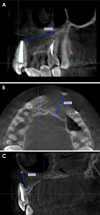
Finally, in stage 3 (at 6 months after the surgical procedure), the 3 maximum dimensions of the surgical site were measured on the CBCT images (Fig. 4). These maximum dimensions and the average blood-flow velocity around the surgical site were measured on the US and color Doppler images in the same way as in stage 1 and 2 (Figs. 3C and 5). The US dimensions were compared with the corresponding dimensions measured on the CBCT images. Data were analyzed using SPSS version 19 (IBM Co., Armonk, NY, USA) by the paired-samples Student t-test and intraclass correlation coefficient (ICC). The level of significance for all tests was set at P<.05.
Results
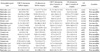
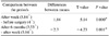
In this study, 21 intraosseous jaw lesions were assessed in 20 patients. Fourteen lesions were located in the mandible, and 7 in the maxilla. Only 2 of the lesions could not be detected using US. The size measurements, distribution of the lesions by histopathological results, and echographic patterns are presented in Table 1. The ICCs between US and CBCT in measuring the 3 dimensions (superoinferior, mesiodistal, and anteroposterior) before surgery were 0.956, 0.957, and 0.917, respectively, indicating almost complete agreement. However, at 6 months after surgery, the ICCs were 0.754, 0.602, and 0.86, respectively, which indicated less agreement. The paired-samples Student t-test between the US and CBCT images before surgery according to the specific dimensions yielded P values for the mesiodistal, anteroposterior, and superoinferior dimensions of .127, .398, and .001, respectively. These results mean that no significant difference existed in the preoperative US and CBCT measurements of the mesiodistal and anteroposterior dimensions, while a significant difference did exist between the preoperative US and CBCT measurements in the superoinferior dimension. However, at 6 months after surgery, the P values for the mesiodistal, anteroposterior, and superoinferior dimensions were .003, .000, and .000, respectively, indicating that significant differences were present between US and CBCT in the measurements of all dimensions (Table 2). When the preoperative and 6-month postoperative measurements were compared by technique and dimension using the paired-samples Student t-test, the P values for the mesiodistal, anteroposterior, and superoinferior dimensions in the US images were .001, .000, and .000, and in CBCT images, the corresponding P values were .036, .002, and .024, respectively. Therefore, there were significant differences in the measurements of all dimensions between before surgery and 6 months after surgery for both techniques (Table 3). Similarly, the blood-flow velocity measurements showed significant differences (P<.05) between before surgery and 1 week after surgery using the paired-samples Student t-test. Significant differences were also found between the values at 1 week and 6 months after surgery (P<.05) (Table 4).
Discussion
Accurate linear measurements on CBCT images are necessary for diagnosis and treatment. Several previous studies showed that measurements made from CBCT images were statistically similar to direct measurements.16171819 Additionally, US is important for evaluating the solid and cystic components of jaw lesions.11 Its advantages are that it is painless, rapid, inexpensive, noninvasive, and easily reproducible.20 Our study used CBCT to examine the accuracy of ultrasound by comparing the dimensions of lesions obtained using US with the corresponding dimensions obtained using CBCT. US was only unable to detect 2 lesions located on the posterior mandible, due to the thick buccal bone plate (Fig. 6). This finding is consistent with several previous studies.112122 As shown in Table 2, no significant differences were found in the mesiodistal and anteroposterior dimensions between US and CBCT images before surgery, while a significant difference was found in the superoinferior dimension. This finding is consistent with that of Goel et al.23 However, this finding is not consistent with that of the study conducted by Mehdizadeh et al. in 2009.21 Their study showed no significant differences in the mesiodistal and superoinferior measurements obtained by panoramic radiography and US.21 These differences might have been due to the projection of an acoustic shadow of the bony edges onto the lateral walls, which made the exact measurements of ultrasound images difficult and subjective.24 Moreover, the buccal bone plate that covers such lesions does not have an even thickness. Therefore, US waves could penetrate this plate in some places, but not in others. These considerations may help explain the difference in the measured lesion dimensions between US and CBCT. Another possible explanation may be that Mehdizadeh et al.21 used panoramic radiography, which was unable to show the third dimension, potentially leading to mistakes in the measurement of the superoinferior dimension due to the overlapping of anatomical structures. Most previous studies compared US with 2D digital and conventional periapical images, but our study compared US with 3D CBCT images. Therefore, our study evaluated the third dimension (anteroposterior dimension) of ultrasound images by comparing it with the same dimension on CBCT images, which was impossible to do with 2D images. We found only 2 studies that compared US images of intraosseous jaw lesions with 3D CBCT images. First, Shahidi et al. in 201525 showed that the ICC between US and CBCT in measuring lesion dimensions was 0.99. This is very similar to the findings of our study, in which the ICC was 0.956 for the superoinferior dimension, 0.957 for the mesiodistal dimension, and 0.917 for the anteroposterior dimension. Second, the study of Bayrakdar et al. in 2018, in agreement with our study, showed no significant differences between US and CBCT in the mesiodistal and anteroposterior dimensions, but a significant difference in the superoinferior dimension.26 As shown in Table 2, the values of all dimensions after 6 months on US images were lower than the values obtained from CBCT images. This can be explained by the ability of US to show healing sites starting only 6 weeks after the beginning of the healing process. In contrast, X-rays need up to 18–22 weeks to show the first signs of healing and bone deposition. 1527 Thus, after 6 months, US would show more bone deposition sites that are not mineralized enough to appear on X-ray images. As seen in Table 3, the measurements in all dimensions obtained after 6 months using both techniques were lower than those before surgery, which is consistent with previous studies.141528 Therefore, US images in this study revealed the healing process more accurately by showing the decrease in lesion size more clearly than CBCT, because US showed a greater decrease in lesion size than CBCT (Table 2). All cases showed increased blood flow at the surgical site at 1 week after surgery compared with the preoperative findings. This increase in blood flow was caused by inflammatory mediators that are present at the surgical site. During the process of healing, inflammation subsides and bloodflow velocity decreases. Therefore, as seen in Table 4, after 6 months, the blood-flow velocity was less than the velocity after 1 week, because of the advanced healing process. This is consistent with the findings reported by Tikuu et al. in 201014 and Mati et al. in 2011.15 In those studies, blood-flow velocity gradually decreased throughout the healing process. Three of the 21 cases showed no evidence of blood flow at the surgical site after 6 months. All those cases were small periapical granulomas, so this most likely occurred because of the very slow baseline flow around these lesions.14
By providing valuable data about the decrease in both the size of the lesion and blood-flow velocity at the surgical site during monitoring periods, our findings indicate that US with color Doppler is an effective tool for monitoring the healing process of jaw lesions after surgery.

 XML Download