

 PDF
PDF ePub
ePub Citation
 Print
Print


Abstract
Purpose
To report a delayed onset of multiple evanescent white dot syndrome in a patient with punctate inner choroidopathy.
Case summary
A 23-year-old female complained about sudden visual loss in the right eye. Best-corrected visual acuity (BCVA) was 20/100 in the right eye and 20/20 in the left eye. In fundus examination and optical coherence tomographic images, subfoveal choroidal neovascularization (CNV) with hemorrhage was observed in the right eye, accompanied by multiple lesions of atrophic pigmentation on the posterior pole in both eyes. We diagnosed the patient as punctate inner choroidopathy (PIC) and CNV in the right eye, and treated her using three monthly intravitreal injections of bevacizumab (Avastin®, Roche, Basel, Switzerland; 1.25 mg/0.05 mL). The CNV regressed and the BCVA improved to 20/20. Two years later, she complained of visual impairment in her left eye. The BCVA was 20/40. Fundus photography revealed numerous small white dots around the posterior pole and optic disc. Disruption of the photoreceptor layer was seen in optical coherence tomography images. Small white dots were observed as multiple hyperfluorescent dots in fluorescein angiography and hypofluorescent spots in indocyanine green angiography. An enlarged blind spot was observed in the visual field. We diagnosed her as multiple evanescent white dot syndrome (MEWDS). One month after systemic steroid treatment, the multiple white dots disappeared and the BCVA improved to 20/20.
References
1. Abu-Yaghi NE, Hartono SP, Hodge DO, et al. White dot abdominals: 20-year study of incidence, clinical features, and outcomes. Ocul Immunol Inflamm. 2011; 19:426–30.
2. Gass JD. Are acute zonal occult outer retinopathy and the white spot syndromes (AZOOR complex) specific autoimmune disease? Am J Ophthalmol. 2003; 135:380–1.
3. Jampol LM, Becker KG. White spot syndromes of the retina: a abdominal based on the common genetic hypothesis of auto-immune/inflammatory disease. Am J Ophthalmol. 2003; 135:376–9.
4. Quillen DA, Davis JB, Gottlieb JL, et al. The white dot syndromes. Am J Ophthalmol. 2004; 137:538–50.

5. Amer R, Lois N. Punctate inner choroidopathy. Surv Ophthalmol. 2011; 56:36–53.
6. Watzke RC, Packer AJ, Folk JC, et al. Punctate inner choroidopathy. Am J Ophthalmol. 1984; 98:572–84.
7. Channa R, Ibrahim M, Sepah Y, et al. Characterization of macular lesions in punctate inner choroidopathy with spectral domain optical coherence tomography. J Ophthalmic Inflamm Infect. 2012; 2:113–20.
8. Turkcuoglu P, Chang PY, Rentiya ZS, et al. Mycophenolate mofetil and fundus autofluorescence in the management of recurrent abdominal inner choroidopathy. Ocul Immunol Inflamm. 2011; 19:286–92.
9. Jampol LM, Sieving PA, Pugh D, et al. Multiple evanescent white dot syndrome. I. Clinical findings. Arch Ophthalmol. 1984; 102:671–4.
10. Bryan RG, Freund KB, Yannuzzi LA, et al. Multiple evanescent white dot syndrome in patients with multifocal choroiditis. Retina. 2002; 22:317–22.
11. Heckenlively JR, Ferreyra HA. Autoimmune retinopathy: a review and summary. Semin Immunopathol. 2008; 30:127–34.
12. Pearlman RB, Golchet PR, Feldmann MG, et al. Increased abdominal of autoimmunity in patients with white spot syndromes and their family members. Arch Ophthalmol. 2009; 127:869–74.
13. Fine L, Fine A, Cunningham ET Jr. Multiple evanescent white dot syndrome following hepatitis a vaccination. Arch Ophthalmol. 2001; 119:1856–8.
14. Baglivo E, Safran AB, Bottuat FX. Multiple evanescent white dot abdominal after hepatitis B vaccine. Am J Ophthalmol. 1996; 122:431–2.
15. Fine HF, Spaide RF, Ryan EH Jr, et al. Acute zonal occult outer abdominal in patients with multiple evanescent white dot syndrome. Arch Ophthalmol. 2009; 127:66–70.
Figure 1.
Multimodal images at the initial visit. (A) Fundus photograph shows multiple atrophic lesions in both eyes and choroidal neovascularization (CNV) with subretinal hemorrhage in right eye. (B) Multiple hyperfluorescent lesions due to increased transmission of both eyes and leakage from CNV of the right eye are observed in fluorescein angiography. (C) Indocyanine green angiography demonstrates multiple hypofluorescent dots. (D) Optical coherence tomography of the right eye shows subfoveal CNV.
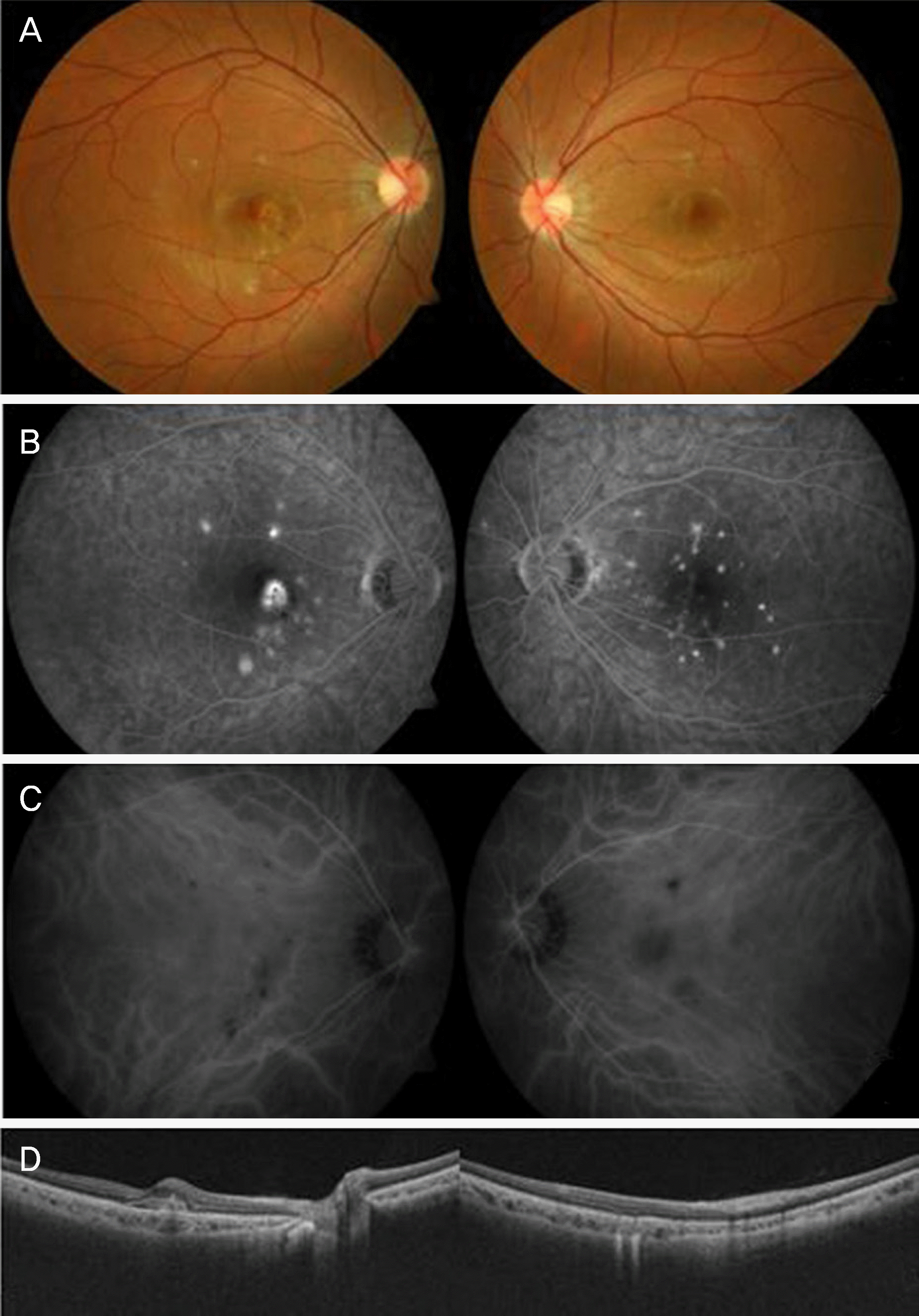
Figure 2.
Multimodal images 2 years later. (A) In the fundus examination, numerous whitish dots are around the posterior pole and optic disk in the left eye. (B) Early and late phase fluorescein angiographies show persistent multiple hyperfluorescent spots. (C) Indocyanine green angiography demonstrates hypofluorescent spots in the left eye. More hypofluorescent spots are observed in the late phase than in the early phase. (D) Disruption of photoreceptor layers of the left eye is detected in the optical coherence tomography. (E) Enlarged blind spot and central scotoma are detected in the automated visual field test.
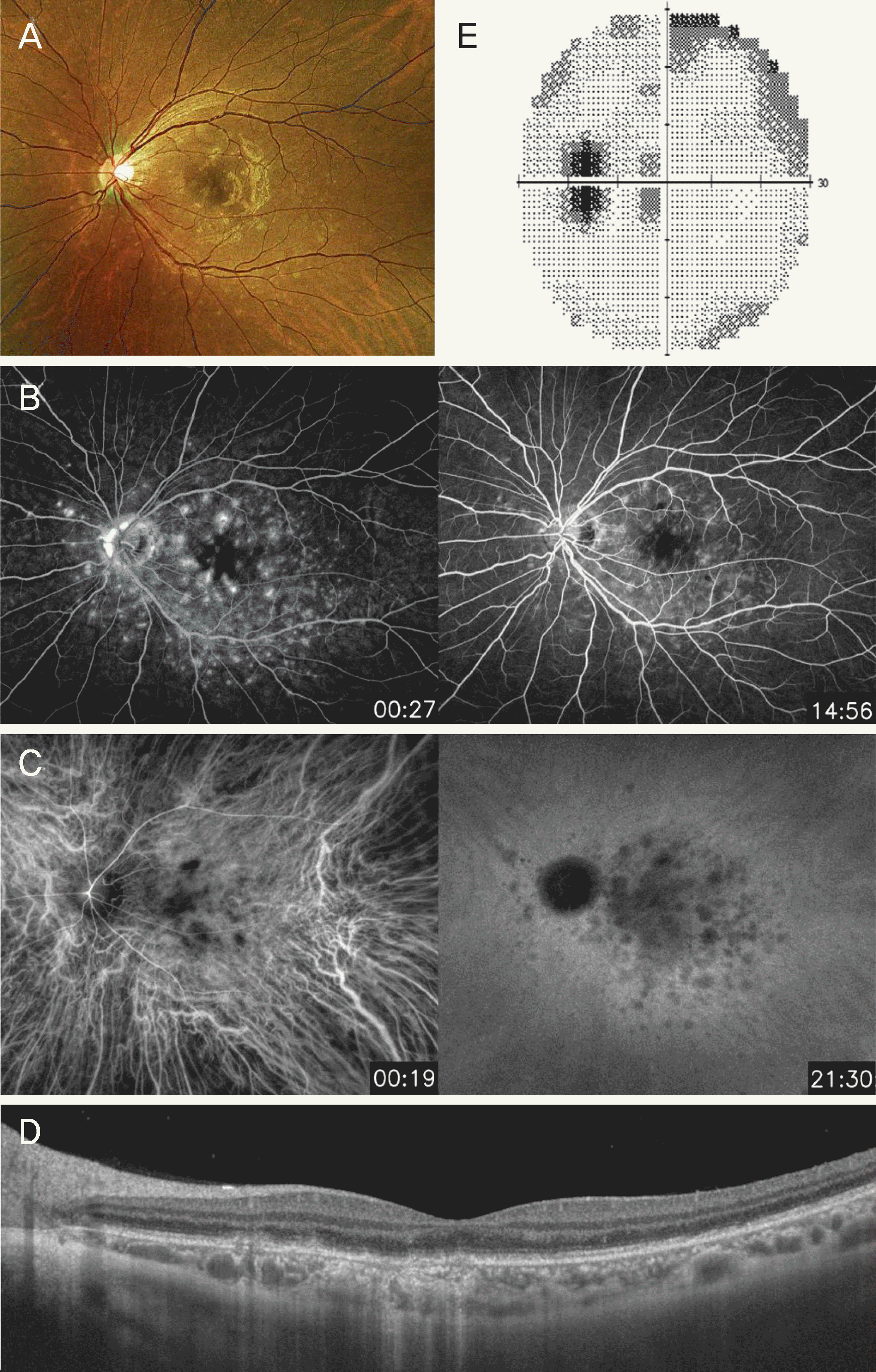
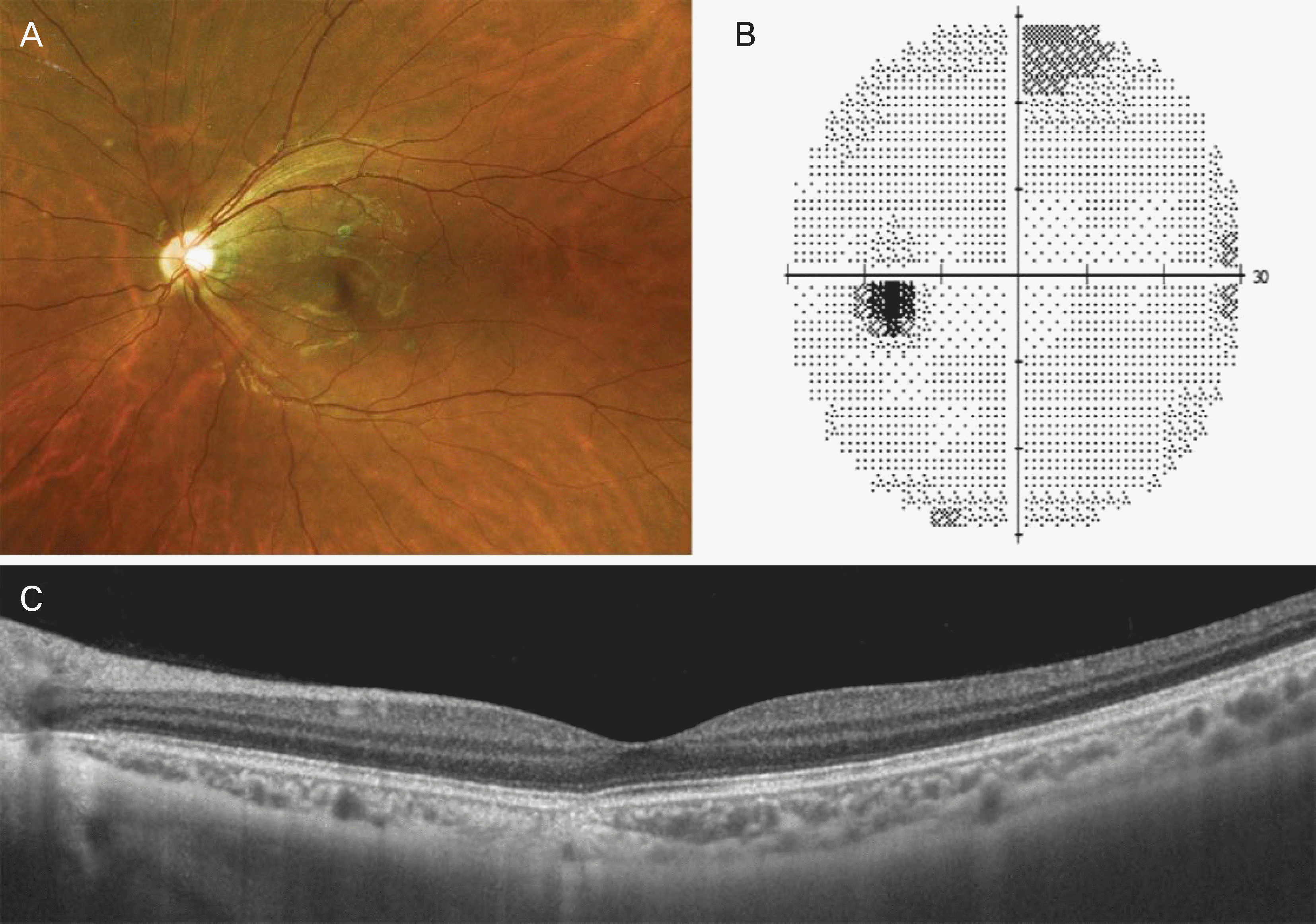
Figure 3.
Multimodal images of the left eye after recovery of multiple evanescent white dot syndrome. (A) Fundus photograph shows disappearance of whitish dots and previous atrophic lesions resulting from punctate inner choroidopathy. (B) Size of blind spot and central scotoma recover. (C) Optical coherence tomography shows intact photoreceptor layers.
 XML Download