

 PDF
PDF ePub
ePub Citation
 Print
Print


Abstract
Purpose
A case of bilateral lacrimal caruncle steatocystoma simplex and sebaceous gland hyperplasia, which rarely occurs in the periocular area, is reported.
Case summary
A 42-year-old female presented with a bilateral caruncular mass, which had been growing for a few months. A physical examination revealed soft, non-tender, rounded masses and no other ophthalmic findings. Under local anesthesia, the masses were completely excised. In the histopathological examination, a cystic mass containing a sebaceous gland in the stratified squamous epithelium cyst wall was found. A hyperplastic sebaceous gland and a dilated sebaceous gland duct were also found. Steatocystoma simplex and sebaceous gland hyperplasia, respectively, were diagnosed. There was no recurrence or complication after excision.
Conclusions
Steatocystoma simplex and sebaceous gland hyperplasia are benign tumors that rarely occur in the lacrimal caruncle. They can be diagnosed and treated by complete excision. Steatocystoma simplex and sebaceous gland hyperplasia should therefore be considered as differential diagnoses of a lacrimal caruncle mass.
Figures and Tables
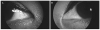
 | Figure 1Preoperative anterior segment photography. Light yellowish masses in the right lacrimal caruncle (arrow) (A) and in the left lacrimal caruncle (arrow) (B).
|
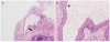 | Figure 2Pathological findings of the mass in the right lacrimal caruncle. (A) Cystic mass lined by non-keratinizing stratified squamous epithelium (arrow) contained sebaceous lobules (asterisk) (hematoxylin and eosin [H&E] stain, ×40). (B) Eosinophilic cuticle lining (arrowhead) of the cyst wall (H&E stain, ×200).
|
References
1. Ishida Y, Takahashi Y, Takahashi E, et al. Steatocystoma simplex of the lacrimal caruncle: a case report. BMC Ophthalmol. 2016; 16:183.

2. Ogawa M, Shinzawa M, Dogru M, et al. Caruncular and pericaruncular sebaceous gland hyperplasia: a report of 2 cases and literature review. Eye Contact Lens. 2018; 44:Suppl 1. S316–S319.
3. Kim NJ, Moon KC, Khwarg SI. Steatocystoma simplex of the caruncle. Can J Ophthalmol. 2006; 41:83–85.
4. Bowyer J, Sullivan T, Whitehead K. Steatocystoma simplex of the caruncle. Br J Ophthalmol. 2003; 87:240–241.
5. Kim SH, Ahn BC. Steatocystoma simplex of the orbit. J Korean Ophthalmol Soc. 2000; 41:2000–2002.
6. Kim YJ, Lee YS, Chi MJ. A case of steatocystoma simplex of the orbit. J Korean Ophthalmol Soc. 2015; 56:1794–1797.
7. Kim TJ, Kim NJ, Park HJ, et al. A case of eyelid steatocystoma. J Korean Ophthalmol Soc. 2012; 53:1027–1029.
8. Procianoy F, Golbert MB, Golbspan L, et al. Steatocystoma simplex of the eyelid. Ophthalmic Plast Reconstr Surg. 2009; 25:147–148.
9. Massry GG, Holds JB, Kincaid MC, Patrinely JR. Sebaceous gland hyperplasia of the caruncle. Ophthalmic Plast Reconstr Surg. 1995; 11:32–36.
10. Shields CL, Shields JA. Tumors of the conjunctiva and cornea. Surv Ophthalmol. 2004; 49:3–24.
11. Kim SY, Paik JS, Yang SW. A case of oncocytoma of the caruncle. J Korean Ophthalmol Soc. 2010; 51:598–600.
 XML Download