

 PDF
PDF ePub
ePub Citation
 Print
Print


Abstract
Purpose
To evaluate the postoperative refractive errors after air tamponade with posterior capsulectomy during combined vitrectomy and cataract surgery.
Methods
Patients who underwent combined vitrectomy, cataract surgery, and air tamponade with or without posterior capsulectomy were reviewed. All patients were followed for 4 months after surgery. Preoperative characteristics such as anterior chamber depth, axial length, and refractive error were analyzed and refractive errors after the surgery were evaluated. The difference between the target refractions and final refractive errors after the surgery according to the biometry method, and intraocular lens power calculations, were observed.
Results
Fourteen eyes of 14 patients who had combined vitrectomy and cataract surgery with posterior capsulectomy and air tamponade were classified as group A, and 10 eyes of 10 patients who had combined vitrectomy and cataract surgery with only air tamponade were classified as group B. The target refraction of group A measured with A-scan biometry using the Sanders-Retzlaff-Kraff/Theoretical (SRK/T) calculation was −0.21 ± 0.22 diopters (D), and the final refractive error at 9.5 (± 2.20) months after the surgery was −0.52 ± 0.54 D. The mean difference between the two was −0.32 ± 0.44 D. The target refraction of group B measured with A-scan biometry using the SRK/T calculation was −0.33 ± 0.29 D, and the final refractive error at 9.5 (±2.20) months after the surgery was −0.27± 0.39 D. The mean difference between the two was 0.06 ± 0.53 D.
Conclusions
Posterior capsulectomy during combined vitrectomy and cataract surgery with air tamponade led to myopic shifts compared with no posterior capsulectomy with air tamponade during combined vitrectomy and cataract surgery. Performing posterior capsulectomy with air tamponade during combined vitrectomy and cataract surgery should, therefore, be carefully considered. J Korean Ophthalmol Soc 2018;59(9):819–826
References
1. Hsuan JD, Brown NA, Bron AJ, et al. Posterior subcapsular and nuclear cataract after vitrectomy. J Cataract Refract Surg. 2001; 27:437–44.

2. Melberg NS, Thomas MA. Nuclear sclerotic cataract after abdominal in patients younger than 50 year of age. Ophthalmology. 1995; 102:1466–71.
3. Thompson JT. The role of patient age and intraocular gas use in abdominal progression after vitrectomy for macular holes and epiretinal membranes. Am J Ophthalmol. 2004; 137:250–7.
4. Grusha YO, Masket S, Miller KM. Phacoemulsification and lens implantation after pars plana vitrectomy. Ophthalmology. 1998; 105:287–94.
5. Toda J, Kato S, Oshika T, Sugita G. Posterior capsule opacification after combined cataract surgery and vitrectomy. J Cataract Refract Surg. 2007; 33:104–7.
6. Bhargava R, Kumar P, Phogat H, Chaudhary KP. Neodymium-yt-trium aluminium garnet laser capsulotomy energy levels for abdominal capsule opacification. J Ophthalmic Vis Res. 2015; 10:37–42.
7. Kim JH, Han SB, Lee SJ, Kim MS. Postoperative refractive errors after posterior capsulectomy during combined vitrectomy and abdominal surgery. J Korean Ophthalmol Soc. 2015; 56:709–14.
8. Suzuki Y, Sakuraba T, Mizutani H, et al. Postoperative refractive abdominal after simultaneous vitrectomy and cataract surgery. Ophthalmic Surg Lasers. 2000; 31:271–5.
9. Ohrloff C, Schalnus R, Rothe R, Spitznas M. Role of the posterior capsule in the aqueous-vitreous barrier in aphakic and abdominal eyes. J Cataract Refract Surg. 1990; 16:198–201.
10. Weinreb RN, Wasserstrom JP, Parker W. Neovascular glaucoma following neodymium-YAG laser posterior capsulotomy. Arch Ophthalmol. 1986; 104:730–1.
11. Poliner LS, Christianson DJ, Escoffery RF, et al. Neovascular abdominal after intracapsular and extracapsular cataract extraction in abdominal patients. Am J Ophthalmol. 1985; 100:637–43.
12. Gimbel HV, Neuhann T. Development, advantages, and methods of the continuous circular capsulorhexis technique. J Cataract Refract Surg. 1990; 16:31–7.
13. Gimbel HV. Posterior continuous curvilinear capsulorhexis and optic capture of the intraocular lens to prevent secondary opacification in pediatric cataract surgery. J Cataract Refract Surg. 1997; 23(Suppl 1):652–6.
14. Galand A, van Cauwenberge F, Moosavi J. Posterior capsulorhexis in adult eyes with intact and clear capsules. J Cataract Refract Surg. 1996; 22:458–61.
15. Jeoung JW, Chung H, Yu HG. Factors influencing refractive abdominals after combined phacoemulsification and pars plana abdominal: Results of a prospective study. J Cataract Refract Surg. 2007; 33:108–14.
16. Wagenfeld L, Hermsdorf K, Stemplewitz B, et al. Refractive abdominal in eyes with intraocular gas tamponade – results of a prospective controlled clinical trial. Clinical Ophthalmology. 2017; 11:993–8.
17. Falkner-Radler CI, Benesch T, Binder S. Accuracy of preoperative biometry in vitrectomy combined with cataract surgery for patients with epiretinal membranes and macular holes: results of a prospective controlled clinical trial. J Cataract Refract Surg. 2008; 34:1754–60.
18. Drexler W, Findl O, Menapace R, et al. Partial coherence abdominal: a novel approach to biometry in cataract surgery. Am J Ophthalmol. 1998; 126:524–34.
19. Haigis W, Lege B, Miller N, Schneider B. Comparison of abdominal ultrasound biometry and partial coherence interferometry for intraocular lens calculation according to Haigis. Graefes Arch Clin Exp Ophthalmol. 2000; 238:765–73.
20. Lim HC, Kim KH, Shin MK, et al. Accuracy of predictive refraction in combined vitrectomy-cataract surgery for epiretinal membrane and macular hole. J Korean Ophthalmol Soc. 2015; 56:219–27.
21. Haigis W. Intraocular lens power calculations. Thorofare: Slack Inc.;2003. p. 41–57.
22. Kim DY, Kim MJ, Kim JY, Tchah H. Comparison of formulas for intraocular lens power calculation installed in a partial coherence interferometer. J Korean Ophthalmol Soc. 2009; 50:523–8.
23. Yi CH, Choi SH, Chung ES, Chung TY. Accuracy of the Haigis abdominal based on axial length and anterior chamber depth. J Korean Ophthalmol Soc. 2011; 52:175–81.
Figure 1.
Clinical intraoperative photograph of the anterior segment showing the center of the posterior capsule removed using vitreous cutter. The posterior capsule was removed cur-vilinearly and completely.

Figure 2.
Comparison of spherical equivalent correction of two different groups. The results of the group A (capsulectomy + air tamponade) (A) and group B (only air tamponade) (B) at last visit (mean postoperative 9.5 ± 2.20) months compared with predictive values from three different biometry devices and formulas combinations. SRK/T = Sanders-Retzlaff-Kraff/Theoretical.
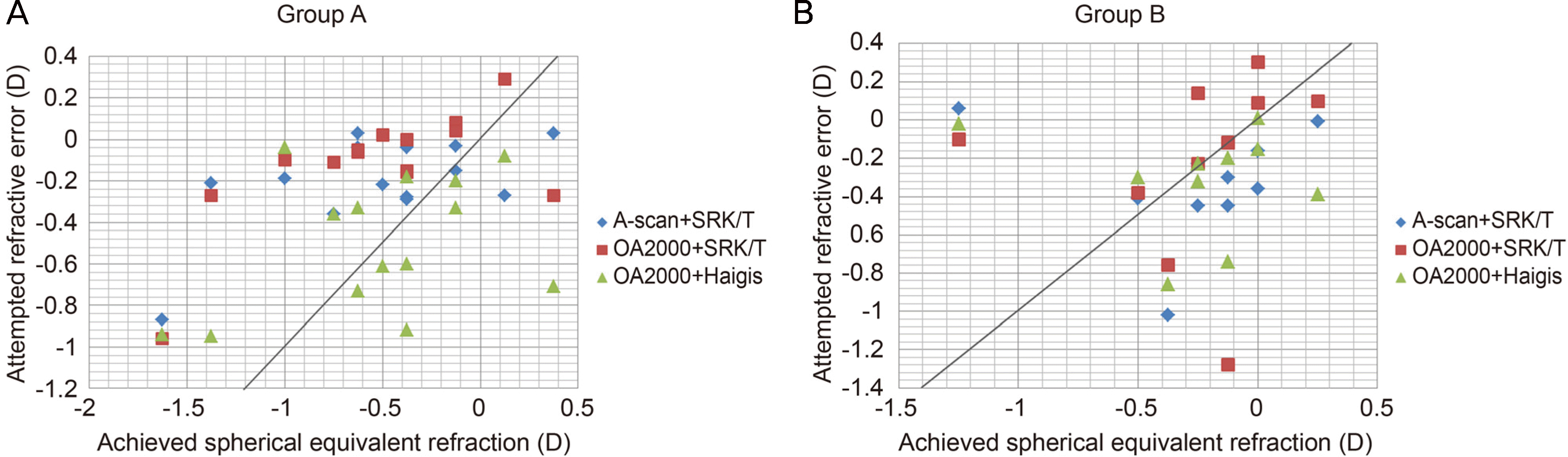
Table 1.
Baseline characteristics of posterior capsulectomy with air tamponade (group A) and only air tamponade (group B)
| Characteristics | Group A (n = 14) | Group B (n = 10) | p-value* |
|---|---|---|---|
| Number of eyes | 14 eyes | 10 eyes | – |
| Gender (male:female) | 4:10 | 3:7 | – |
| Age (years) | 67.6 ± 5.8 | 64.9 ± 5.8 | 0.378 |
| Duration of follow-up (months) | 6.37 ± 1.98 | 8.27 ± 4.26 | 0.349 |
| Preoperative UCVA (logMAR) | 0.56 ± 0.19 | 0.54 ± 0.26 | 0.662 |
| Preoperative BCVA (logMAR) | 0.47 ± 0.21 | 0,43 ± 0.26 | 0.389 |
| Central macular thickness (μ m) | 379.7 ± 79.8 | 414.5 ± 65.1 | 0.235 |
| Preoperative sphere (diopter) | 0.35 ± 1.02 | 1.34 ± 1.54 | 0.051 |
| Preoperative cylinder (diopter) | –1.07 ± 0.71 | –1.43 ± 0.47 | 0.112 |
| Preoperative spherical equivalent (diopter) | –0.17 ± 1.13 | 0.62 ± 1.68 | 0.161 |
| K1 (keratometry, diopter) | 44.3 ± 1.4 | 44.2 ± 1.8 | 0.953 |
| K2 (keratometry, diopter) | 44.4 ± 1.5 | 43.9 ± 1.9 | 0.428 |
| Corneal astigmatism (diopter) | –0.86 ± 0.5 | –0.81 ± 0.41 | 0.929 |
| Anterior chamber depth (mm) | 3.054 ± 0.28 | 3.18 ± 0.42 | 0.364 |
| Axial length (mm) | 23.53 ± 0.65 | 23.08 ± 0.91 | 0.169 |
Table 2.
Predicted postoperative refraction values measuring with different biometry machines and formula, and actual inserted intraocular lens (IOL) power in posterior capsulectomy with air tamponade (group A) and only air tamponade (group B)
| Group A (n = 14) | Group B (n = 10) | p-value* | |
|---|---|---|---|
| Preoperative target diopter using A-scan | –0.21 ± 0.22 | –0.33 ± 0.29 | 0.151 |
| (IOL calculation formula: SRK/T) | |||
| Preoperative target diopter using OA-2000 | –0.12 ± 0.28 | –0.23 ± 0.51 | 0.992 |
| (IOL calculation formula: SRK/T) | |||
| Preoperative target diopter using OA-2000 | –0.49 ± 0.32 | –0.35 ± 0.29 | 0.357 |
| (IOL calculation formula: Haigis) | |||
| IOL power (diopter) | 22.2 ± 1.91 | 22.5 ± 1.79 | 0.563 |
Table 3.
Actual postoperative refraction values measuring at 1,2,4,6 months in phacovitrectomy and posterior capsulectomy with air tamponade (group A) and phacovitrectomy with only air tamponade (group B)
| Group A (n = 14) | Group B (n = 10) | p-value* | |
|---|---|---|---|
| Postoperative SE (diopter) | |||
| 1 month | –0.59 ± 0.54 | –0.28 ± 0.67 | 0.086 |
| 2 months | –0.41 ± 0.41 | –0.37 ± 0.52 | 0.837 |
| 4 months | –0.44 ± 0.51 | –0.41 ± 0.39 | 0.993 |
| 6 months | –0.51 ± 0.67 | –0.31 ± 0.43 | 0.506 |
| Final | –0.52 ± 0.54 | –0.27 ± 0.39 | 0.141 |
Table 4.
Preoperative and postoperative mean central macular thickness (CMT) measurements of the patients in two groups
| Group A (n = 14) | Group B (n = 10) | p-value* | |
|---|---|---|---|
| preoperative mean CMT (μ m) | 379.7 ± 79.8 | 414.5 ± 65.1 | 0.235 |
| postoperative mean CMT (μ m) at 6 months | 320.5 ± 70.1 | 357.8 ± 57.1 | 0.084 |
| p-value† | 0.04 | 0.028 |
Values are presented as mean ± SD unless otherwise indicated. No significant differences related to the mean CMT value was seen between group A and group B, preoperatively or postoperatively (p > 0.05, in both). In both groups, the mean CMT was significantly reduced at 6 months visit compared with preoperative values (p < 0.05, in both).
Table 5.
Average difference of preoperative target diopter and actual postoperative refraction values (group A) and (group B)
| Average difference (diopter) | Group A (n = 14) | Group B (n = 10) | p-value* |
|---|---|---|---|
| Refractive error using A-scan, SRK/T | –0.32 ± 0.44 | 0.06 ± 0.52 | 0.026 |
| (Postop SE – Target diopter) | |||
| Refractive error using OA2000, SRK/T | –0.41 ± 0.41 | –0.06 ± 0.61 | 0.036 |
| (Postop SE – Target diopter) | |||
| Refractive error using OA2000, Haigis | –0.03 ± 0.51 | 0.01 ± 0.55 | 0.886 |
| (Postop SE – Target diopter) |
Values are presented as mean ± SD unless otherwise indicated. Average difference of preoperative target diopter and actual postoperative refraction values measuring at last visit 9.5 (±2.20) months in phacovitrectomy and posterior capsulectomy with air tamponade (group A) and phacovitrectomy with only air tamponade (group B).
Table 6.
Comparison in A-scan ocular biometry and OA2000 of phacovitrectomy and posterior capsulectomy with air tamponade (group A) and phacovitrectomy with only air tamponade (group B)
| Comparison in ocular biometry | Group A (n = 14) | Group B (n = 10) |
|---|---|---|
| Refractive error using A-scan, SRK/T | –0.32 ± 0.44 | 0.06 ± 0.52 |
| (Postop SE – Target diopter) | ||
| Refractive error using OA2000, SRK/T | –0.41 ± 0.41 | –0.06 ± 0.61 |
| (Postop SE – Target diopter) | ||
| p-value* | 0.655 | 0.653 |
Table 7.
Comparison in SRK/T and Haigis formula of phacovitrectomy and posterior capsulectomy with air tamponade (group A) and phacovitrectomy with only air tamponade (group B)
| Comparison in IOL power calculation formulas | Group A (n = 14) | Group B (n = 10) |
|---|---|---|
| Refractive error using OA2000, SRK/T | –0.41 ± 0.41 | –0.06 ± 0.61 |
| (Postop SE – Target diopter) | ||
| Refractive error using OA2000, Haigis | –0.03 ± 0.51 | 0.01 ± 0.55 |
| (Postop SE – Target diopter) | ||
| p-value* | 0.087 | 0.787 |
 XML Download