

 PDF
PDF ePub
ePub Citation
 Print
Print


Introduction
Minimal deviation adenocarcinoma (MDA) of the uterine cervix, also called adenoma malignum, is a rare variant of cervical adenocarcinoma. It accounts for 1–3% of all cervical adenocarcinoma cases [1]. MDA demonstrates an endophytic and not an exophytic growth pattern. It resembles multiple benign nabothian cysts on transvaginal ultrasonography. Routine screening methods for the uterine cervix including the Papanicolaou (Pap) and the human papillomavirus (HPV) tests [2] and even invasive diagnostic tools (e.g., punch biopsy and cervical conization) often misdiagnose MDA before performing definitive surgery. This could lead to an incidental diagnosis of MDA following a simple hysterectomy for other benign conditions. The prognosis of MDA is known to be relatively poor [34]. Reportedly, the mean survival time in patients with MDA is approximately 60, 38, 22.8, and 5.4 months for stage I, II, III, and IV disease, respectively [1].
MDA is often associated with the Peutz-Jeghers syndrome (PJS), a rare autosomal dominant disorder caused by germline mutations of the serine/threonine kinase gene (STK11) located on chromosome 19p13.3 [5]. It implicates the management of cancer and cancer risk for patients and their families in the era of precision medicine.
No standard treatment is available for MDA owing to the rarity of this condition and the difficulty with accurately diagnosing it. The objective of the present study was to determine the clinicopathological features of MDA and to analyze its prognostic factors at a single hospital.
Materials and methods
We retrospectively reviewed the medical charts of patients who were diagnosed with MDA (based on histopathological findings) between January 2005 and December 2015, at the Comprehensive Gynecologic Cancer Center of CHA Bundang Medical Center. We analyzed medical records and obtained information regarding clinical characteristics, surgical findings, and oncologic outcomes.
We used the 2014 World Health Organization (WHO) classification for the histopathological classification of cervical adenocarcinoma [6]. The histopathological definition and characteristics of MDA are: 1) a well-differentiated mucinous adenocarcinoma in which most glands are indistinguishable histologically from normal endocervical glands, 2) a lesion showing cytologically bland glands of varying sizes and shapes, 3) a lesion showing increased mitotic activity, 4) a lesion with hyperplastic glands at the surface and, 5) lesions showing an increased number of glands deeper than the lower level in normal endocervical glands. All patients diagnosed with MDA underwent examination by a gynecologic pathologist at our hospital. Staging of tumors was performed based on the criteria recommended by the International Federation for Obstetrics and Gynecology. Treatment was initiated with surgery with or without adjuvant therapy except in 3 patients who underwent neoadjuvant chemotherapy. Adjuvant therapy was classified into 3 types: exclusive chemotherapy, exclusive radiotherapy, and concurrent chemoradiation therapy (CCRT). This study was approved by the Institutional Review Board of the CHA Bundang Medical Center (CHA IRB 2017-09-012). Ethical approval was provided by the Ethics Committee.
The survival time for each patient was calculated from the date of diagnosis to the date of death with right-censoring on the date of the last follow-up among survivors. Cumulative survival probabilities of patients were estimated using the life table method (commonly referred to as the Kaplan–Meier method). The log-rank test was used to compare the 2 survival curves. Multivariate analysis was performed using Cox regression analysis. Hazard ratios (HRs) with 95% confidence intervals (CIs) were reported. The Statistical Package for the Social Sciences (SPSS Inc., Chicago, IL, USA) ver. 16.0 was used for all statistical analyses. A P-value <0.05 was considered statistically significant.
Results
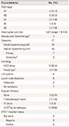
We identified 17 patients for whom clinicopathological information was available. Clinical characteristics of these patients are summarized in Table 1. Their mean age was 47.7 years. Chief complaints at the time of diagnosis were vaginal spotting (6/17, 35.3%) and vaginal discharge (5/17, 29.4%). The Pap test performed in 13 patients revealed negative findings in 3 (3/13, 23.1%) and abnormalities in 10 patients (10/13, 76.9%). Atypical glandular cells were observed in 9 of the 10 patients showing cytological abnormalities. The remaining patient showed an adenocarcinoma. HPV infection was detected in 1 patient (1/13, 7.7%). The Pap or HPV tests were not performed in 4 patients. Tumor markers were within the reference range in most patients except 3. Presumed diagnoses before performing a hysterectomy were benign lesions (4/17, 23.5%), nabothian cysts (2/17, 11.8%), adenocarcinoma in situ (4/17, 23.5%), and MDA (7/17, 41.2%). The biopsy-proven diagnostic rate of MDA before surgery was 41.2% (punch biopsy: 5/17, conization: 2/17).
Table 1
Clinical characteristics of patients (n=17)

Stage IB disease was observed in 12 (12/17, 70.6%), stage II in 3 (3/17, 17.7%), and stage III in 2 patients (2/17, 11.8%) (Table 2). The mean tumor diameter was 4.01 cm. Three patients diagnosed with MDA before undergoing the staging operation received 3 cycles of neoadjuvant chemotherapy using cisplatin and etoposide. Ten patients underwent simple hysterectomy. After the diagnosis of MDA was confirmed in incidentally diagnosed lesions following simple hysterectomy, subsequent staging operations including radical parametrectomy, bilateral salpingo-oophorectomy, and pelvic and/or para-aortic lymph node dissection were performed in 3 patients. Radical hysterectomy was initially performed in 7 patients diagnosed with MDA. Fourteen patients (14/17, 82.4%) showed only MDA, whereas 3 (3/17, 17.6%) showed MDA and concomitant mucinous adenocarcinoma. Among the 9 patients who underwent lymph node dissection, lymph node metastasis was detected in 2. Lymphovascular space invasion (LVSI) was positive in 6 patients including 2 with lymph node metastasis. Thirteen patients (13/17, 76.5%) received adjuvant treatment after the initial surgery. Among those who received adjuvant therapy, 2, 1, and 10 patients underwent exclusive chemotherapy, exclusive radiotherapy, and CCRT with or without chemotherapy, respectively. Three patients underwent genetic testing for the STK11 germline mutation to detect PJS, and all patients showed negative findings.
Table 2
Histopathological findings and treatment strategies used (n=17)
CCRT, concurrent chemoradiation therapy; FIGO, International Federation for Obstetrics and Gynecology; LND, pelvic and/or para-aortic lymph node dissection; LVSI, lymphovascular space invasion; MDA, minimal deviation adenocarcinoma; RT, radiation therapy; STK11, serine/threonine kinase gene.
a)Cisplatin and etoposide combination chemotherapy; b)Radical parametrectomy after confirmation of MDA following simple hysterectomy; c)Mixed with mucinous adenocarcinoma; d)Weekly cisplatin administration.
At the time of the last follow-up, 11 patients (11/17, 64.7%) showed no evidence of disease, 2 (2/17, 11.8%) developed recurrence, and 4 (4/17, 23.5%) showed disease persistence. All patients showing recurrence or disease persistence died of the disease except 1 patient who is currently undergoing chemotherapy after recurrence (Table 3).
Table 3
Oncological outcomes

| Characteristics (n=17) | No. (%) | |
|---|---|---|
| Recurrence or persistence | 6 (35.3) | |
| Died of disease | 5 (29.4) | |
| Alive with disease | 1 (5.9) | |
| No evidence of disease | 11 (64.7) | |
| Median follow-up (mon) | 33.6 (range: 7–99) | |
The median follow-up was 33.6 months (range, 7–99 months). Univariate analysis showed that advanced stage disease (P=0.016) and LVSI (P=0.002) were associated with poor overall survival (OS) rates (Fig. 1); however, other parameters including age (P=0.069) and radicality of hysterectomy (P=0.217) were not significantly associated with OS. Multivariate analysis showed that advanced stage disease was significantly associated with a poor OS rate (HR, 2.92; 95% CI, 1.097–7.746; P=0.032). However, no statistically significant differences in OS were observed with respect to age, type of hysterectomy performed, and LVSI (Table 4).
Fig. 1
(A) Kaplan-Meier curve showing the overall survival rate based on stages of minimal deviation adenocarcinoma (P=0.016). (B) Kaplan-Meier curve showing the overall survival rate based on lymphovascular space invasion (P=0.002).
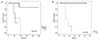
Table 4
Univariate and multivariate analysis of clinicopathological factors associated with overall survival rate

Discussion
Most cervical cancers are squamous cell carcinomas. Adenocarcinomas represent 20–25% of all cervical carcinomas [7]. MDA is a rare subtype of cervical mucinous adenocarcinoma accounting for only 1–3% of cervical adenocarcinomas [8]. Adenoma malignum of the uterine cervix was first described in 1870 [1]. The term MDA was proposed by Silverberg and Hurt [9] in 1975 to indicate its resemblance to normal endocervical glands and the lack of cellular features of malignancy [10]. Recently, MDA has been reclassified by the WHO as a subcategory of gastric-type mucinous cervical adenocarcinoma. This categorization is reserved only for the extremely well-differentiated cell types [6].
Based on the literature reviews and meta-analysis of 347 cases of MDA, the mean age at diagnosis is 45 years (range 20–78 years). The major clinical manifestations are profuse vaginal discharge (60.4%) and irregular vaginal bleeding (50.0%) [1]. In this study, the mean age of patients diagnosed with MDA was 47.7 years. Most patients (64.7%) presented with typical symptoms of vaginal discharge or bleeding (Table 1).
Pap and HPV tests serve as routine screening tests for cervical cancer [211]. Colposcopically directed punch biopsy or cervical conization can be performed on suspicious cervical lesions. The Pap test used as a diagnostic tool for MDA has shown a limited detection rate (32.7%, 37/113). Literature reviews have shown that cervical biopsies performed in 185 patients demonstrated a detection rate of 50.7%, and cervical conization performed in 14 patients demonstrated a detection rate of 100% [1]. In the present study, although a direct comparison was difficult, the detection rates of MDA using cervical cytology, punch biopsy, and conization were 7.6% (1/13), 62.5% (5/8), and 40% (2/5), respectively (Table 1). Adenocarcinoma in situ was detected even after a secondary conization in 2 of 5 patients who underwent conization indicating that MDA is usually located deep within the endocervix and that it shows an endophytic and not an exophytic growth pattern, unlike cervical squamous cell carcinoma. Therefore, it is difficult to detect this lesion using conventional tools for cervical cancer screening. Notably, MDA was incidentally diagnosed in 6 patients (35.3%) in this study after simple hysterectomy was performed for presumed benign gynecological conditions.
The optimal treatment for MDA has not been well-established. Surgical treatment is the most successful option for MDA. The surgical approach resembles that used for routine cervical cancer operations and includes a radical hysterectomy, bilateral pelvic lymphadenectomy, and salpingo-oophorectomy at an early stage. Chemotherapy and/or radiotherapy are reserved for treatment of advanced stage disease.
The prognosis of MDA is controversial, although it is known to be relatively poor, which is attributable to the likelihood of lymph node metastasis and early peritoneal carcinomatosis in contrast to localized metastases observed in patients with squamous cell carcinomas [12]. The poor prognosis of MDA could be attributed to clinical under-staging, misdiagnosis, and under-treatment. In the present study, 6 patients were incidentally diagnosed following simple hysterectomy for benign diseases. Three of these 6 underwent subsequent staging operations to define the exact tumor status and received adjuvant therapy based on surgical outcomes. The other 3 underwent adjuvant therapy without secondary surgery. Of note, a few studies have reported that prognosis in patients with MDA is not necessarily poor [113] and that diagnosis of MDA at an early stage could be associated with a favorable prognosis. Our study also showed that advanced stage MDA was significantly associated with poor prognosis (Fig. 1A). Four of 5 patients with advanced stage disease died of the disease and 2 of 12 patients with stage I disease showed recurrence (1 patient with recurrence is currently receiving treatment). A significant prognostic factor observed in the present study was LVSI. Univariate analysis using the Kaplan–Meier curve showed LVSI to be a statistically significant factor (Fig. 1B); however, multivariate analysis did not show a statistically significant association between LVSI and OS rates. All 6 patients with LVSI showed recurrence or persistence of the disease and 5 of them died of the disease. One of the 5 patients demonstrated advanced stage disease (stage II) without LVSI and no evidence of recurrence for 34 months. Previous studies have not conclusively determined the association between LVSI and OS rates; thus, it is difficult to compare their results with those of the present study. Multivariate analysis demonstrated that other factors including type of hysterectomy and age did not show a statistically significant association with the OS rate (Table 4).
The etiopathogenesis of MDA remains unclear. Previous studies have revealed no significant association between MDA and the HPV virus [141516] — an important distinguishing feature between MDA and common cervical cancer given that a significant association is observed between HPV infection and carcinogenesis of the uterine cervix [17]. HPV infection was detected in only 1 patient (1/13, 7.7%) in the present study. A few studies have demonstrated a close link between MDA and gastric metaplasia or endocervical glandular hyperplasia [18]. McGowan et al. [19] have reported that PJS may complicate MDA. PJS is an autosomal dominant disorder characterized by gastrointestinal hamartomatous polyps and mucocutaneous pigmentation. It could possibly trigger the development of MDA secondary to a mutation of the responsible tumor suppressor gene (STK11) [2021]. A previous study has shown that 4 of 27 women (14.8%) with PJS developed MDA with lobular endocervical hyperplasia [22]. The prognosis of patients with MDA associated with PJS is usually poor [323]. There was no comparable result of MDA with PJS based on family history, clinical, or gastrointestinal endoscopic findings in the present study. Three recently diagnosed patients with MDA underwent genetic testing for the STK11 gene, and no patient demonstrated a genetic mutation.
Limitations of our study: 1. This is a single-institution study involving a small number of patients. 2. The retrospective study design serves as a limitation because we could not accurately determine several parameters. 3. An absence of a uniform therapeutic protocol was an important drawback. 4. Comparative analysis between MDA and other benign lesions was not performed.
In conclusion, early diagnosis is important to manage MDA. However, early diagnosis of MDA is challenging for gynecologic oncologists in the absence of appropriate diagnostic methods. Therefore, clinicians should consider MDA among the differential diagnoses in patients with a clinically suspicious presentation including profuse vaginal discharge and multilobular cysts at the uterine cervix detected by transvaginal ultrasonography, even in those with a negative cervical Pap test. Because of the rarity of MDA, future research should involve nationwide studies. Data collection, information sharing, and inter-institutional consultations are necessary to define the nature of MDA to establish appropriate therapeutic guidelines and provide optimal advanced therapy in the era of precision medicine.
 XML Download