

 PDF
PDF ePub
ePub Citation
 Print
Print


Introduction
Tamoxifen has been used as adjuvant therapy for breast cancer in postmenopausal women, but has been implicated in endometrial changes. Therefore, gynecologic surveillance of asymptomatic women is needed.1 Endometrial pathology has been identified in up to 35.5% of postmenopausal breast cancer tamoxifen-treated patients.2
Benign endometrial polyps are the most common pathology described in these patients, with an incidence of 8% to 36%.3 Some women develop recurrent polyps with an incidence of malignancy of up to 10.7%.4 There is therefore an urgent need for a long-term investigation on the incidence of endometrial pathologies among large cohorts of postmenopausal breast cancer patients following continuous tamoxifen therapy for 5 years.
Because of the estrogenic effects on the endometrium, it is necessary to screen postmenopausal patients taking tamoxifen, especially those with no gynecologic symptoms.5
Transvaginal sonography (TVS) is considered a simple, accurate, noninvasive procedure for surveillance of endometrial changes, although false-negative diagnoses of small polyps, localized areas of atypical hyperplasia, or endometrial cancer have been reported.6
Some investigators consider TVS endometrial screening to have limited prognostic value in these patients because of excessive false-positive results and have proposed additional diagnostic procedures.78
Hysteroscopy directly visualizes the endometrium and allows biopsy tissue to be precisely sampled.5
We performed hysteroscopy to evaluate the effect of prolonged tamoxifen therapy on endometrium of postmenopausal patients with breast cancer. In asymptomatic women, endometrial thickness on TVS was the indication for hysteroscopy.
Materials and Methods
We reviewed clinical records of 46 postmenopausal patients, referred to our institution between January 2008 and December 2017. These women had been receiving tamoxifen for at least 12 months for breast cancer. Fifteen were experiencing abnormal uterine bleeding (AUB), but the others were asymptomatic. Each patient underwent TVS. Hysteroscopy was only performed in patients with AUB or with endometrial thickness ≥4 mm. All patients were asymptomatic at the beginning of therapy and none had a pretreatment endometrial evaluation.
1. Ultrasound evaluation
A Hitachi Aloka (Hitachi Aloka Medical, Ltd., Tokyo, Japan) instrument with a 50/60 Hz transvaginal probe was used. Maximum endometrial thickness was measured in a longitudinal section including both endometrial layers. The measurement of free fluid in the endometrial cavity was subtracted from the total.
2. Hysteroscopic technique
Hysteroscopy was performed with a new continuous-flow, 5 mm rod lens, operating office hysteroscope (Karl Storz, Tuttlingen, Germany). To reduce discomfort and pain, intravenous sedation was performed using diazepam and pethidine. The uterine cavity was distended with normal saline solution, and intrauterine pressure was automatically controlled by an electronic irrigation-suction device (Endomat; Karl Storz). Intrauterine pressure was set at 45 mmHg, resulting in balanced irrigation flow of about 200 mL/min with a vacuum of 0.2 bars. The procedure was considered satisfactory when both tubal ostia and the entire cavity could be seen.
Hysteroscopy was only performed by experienced operators. Directed biopsy samples were taken with a new 5-Fr crocodile biopsy forceps that collected about 4 mm3 of endometrial tissue. Endometrial polyps were resected at the time of diagnosis using the hysteroscopic operative features.
Results
The non-AUB group included 31 patients, with 15 in the AUB group. Endometrial thickness by TVS was > 4 mm in 45 patients (97.8%). One patient had an endometrial thickness < 4 mm but also had AUB. The mean age of those who underwent TVS and hysteroscopy with direct biopsy sampling was 57.3 years old (age range, 42–68 years, mean ± standard deviation [SD] 51 ± 7.6 years).
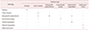
Patients had been taking tamoxifen for 13 to 68 months (mean ± SD = 27.1 ± 12.9). Hysteroscopic examination in 31 patients in the non-AUB group revealed 5 (16.1%) with atrophic endometrium, 3 (9.7%) with glandular-cystic atrophy, 8 (25.8%) wi th nonspecific endometrium, 7 (22.6%) wi th polyps, 6 (19.4%) with endometrial hyperplasia, and 2 (6.5%) with areas suspicious for adenocarcinoma.
Histologic examination of biopsy specimens confirmed the hysteroscopic diagnoses. Two cases of apparent endometrial hyperplasia on hysteroscopy were histologically diagnosed as non-specific endometrium. One case with 2 adenocarcinomas was changed to atypical hyperplasia on histological examination (Table 1).
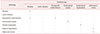
Hysteroscopic examination in the AUB group revealed 3 (20%) with atrophic endometrium, 2 (13.3%) with nonspecific endometrium, 5 (33.3%) with polyps, 2 (13.3%) with hyperplasia, and 3 (20%) with areas suspicious for adenocarcinoma.
One of the 3 suspicious adenocarcinomas was found to be atypical hyperplasia on histologic examination (Table 2). When the 2 groups were compared, the incidence of malignancy was high in cases with bleeding. However, more research is needed because of the small sample size.
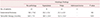
Hysteroscopy had a sensitivity of 0.85, specificity of 0.83, positive predictive value of 0.79, and negative predictive value of 0.87. A significant difference was found between the presence or absence of endometrial pathology and duration of tamoxifen therapy (in months; P < 0.05), with longer therapy associated with more pathologic findings on histology (Table 3).
Discussion
Tamoxifen is a nonsteroidal selective estrogen receptor modulator that is used primarily for adjuvant treatment of estrogen receptor-positive breast cancer in premenopausal women, and in some postmenopausal women.9 It is also used for chemoprevention in women at increased risk of breast cancer. Tamoxifen is associated with increased risks of uterine pathology, including endometrial polyps, endometrial carcinoma, hyperplasia, uterine sarcoma, and uterine carcinosarcoma.
Ozşener et al.10 showed that tamoxifen use increases the risk of endometrial cancer and premalignant change. They also noted a significant association between endometrial thickness and duration of tamoxifen treatment (P = 0.025). Hann et al.11 reported abnormal endometrial biopsies in 44% of women treated with tamoxifen for less than 5 years, whereas 58% of endometrial biopsies revealed abnormal results when duration of tamoxifen treatment was > 5 years.
Cohen et al.12 showed that 28.6% of patients on tamoxifen had endometrial pathology. The incidence was significantly more in symptomatic patients. Seoud et al.13 concluded that the value of routine screening for endometrial pathology in patients on tamoxifen is controversial. They found that all patients who developed an abnormal endometrium had abnormal vaginal bleeding.
Clinical trials confirm that long-term tamoxifen therapy for breast cancer for at least 5 years, is more effective than short-term treatment (< 2 years).314 During that time, tamoxifen acts on the endometrium as an estrogen receptor agonist. Its effects vary depending on dosage, duration of treatment, and patient age and menopausal status.15
The frequency of endometrial cancer was reported to double in trials of 1 or 2 years of tamoxifen and approximately quadrupled in trials of 5 years of therapy.14 Therefore, screening of postmenopausal asymptomatic patients taking tamoxifen is necessary to identify those who may develop significant endometrial pathology.15
In these patients, endometrial pathology may be diagnosed earlier because they are likely to have symptoms such as AUB;16 however, it is not known whether the increase in endometrial carcinoma represents a true increase. TVS is considered accurate for screening of these patients for endometrial changes and is used to suggest additional diagnostic evaluations.15
TVS may fail, however, due to the echogenic, irregular, cystic effect produced by tamoxifen on endometrial stroma and on the myometrium, without necessarily causing epithelial disease.17 Therefore endometrial thickness cannot be used in these patients to define abnormalities.18 A high prevalence of polyps (40%) in patients with thickened endometrium was reported in another study and could correlate with duration of tamoxifen intake.19 It does not seem likely that endometrial polyps are premalignant lesions, but malignancies in polyps of women taking tamoxifen were recently reported.20
Women on tamoxifen therapy with AUB or a thick endometrium require evaluation for uterine pathology. The approach to evaluation differs between pre- and postmenopausal women.
The evaluation typically includes TVS and/or endometrial sampling. American College of Obstetricians and Gynecologists (ACOG) guidelines advise that the initial test used to evaluate postmenopausal bleeding in average-risk women may be either endometrial sampling or TVS.21
Endometrial thickness ≤4 mm on TVS in postmenopausal women at average risk has been demonstrated to be an effective test to exclude endometrial cancer. The ACOG also advises that blind endometrial biopsy is most effective for detecting global (pathology occupies at least 50% of the surface area of the endometrial cavity), but may miss focal pathology.22
If focal pathology is suspected, a saline infusion sonogram or hysteroscopy should be performed. For postmenopausal women on tamoxifen, most experts advise endometrial biopsy rather than TVS alone; however, some find expectant management in the setting of a thin endometrial echo (≤4 mm) to be acceptable. On the other hand, as noted, endometrial biopsy also has limitations, since it may miss focal pathology. For these reasons, we perform both hysteroscopic endometrial biopsy and TVS.
Conclusion
Our experience indicates that hysteroscopic biopsy can get accurate information on endometrial thickening by tamoxifen. We believe that all women undergoing tamoxifen therapy for breast cancer should have regular TVS assessment of the endometrium. TVS is helpful for screening patients receiving long-term therapy. For early diagnosis of endometrial abnormalities, hysteroscopy is required if patients become symptomatic or if TVS reveals thickened endometrium.
 XML Download