

 PDF
PDF ePub
ePub Citation
 Print
Print


INTRODUCTION
Fewer than 20% of patients diagnosed with pancreatic ductal adenocarcinoma (PDAC) involving the head of the pancreas present with resectable disease.1 An additional 5–10% of patients present with borderline resectable pancreatic cancer (BRPC) and are likely to benefit from resection after neoadjuvant therapy (NAT).12 Pancreatic resection with simultaneous resection of the portomesenteric venous axis improves resectability and consequently survival in patients with PDAC with operative morbidity and mortality comparable to standard resections.3456 The benefit is clear and consistent when compared to survival after surgical bypass procedures.37 It yields maximal benefit in patients with short segment venous involvement8 and those without histologically demonstrable vein wall infiltration.69 However, a few reviews have raised concerns that venous involvement by PDAC indicates advanced disease stage45 and questioned the rationale for this procedure unless the vein wall was free of tumor and R0 resection was achieved.9
Conventional preoperative imaging is unreliable in predicting vein wall infiltration and determining the need for vein resection (VR).910 Differentiation between tumor adhesion and infiltration of the vein is unreliable even during surgery. Only about 40% (range 17 to 78%) of patients who undergo VR during pancreatic resection manifest true vein wall infiltration.6 Nonetheless, Delpero et al.5 while reporting the findings of a survey from the Association Francaise de Chirurgie recommended NAT for all patients scheduled for VR during pancreatic resection, including those with venous involvement in the form of impingement, abutment, narrowing, thrombosis or occlusion observed in cross-sectional imaging studies.9
This study presents the experience of the authors with VR in patients with PDAC of the head of pancreas.
MATERIALS AND METHODS
A retrospective analysis of patients undergoing resection for proximal pancreatic cancer between July 2010 and July 2016 was performed. The study group consisted of all patients with histologically confirmed PDAC following resection. The cohort was divided into two subgroups based on whether or not they underwent VR. Patients who underwent standard pancreatic resection constituted Group SR. Those who underwent an additional VR were included in Group VR. The preoperative patient characteristics, radiological imaging, operative findings, hospital stay, morbidity and 90-day mortality, tumor pathology including TNM stage, margin status and histological vascular invasion, were recorded and compared between the groups.
All patients underwent MDCT with pancreatic protocol. Patients with BRPC as per the MD Anderson Cancer Center definition (‘short segment occlusion of the portal vein (PV), superior mesenteric vein (SMV) or splenoportal junction with reconstructable, healthy vein proximal and distal to the area of tumor involvement’)129 were advised NAT. For purposes of this study, computed tomography (CT) scans were reviewed and venous involvement was further assessed using the Ishikawa et al.11 classification (Table 1). Patients diagnosed with BRPC underwent endobiliary stenting and endoscopic ultrasonography (EUS)-guided biopsy of the tumor, followed by NAT as advised by a medical oncologist. Preoperative biopsies and endobiliary stenting were performed selectively in patients who did not undergo NAT. Laparoscopy was performed on all patients prior to laparotomy.
All patients were treated with classical Whipple's pancreaticoduodenectomy and standard lymphadenectomy. A combined posterior and uncinate dissection along the superior mesenteric artery (SMA) was used to completely mobilise the head of pancreas prior to transection of the pancreatic neck.12 An isolated Roux loop was used to create an infracolic gastrojejunostomy to the left of the middle colic vessels. The pancreatic and biliary reconstruction was performed on a single jejunal loop. The preferred pancreatic anastomosis was a duct-to-mucosa pancreaticojejunostomy. VR was performed when a trial dissection demonstrated venous involvement. Reconstruction was either by direct end-to-end anastomosis of the vein or by interposition with autologous vein graft (left internal jugular vein) or 10 mm polytetrafluoroethylene (PTFE). The splenic vein (SV) was reimplanted when possible. Portal flow was documented after reconstruction with intraoperative Doppler ultrasound. Anticoagulation with low molecular weight heparin for 2 weeks was advised only for patients with PTFE interposition grafts.
Drain fluid amylase was monitored routinely on day 3, prior to removal of drains, and as needed, based on clinical suspicion of a pancreatic leak. International Study Group definitions were used to diagnose pancreatic leaks,13 haemorrhage14 and delayed gastric emptying (DGE).15 Chylous leaks were diagnosed when drain fluid amylase was normal and drain fluid triglycerides were elevated on a normal diet.
All patients were recommended adjuvant chemotherapy. Adjuvant radiotherapy was not prescribed for any patient.
Pancreaticoduodenectomy specimens were evaluated as per the Leeds Protocol16 using serial axial specimen slicing after inking the different margins. A positive margin was defined as one in which tumor cells were within 1mm of the inked surface of the specimen.
IBM SPSS Statistic v.20 for Windows (IBM, Armonk NY) was used for data entry and analysis. Continuous data were evaluated using medians, ranges and Mann-Whitney U test. Categorical data were evaluated by Fisher's exact test. Multivariate analysis was performed using multiple regression to determine the factors predicting 90-day survival. A p-value below 0.05 was considered statistically significant for all tests of comparison. Survival was estimated using Kaplan-Meier method and compared using the Log rank test.
RESULTS
We performed 94 pancreaticoduodenectomies between July 2010 and July 2016, 41 of which were indicated for PDAC and constituted the study group. An additional 7 patients with PDAC did not proceed to resection due to metastatic disease diagnosed at laparoscopy or laparotomy. The median age of the patients in the study group was 59 years (range: 42–79 years) and included 17 women.
Of these, 5 patients were diagnosed with BRPC and received NAT. Four patients received a gemcitabine-capecitabine combination, and one received FOLFIRINOX (5-fluorouracil, leucovorin, irinotecan, and oxaliplatin) regimen. Three patients also underwent preoperative radiation therapy. None experienced down-sizing of the tumor with NAT.
Operative details of vascular resection
The extent of vein involvement by Ishikawa Classification is described in Table 1.
Segmental resection was performed in all but one patient who underwent sleeve resection. Reconstruction was carried out via primary anastomosis in 12 patients, interposition autologous left internal jugular vein graft in 10, and interposition PTFE graft in 3 patients. Of the 11 patients with tumor involvement of the splenoportal junction, the SV was ligated in 6, reconstructed to the neo portal vein in 3, and 2 patients underwent total pancreatectomy with splenectomy.
Arterial reconstruction was performed in a patient with limited involvement of the common hepatic artery adjacent to the gastroduodenal artery.
Morbidity, mortality and hospital stay
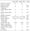
Of the 4 pancreatic fistulae, 3 were Grade A, and 1 was Grade B requiring percutaneous drainage of an intra-abdominal collection. Of the 5 patients with DGE, all from group VR, 3 were Grade A, and 1 each Grade B and Grade C. One patient underwent re-exploration for bleeding from the mesentery on day 1 after resection in the VR group. Details are provided in Table 2.
All three deaths occurred in the VR group. One death was attributed to hepatic ischemia in a patient with inadequate portal flow following prolonged PTFE reconstruction with compromised hepatic arterial flow. Another patient died from pulmonary sepsis following re-suturing of her abdominal wound for wound dehiscence. She also had Grade C DGE. The third patient also died from sepsis, the source of which is unclear. However, drain amylase was normal and CT scans did not reveal any intra-abdominal collections. Of the three deaths, two occurred in patients who underwent PTFE reconstruction.
Histopathology
The median maximum tumor diameter of the entire group was 3.6 cm (range 1.5–7cm). The median number of lymph nodes excised was 23 (range 1–77). Twenty-eight patients (68.3%) had lymph node metastases. The lymph node ratio (LNR) for the entire cohort was 19.25%.
Positive microscopic resection margins were noted in 20 (48.8%) patients. The superior mesenteric artery margin (39%) was the most frequently involved followed by the pancreatic transection margin (27%). Forty per cent of patients with positive margins showed multiple margin involvement. Patients with Ishikawa grade III and IV vein involvement were more likely to have a positive SMA margin than those with lesser degrees of involvement (p=0.04). Tumour infiltration of the splenoportal junction was associated with a significantly greater risk of pancreatic transection margin involvement (p=0.03). Four of 5 patients who received NAT had R0 resections, despite Ishikawa grade IV and V venous involvement. Details are presented in Table 1 and 2.
Histological venous invasion was demonstrated in 13 patients (50%), and was significantly more common in patients with Ishikawa III and IV grade venous involvement (p=0.04); it was associated with increasing Ishikawa type (Table 2).
Survival
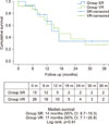
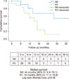
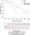
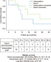
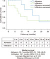
The median survival for the entire group of patients was 17 months (95% CI 13.3–20.7) and was not different between Groups SR (14 months, 95%CI 8.7–19.3) and VR (17months, 95%CI 7.1–26.9), (Log Rank p=0.91) (Fig. 1). Median survival was significantly better in patients without lymph node metastases (44 months vs. 15 months, Log Rank p=0.05) (Fig. 2) and worse in patients with histologically demonstrable vein wall infiltration by tumor (22 months vs. 11 months, Breslow Wilcoxon p=0.04, Log Rank 0.14) (Fig. 3). No statistically significant difference was found in survival between patients who underwent R0 or R1 resections (22 months vs. 12 months, p=0.28) (Fig. 4). Survival was not significantly better in those who underwent direct venous reconstruction, indicative of a shorter resection, compared to those who required an interposition graft (28 months vs 12 months, p=0.12) (Fig. 5).
Multivariate analysis revealed that the 90-day survival was not significantly affected by age, tumor size, lymph node ratio, completeness of resection (R0 or R1), complications (DGE, pancreatic leak) or concomitant vascular resection. However, this finding may be attributed to the small sample size.
DISCUSSION
Early reports extolling the benefits of VR along with pancreaticoduodenectomy found that vein involvement was a function of tumor location rather than an indication of aggressive tumor.17 This observation was supported by the latest relevant meta-analysis of 9005 patients from 27 selected trials.6 However two large, recently published series from the Association Francaise de Chirurgie5 and the Japanese Multicentre Study group for Hepatobiliary Surgery4 state that tumors requiring VR were more likely to be stage T4,45 poorly differentiated,5 and have a greater incidence of lymph node metastases compared with those undergoing standard resections.45 The present series supports the meta-analysis data. Tumor size and lymph node involvement did not correlate with venous involvement. Small tumors measuring 1.5 cm in maximal diameter required VR whereas others as large as 7 cm in maximal diameter did not. Lymph node metastases were more common and the LNR higher in patients undergoing standard resections (SR). Patients with lymph node metastases showed significantly poorer survival after resection in the present series. Improved survival in the VR group therefore likely reflects the lower LNR rather than any benefit from VR itself.
In a meta-analysis, Giovinazzo et al.6 reported significantly greater overall morbidity (OR 1.34), reoperations (OR 1.4) and postoperative bleeding (OR 1.61) in patients undergoing VR compared with standard resections. The incidence of pancreatic fistula and DGE was not increased after VR. The mean operative mortality in their analysis was 3.9% in patients undergoing VR and 3% in those undergoing standard resections (p=0.02). Although the overall morbidity in the present series was greater following VR, all the complications in the SR group, and all but one in the VR group were minor. The difference in morbidity was wholly accounted for by the 3 postoperative deaths in the VR group, 2 of which occurred in the 3 patients who underwent PTFE reconstruction. Since autologous vein graft was the preferred mode of reconstruction by the authors, the use of PTFE may reflect either the need for an unplanned or uncontrolled reconstruction that did not provide enough time for harvesting an autologous vein, or a very long reconstruction. These situations are known to be associated with increased operative mortality.818
Factors associated with poor survival post VR include the need for reconstruction longer than 3 cm,8 true vein wall invasion on histology,69 and R1 resection.9 Although shorter venous reconstructions, as indicated by the ability to perform primary reconstructions were associated with improved survival compared with those requiring interposition grafts, the difference was not significant in the present series. True vein wall invasion was associated with significantly poorer early survival as indicated by the Breslow Wilcoxon test (p=0.04) though not the Log Rank test, and may be predicted reliably in patients with Ishikawa types 3, 4 and 5. VR may thus be beneficial in those patients with higher grades of radiological portomesenteric venous tumor involvement (Ishikawa type 3 and above). This finding together with the 48% R1 resection rate in the present series suggest that all patients planned for VR are recommended NAT despite the lack of reliable evidence suggesting that R0 resections prolong survival compared with R1 resections either in the present series or in other studies.19
The risk of isolated local recurrence in patients with R1 resections is less than 10%.20 Evolving consensus suggest that margin involvement might be more indicative of the quality of pathological examination of the resected specimen21 and tumor biology.22 A review of specimen histology using the axial slicing technique described by Verbecke et al.16 revised the R1 resection rate from 14% to 76%,23 and 53% to 85%16 respectively, in 2 large independent series even in patients without VR. The importance of tumor biology in the present series is supported by the high rate of R1 resection in the SR group, the fact that 40% of patients with R1 resections had more than one margin involved by tumor, and that survival advantage from R0 resection was not apparent even with tumor-free margins of 1 mm.
SMA margin is the most frequently (15 to 45%) involved margin after VR:,6 and was involved in 39% of patients in the present series. SMA-first dissection12 and periadventitial dissection of the SMA24 are recommended in order to improve the likelihood of R0 resections along this margin, without clear evidence to support.25 The other commonly involved margin is the pancreatic transection margin (27% in the present series) especially in patients with tumor overlying the splenopancreatic junction.26 Pancreatic transection to the left of the SMV26 (at the splenic artery origin) in such patients, as well as additional resections based on frozen section biopsy of the margin are desirable to ensure clear resection margins. Although the ‘Whipple At The Splenic Artery’ or ‘WATSA’,26 advocated by Strasberg, appears sensible and also permits better control of the splenic vein in such patients, the role of additional resection dependent on frozen section biopsy is disputed. Kooby et al.,27 and others,19 determined that involvement of this margin reflected adverse pathological factors such as tumor size, lymph node involvement and perineural infiltration, with an overwhelming effect on survival despite additional pancreatic resection with clear margins.
This study suffers from being retrospective in nature. Therefore, additional details such as the true length of vein resection, the use of SMA first dissection and periadventitial resection of the SMA, the proportion of patients completing adjuvant therapy and the nature of recurrence (local or distant) are not available. The numbers are relatively small, although the proportion of patients undergoing VR is high, reflecting a referral bias.
In conclusion, vein involvement by proximal PDAC is indicative of location rather than tumor biology. VR benefits patients with tumor adhesion to the portomesenteric venous axis despite the risk of R1 resection and operative mortality. Primary reconstruction or interposition of autologous vein grafts appears to yield better outcomes compared with PTFE. Increased use of NAT in patients with high preoperative suspicion for vein resection may be beneficial, although the survival benefit derived from R0 resection is not clearly established in the present series.
 XML Download