

 PDF
PDF ePub
ePub Citation
 Print
Print


INTRODUCTION
Total hip arthroplasty, which began to become popular at around 1960, can dramatically improve the quality of human life and is regarded as one of the best surgeries developed in the last 100 years1).
Initially, the bone cement fixation method developed by Sir John Charnley was mainly used23). However, as the bone cement was mistakenly believed to play an important role in the pathogenesis of aseptic loosening4), cementless implants with biological fixation started to be developed in the early 1980s and are widely used up to date5).
In Korea, the first total hip arthroplasty was performed by Ahn et al.6) in the late 1960s. In 1988, Kim et al.7) started to use a porocoated femoral stem of his own design and reported satisfactory results with this stem. The efforts to develop and manufacture artificial hip joints in Korea began in the 1990s and a few developments were trialed clinically but were unsuccessful. The COREN hip system (Corentec, Seoul, Korea), which was developed in the early 2000s and commercialized in 2006, is recognized as the first successful artificial hip joint system developed in Korea.
The COREN system has been used at our institution since February 2007. We evaluated the clinical and radiological outcomes of total hip arthroplasty using this system after a follow-up of at least 5 years.
MATERIALS AND METHODS
This study was approved by the institutional review board of our hospital (H-1607-049-774). The first 200 primary total hip arthroplasties performed in 169 patients using the COREN hip system between February 2007 and April 2011 were included. Six patients (6 hips) died within 5 years, and 12 patients (13 hips) were lost to follow-up. The remaining 151 patients (181 hips) were evaluated with a minimum follow-up of 5 years (average, 87 months; range, 60 to 118 months).
The study group included 93 men and 58 women. The mean age at the index operation was 47 years (range, 19–69 years). The primary diagnoses included osteonecrosis of the femoral head (108 cases), degenerative arthritis secondary to acetabular dysplasia (28 cases), sequelae of hip joint infection (10 cases), Legg-Perthes disease (9 cases), rheumatoid arthritis (6 cases), nonunion of a femoral neck fracture (4 cases), ankylosing spondylitis (2 cases) and degenerative arthritis due to other causes (14 cases). The senior author (HJK) performed all of the procedures. Total hip arthroplasty was performed in the lateral decubitus position through a posterolateral approach in 176 cases and a transtrochanteric approach in 5 cases. In all cases, an alumina-alumina bearing system (Biolox Forte; CeramTec, Plochingen, Germany) was used. A 28-mm diameter head was used in 131 cases and a 32-mm diameter head in 55 cases.
Postoperatively, toe-touch non-weight-bearing crutch walking was recommended for the first 6 weeks followed by tolerable weight bearing to full weight bearing at 10 weeks. Patients were invited for follow-up evaluation at 6 weeks, 3 months, 9 months and then annually after the surgery.
Clinical outcomes were assessed with the use of the Harris hip score and a questionnaire that included items on thigh pain and noise; the questions were asked directly at the follow-up visit (142 cases) or by means of a telephone interview (39 cases).
Radiological evaluation was possible only in 142 cases for which follow-up radiographs for ≥5 years were available. The last radiographic evaluation was performed at a mean of 86 months (range, 60 to 118 months) after the surgery. Serial radiographs were examined with respect to component stability8), radiolucent lines, osteolysis, evidence of stress shielding8), migration of the components, and loosening. Zones described by Gruen et al.9) and DeLee and Charnley10) were used to assess the location and extent of radiolucent lines and osteolysis. Osteolysis was defined as a periprosthetic cystic or scalloped lesion with a diameter exceeding 2 mm that had not been present on the immediate postoperative radiographs1112). When heterotopic ossification was observed, it was graded by the criteria of Brooker et al13).
Other complications such as dislocation, infection, deep vein thrombosis, and ceramic fracture were also examined.
RESULTS
The mean Harris hip score improved considerably from 59.4 (range, 30 to −69) preoperatively to 97.2 (range, 84 to 100) at the last follow-up. Noise in the hip-joint area, which was occasional and transient, was reported in 7 cases (3.9%), including squeaking noise in 3 cases (1.7%) out of them14). No patient complained of thigh pain.
According to the criteria for implant fixation described by Engh et al.8), all acetabular cups and femoral stems were stably fixed with bony ingrowth. No changes in implant position such as stem subsidence were detected on follow-up radiographs. There was no evidence of implant loosening and no cases of implant revision up to the last follow-up.
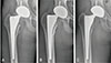
Eleven hips (7.7%) had a radiolucent line around the femoral stem. It was located in Gruen zone 7 in 10 cases and in zones 1 and 7 in one case (Fig. 1). Generally, the lines appeared 3 to 9 months after surgery and then remained unchanged with a gradual sclerotic change of the outer margin. No radiolucent lines were detected around the metal shells.
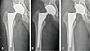
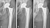
Focal osteolytic area was detected in 3 cases. In one case, it was detected in Gruen zone 1 at postoperative year 1 and remained until 7 years after the operation with no increase in size (Fig. 1). In the other 2 cases, osteolysis was detected in zones 1 and 7 at postoperative years 5 (Fig. 2) and 9 (Fig. 3), respectively. These two lesions needed further follow-up to evaluate temporal changes.
Stress shielding was observed in 80.3% of cases. It was first degree of Engh et al.8) in 51 hips (35.9%) and second degree in 63 hips (44.4%); there were no cases of third or fourth degree stress shielding.
Heterotopic ossification was identified in 25 cases (17.6%). Thirteen cases were grade I and 12 cases were grade II.
Femoral fractures occurred in 5 cases (2.7%) during stem implantation. Four were localized to the proximal femoral end and one was extended distally below the middle of the stem. All were treated with circumferential wiring and healed without any problem.
Femoral nerve palsy occurred in one case as a surgical complication. It gradually partially recovered during the follow-up period. However, residual palsy did not markedly disturb daily activity.
In one case, bacterial infection developed immediately postoperatively. It was controlled by surgical drainage with debridement, which was performed three times, and there was no recurrence until postoperative year 9. In the same case, posterior dislocation occurred 3 months postoperatively (1 month after the last surgical drainage) and was treated conservatively.
DISCUSSION
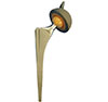
In the COREN hip system, the femoral stem and acetabular cup are made of titanium alloy (Ti6Al4V). The femoral stem is straight, double wedged, and tapered with a rectangular cross-section (Fig. 4). There are anterior, posterior and lateral vertical ribs on its proximal end. Its surface is grit blasted and then further treated by micro-arc oxidation after15161718).
In this study, all femoral stems were fixed stably with bony ingrowth without position change or subsidence, and the fixation was well maintained up to the latest follow-up. Other studies have reported similar results15171819). In contrast, early postoperative subsidence of the stem has been reported in many studies of the cementless Spotorno stem (CLS stem; Zimmer, Warsaw, IN, USA), whose design is similar to that of the COREN prosthesis202122). The better early fixation might be the result of three proximal vertical pins for rotational stability and micro-arc oxidation treatment to maximize the contact surface with bone16).
Unlike in other reports15171819), 11 cases (7.7%) showed a radiolucent line in Gruen zone 1 or 7. The thickness of these lines did not exceed 2 mm. They did not expand or affect the stability of the stem (Fig. 1).
We found rounding of the most proximal medial edge of the cut femoral neck (first degree) in most cases and some reduction in the proximal medial cortical density (second degree of stress shielding by Engh et al.8)) in more than half of the cases (Fig. 1). We found no cases of severe reduction in the proximal medial cortical density or extensive resorption. These results are quite different from those of Kim et al.17), who observed stress shielding only in 19.7% of cases. There is also big difference between results of Schramm et al.21) (37%) and of Yamasaki et al.23) (1.5%), who used the CLS stems. These discrepancies might be due to the differences in subjective interpretation of the change in cortical density.
No patient complained of thigh pain in the present study. Most studies171819) that used the COREN stems reported a very low incidence of thigh pain, similar to studies that used the CLS stems, which have similar design and surface treatment2021).
The surface of the COREN acetabular cup is coated with porous titanium by a plasma-spray method. In all cases, the acetabular cup was fixed stably by bone ingrowth no change in position and no radiolucent line detected in any case. Very similar results were observed in another study that used an acetabular cup made of the same material using the same surface treatment24).
It was a retrospective study and radiological evaluation was not possible in all cases. However, it was a series of consecutive cases, and we believe that the number of cases and the follow-up rate were good enough for objective data analysis.
CONCLUSION
The clinical and radiological results of total hip arthroplasty using the COREN hip system were very satisfactory after a follow-up of at least 5 years. All implants were stably fixed with bone ingrowth and no aseptic loosening occurred during the follow-up period. No patients needed revision surgery for any reason.
 XML Download