

 PDF
PDF ePub
ePub Citation
 Print
Print


Yu-Hun Jeong, M.D., Young-Sang Lee, M.D. , Dong-Chan Eun, M.D., Chan-Woong Byun, M.D.
, Dong-Chan Eun, M.D., Chan-Woong Byun, M.D.
Abstract
Neurocysticercosis (NCC) by Taenia solium is the most common parasitic infection of the central nervous system involving the cerebrum. However, spinal involvement of NCC is rare. Spinal NCC can cause radiculopathy, myelopathy, cauda equina syndrome, and even paraparesis, depending on its location and size. Spinal NCC may require surgical treatment as a first-line treatment because medical therapy can further aggravate the inflammation due to dead cysts, resulting in clinical deterioration. The current standard therapy for spinal NCC is surgical decompression followed by medical therapy. We experienced a case of widespread thoracolumbar intradural extramedullary cysticercosis involving the spinal canal with cerebral cysticercosis. We report this rare case with literature review.
Figures and Tables
Figure 1
(A) Magnetic resonance imaging demonstrated variable sized cystic lesions in the intradural, extramedullary space and a huge, septated cystic lesion with diffuse enhancement and adhesion of arachnoid at the T12–L3 level. (B) Diffuse enhancement and adhesion of arachnoid with empty sac sign (arrows) in the lumbar spine.
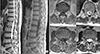
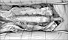
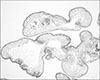
Figure 2
After careful cystic wall incision, clear cystic fluid gushed out, which was sucked using a suction. Multiple small grape-like whitish masses (arrows) within the cyst were found and removed in a piecemeal fashion. Massive irrigation with hypertonic saline was performed.

References
1. Martinez HR, Rangel-Guerra R, Arredondo-Estrada JH, Marfil A, Onofre J. Medical and surgical treatment in neurocysticercosis a magnetic resonance study of 161 cases. J Neurol Sci. 1995; 130:25–34.

2. Kim SW, Lee SM. Sacral intradural cysticercosis misdiagnosed as brain tumor metastasis. J Korean Neurosurg Soc. 2005; 37:67–69.
3. Chang GY, Keane JR. Visual loss in cysticercosis: analysis of 23 patients. Neurology. 2001; 57:545–548.
4. Kishore LT, Gayatri K, Naidu MR, Mateen MA, Dinakar I, Ratnakar KS. Intermedullary spinal cord cysticercosis: a case report and literature review. Indian J Pathol Microbiol. 1991; 34:219–221.
5. Colli BO, Assirati Júnior JA, Machado HR, dos Santos F, Takayanagui OM. Cysticercosis of the central nervous system. II. Spinal cysticercosis. Arq Neuropsiquiatr. 1994; 52:187–199.
6. Paterakis KN, Kapsalaki E, Hadjigeorgiou GM, Barbanis S, Fezoulidis I, Kourtopoulos H. Primary spinal intradural extramedullary cysticercosis. Surg Neurol. 2007; 68:309–311. discussion 312.
7. Alsina GA, Johnson JP, McBride DQ, Rhoten PR, Mehringer CM, Stokes JK. Spinal neurocysticercosis. Neurosurg Focus. 2002; 12:e8.
8. Rahalkar MD, Shetty DD, Kelkar AB, Kelkar AA, Kinare AS, Ambardekar ST. The many faces of cysticercosis. Clin Radiol. 2000; 55:668–674.
9. Holtzman RN, Hughes JE, Sachdev RK, Jarenwattananon A. Intramedullary cysticercosis. Surg Neurol. 1986; 26:187–191.
10. Schnepper GD, Johnson WD. Recurrent spinal hydatidosis in North America. Case report and review of the literature. Neurosurg Focus. 2004; 17:E8.
 XML Download