

 PDF
PDF ePub
ePub Citation
 Print
Print


Changsu Kim, M.D. , Daemoo Shim, M.D.*, Seokjoong Lee, M.D.†, Youngha Woo, M.D.‡, Samuel Baek, M.D., Haksun Chung, M.D.
, Daemoo Shim, M.D.*, Seokjoong Lee, M.D.†, Youngha Woo, M.D.‡, Samuel Baek, M.D., Haksun Chung, M.D.
Abstract
Purpose
The purpose of this study was to compare accuracy of proper needle insertion angle between magnetic resonance imaging (MRI) and ultrasonography during lumbar medial branch nerve block procedure.
Materials and Methods
Between January 2015 and June 2016, 80 people who underwent MRI in the past 3 months with improved lumbar pain after sono-guided medial branch nerve block were enrolled for analysis (male, 39; female, 41; average age, 63.3 years). The insertion angle and depth between the spinous process and needle at each target points were measured at various levels (superior, inferior segment of each facet joints from L2–3 to L5–S1). The needle was positioned 1 cm apart from both lateral sides of the probe, locating spinous process in the middle. A comparative analysis was performed between an ultrasonography and an MRI. We determined the statistical correlation between the two methods.
Results
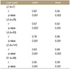
The average differences with respect to the distance between each level on a sono-guided medial branch nerve block were 1.28±1.07 mm in L2 (7 cases), 1.27±4.26 mm in L3 (25 cases), 1.63±5.89 mm in L4 (93 cases), 1.99±4.12 mm in L5 (141 cases), and 1.51±3.87 mm in S1 (66 cases). The average differences regarding the angle of each level were 1.69°±1.34° in L2 (7 cases), 2.03°±5.35° in L3 (25 cases), 1.49°±3.42° in L4 (93 cases), −1.55°±3.67° in L5 (141 cases), and 1.86°±4.83° in S1 (66 cases). All measurements followed a normal distribution (p<0.05), showing statistical correlation without significant difference (p<0.05).
Figures and Tables
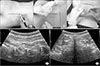 | Figure 1(A) When longitudinal axis is a line along the spinous process (A-1) and center of probe is on the spinous process (A-2), entrance points of the needle are 1 cm from both lateral sides of the probe (A-3). (B) Facet joint seen at a longitudinal view; the exact point is set by counting the interface between spinous process and facet joint and upper portion of sacrum (B-1) facet joint and spinous process are checked after rotating the probe 90° for a horizontal view (B-2). S, spinous process; F, facet; T, transverse process.
|
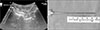 | Figure 2Needle is inserted through the index at each level, needle's depth and angle between the needle and spinous process are measured when the needle touches the target point of the medial branch block.
|
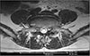 | Figure 3On a magnetic resonance imaging, the length (depth) and angle of the needle at each level are measured using the distance between the entrance point of needle insertion and the target point when interface between the transverse process and superior articular process is observed on the axial view.
|


References
1. Mooney V, Robertson J. The facet syndrome. Clin Orthop Relat Res. 1976; (115):149–156.

2. Frymoyer JW. Back pain and sciatica. N Engl J Med. 1988; 318:291–300.
3. Bogduk N. International spinal injection society guidelines for the performance of spinal injection procedures. Part 1: zygapophysial joint blocks. Clin J Pain. 1997; 13:285–302.
4. Gangi A, Dietemann JL, Mortazavi R, Pfleger D, Kauff C, Roy C. CT-guided interventional procedures for pain management in the lumbosacral spine. Radiographics. 1998; 18:621–633.
5. Saal JS. General principles of diagnostic testing as related to painful lumbar spine disorders: a critical appraisal of current diagnostic techniques. Spine (Phila Pa 1976). 2002; 27:2538–2545.
6. Saranteas T, Paraskeuopoulos T, Anagnostopoulou S, Kanellopoulos I, Mastoris M, Kostopanagiotou G. Ultrasound anatomy of the cervical paravertebral space: a preliminary study. Surg Radiol Anat. 2010; 32:617–622.
7. Ha DH, Shim DM, Kim TK, Kim YM, Choi SS. Comparison of ultrasonography- and fluoroscopy-guided facet joint block in the lumbar spine. Asian Spine J. 2010; 4:15–22.
8. Shim JK, Moon JC, Yoon KB, Kim WO, Yoon DM. Ultrasound-guided lumbar medial-branch block: a clinical study with fluoroscopy control. Reg Anesth Pain Med. 2006; 31:451–454.
9. Pedersen HE, Blunck CF, Gardner E. The anatomy of lumbosacral posterior rami and meningeal branches of spinal nerve (sinu-vertebral nerves); with an experimental study of their functions. J Bone Joint Surg Am. 1956; 38:377–391.
10. Hirsch C, Ingelmark BE, Miller M. The anatomical basis for low back pain. Studies on the presence of sensory nerve endings in ligamentous, capsular and intervertebral disc structures in the human lumbar spine. Acta Orthop Scand. 1963; 33:1–17.
11. Kellgren JH. Referred pains from muscle. Br Med J. 1938; 1:325–327.
12. Mooney V. The syndromes of low back disease. Orthop Clin North Am. 1983; 14:505–515.
13. Rees WS. Rhysolysis of the nerves of the zygoapophyseal joints. Spine (Phila Pa 1976). 1983; 8:118–120.
14. Shealy CN. Facet denervation in the management of back and sciatic pain. Clin Orthop Relat Res. 1976; 115:157–164.
15. Silvers HR. Lumbar percutaneous facet rhizotomy. Spine (Phila Pa 1976). 1990; 15:36–40.
16. Alhelail M, Al-Salamah M, Al-Mulhim M, Al-Hamid S. Comparison of bupivacaine and lidocaine with epinephrine for digital nerve blocks. Emerg Med J. 2009; 26:347–350.
17. Birkenmaier C, Veihelmann A, Trouillier HH, Hausdorf J, von Schulze Pellengahr C. Medial branch blocks versus pericapsular blocks in selecting patients for percutaneous cryodenervation of lumbar facet joints. Reg Anesth Pain Med. 2007; 32:27–33.
18. Jeon CH, Lee WI, Kang SY. Intra and extraspinal infected synovial cyst of the lumbar spine: case report. J Korean Soc Spine Surg. 1997; 4:357–364.
19. Moon SH, Lee S, Kim KH, et al. Effect of ultrasound-guided lumbar medial branch block in chronic low back pain. J Korean Orthop Res Soc. 2012; 15:54–61.
20. Greher M, Scharbert G, Kamolz LP, et al. Ultrasound-guided lumbar facet nerve block: a sonoanatomic study of a new methodologic approach. Anesthesiology. 2004; 100:1242–1248.
21. Galiano K, Obwegeser AA, Walch C, Schatzer R, Ploner F, Gruber H. Ultrasound-guided versus computed tomography-controlled facet joint injections in the lumbar spine: a prospective randomized clinical trial. Reg Anesth Pain Med. 2007; 32:317–322.
 XML Download