Journal List > J Korean Orthop Assoc > v.53(4) > 1100517

임, 류, 손, Lim, Ryu, and Shon: 슬관절 주위 절골술: 적응증 및 수술 전 계획

Abstract

슬관절 골관절염과 하지 정렬 이상이 동반된 경우 슬관절 주위 절골술을 고려할 수 있다. 슬관절 내반 변형의 경우 근위 경골 절골술을, 슬관절 외반 변형의 경우 원위 대퇴골 절골술을 선택하여 교정을 하는 것이 절골술의 기본적인 원칙으로 알려져 있다. 그러나 단일 절골술만으로 불가능할 경우 이중 절골술도 고려해볼 수 있다. 본 종설에서는 슬관절 주위 절골술을 시행함에 있어 환자의 선택 및 수술 전 계획의 원칙과 최신 지견에 대해 살펴보고자 한다

초록

Osteotomy around the knee is a widely considered surgical procedure for osteoarthritis with lower extremity malalignment. High tibial osteotomy (HTO) is performed for varus deformity, while distal femur osteotomy (DFO) is performed for valgus deformity. However, if the correction is insufficient, double osteotomy can also be considered. This report included the basic principles and current concepts of patient selection and preoperative planning in osteotomy around the knee.

서론

슬관절은 고관절과 족관절 사이에 위치하며 정상적으로 중력에 의한 체중 부하선이 관절의 중심을 지나가게 된다. 만약 하지의 정렬이 정상범위를 벗어나게 되면 슬관절의 특정 구획에 과도한 부하를 유발하게 되어 통증과 기능 저하를 유발할 뿐 아니라 결국 퇴행성 변화를 일으키게 된다.1) 교정 절골술은 하지의 정렬 이상과 슬관절의 퇴행성 변화를 보이는 환자에서 비정상적인 역학적 축을 교정함으로써 병변부에 가해지는 하중을 분산시키는 수술 방법으로 인공관절 치환술과는 달리 수술을 통해 환자의 통증을 줄이고 퇴행성 변화를 지연시켜 슬관절을 보존하여 수명을 연장시킨다는 차이점이 있다. 이러한 장점 때문에 나이가 비교적 젊은 환자에서 슬관절 주위 단일 구획 관절염의 치료에 근위 경골 절골술과 원위 대퇴골 절골술이 사용되고 있고, 좋은 결과를 보고하고 있다.23456789)
슬관절 변형의 대부분을 차지하는 내반 변형의 경우 근위 경골절골술을, 상대적으로 드문 슬관절 외반 변형의 경우 원위 대퇴골 절골술을 선택하여 교정을 하는 것이 절골술의 기본적인 원칙으로 알려져 있다.1011) 하지만 교정각이 너무 크거나 경골 및 대퇴골에 모두 변형이 있어 근위 경골 절골술이나 원위 대퇴골 절골술 어느 하나만으로는 적절한 교정을 얻지 못할 경우, 이중 절골술 역시 고려해 볼 수 있다.1213) 그러나 환자 선택 및 교정각도가 적절하지 못한 절골술을 시행할 경우, 오히려 나쁜 결과를 초래할 수 있다. 본 종설에서는 슬관절 주위 절골술을 올바르게 시행하기 위한 환자의 선택 및 수술 전 계획의 원칙과 최신 지견 등에 대해 살펴보고자 한다.

본론

1. 하지의 축

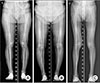
하지의 역학적 축은 기립 전후면 하지 전장 방사선 사진에서 대퇴골 두의 중심점부터 거골 원개의 중심점까지 연결한 직선으로 정상적으로 경골의 중심점을 지나가게 된다.14) 정상 슬관절에서는 내측 구획에 60%–70%의 체중이 부하되어 내측 구획이 외측 구획보다 골관절염의 진행이 더 많다.15) 양측 고관절 사이의 간격이 슬관절 사이의 간격보다 넓기 때문에 하지의 역학적 축은 지면에 수직인 선에 대하여 3도 내반되어 있다.161718) 하지의 역학적축이 슬관절의 중심을 지나지 않고 내측으로 통과하는 경우를 역학적 내반 정렬이라 하고, 외측으로 통과하는 경우를 역학적 외반 정렬이라 한다(Fig. 1).14)
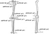
대퇴골의 역학적 축은 대퇴골 두의 중심부터 과간 절흔의 중심점까지 연결한 선이며 경골의 역학적 축은 경골 고평부의 중심부터 경골 천장부의 중심까지 연결한 선이다.19) 대퇴골과 경골의 해부학적 축은 각각 대퇴골과 경골 간부의 축으로 평균 6도의 외반을 이루고 있다. 대퇴골의 역학적 축과 해부학적 축은 평균 6도의 각을 이루고 있으며, 경골의 역학적 축과 해부학적 축은 거의 일치한다.20) 대퇴골의 내과와 외과의 관절면을 이은 선과 대퇴골의 역학적 축이 이루는 각인 역학적 외측 원위 대퇴각은 평균 88도를 이루고 있고, 대퇴골의 관절면을 이은 선과 대퇴골의 해부학적 축이 이루는 각인 해부학적 외측 원위 대퇴각은 평균 81도를 이룬다.21) 경골의 고평부선과 경골의 역학적 축이 이루는 경골의 역학적 내측 근위 경골각은 정상적으로 평균 87도의 각을 갖고 족관절선과 경골이 이루는 역학적 외측 원위 경골각은 평균 89도를 이룬다(Fig. 2).1122)
하지의 정렬 이상에서는 이와 같은 하지의 생리적 축과 생리적각들이 정상 범위를 갖지 않고, 정렬 이상의 종류에 따라 비정상적인 값을 갖는다. 수술 전 방사선 사진을 통한 정확한 평가를 통해 정렬 이상의 원인을 찾아 변형이 있는 부위에서 절골술을 시행하는 것을 원칙으로 교정 부위와 교정 각도 등의 술 전 계획을 세울 수 있다.12)

2. 근위 경골 절골술

1) 환자의 선택

(1) 나이

현재 환자 나이에 따른 근위 경골 절골술의 결과에 대해 합의된 결과는 없는 실정이다. 일부 보고들에 따르면 65세 이상으로 나이가 많을 경우 10년 생존율이 감소한다고 하지만,23242526) 나이가 생존율에 영향을 미치지 않는다는 반대되는 보고도 존재한다.272829) 일반적으로 술자들이 나이 많은 환자군에서 실패율이 높을 것이라고 믿는 경향이 있기 때문에 적응증을 좀 더 좁게 잡는 경향이 있는 것으로 보인다.924) 이처럼 저자들에 따라 결과의 차이가 있기 때문에 나이의 적응증을 잡을 때, 실제 나이보다는 생리학적 나이를 고려하는 것이 더 중요하다고 생각된다.

(2) 활동성

근위 경골 절골술은 단일 구획 골관절염이 있는 환자에서 교정 후 가장 높은 정도의 활동성을 보장해 준다.3031) Fu 등32)에 따르면, 단일 관절 치환술을 시행한 군보다 근위 경골 절골술을 시행한 군에서 술 후 더 빨리 걷는 것이 가능하다고 보고하였다. 물론 근위 경골 절골술을 시행한 군이 더 오랜 재활기간을 거쳐야 한다는 단점이 존재한다.33) 단일 관절 치환술과 근위 경골 절골술의 치환물 생존율과 인공 관절 전치환술로의 전환에 대해서는 저자에 따라 다른 결과를 보고하고 있다.22343536)

(3) Osteoarthritis 등급

근위 경골 절골술에서 내측 구획 골관절염의 정도는 술 후 결과에 영향을 미치는 중요한 요소 중 하나이다. 연골손상이 내측에 국한되어 있을 때 술 후 좋은 결과를 기대할 수 있지만, 심한 골관절염에서는 수술 후에도 내측에 통증이 지속될 수 있다.3738) 특히 슬개 대퇴관절염이 있거나 삼중 구획 골관절염까지 진행된 경우는 절골술의 금기증에 해당한다.1839) Ahlbäck40)이 제안한 골관절염의 분류에 따르면, 3등급 이상인 경우 결과가 좋지 않고,23) 0등급일 때 보다 나은 결과를 보인다고 하였다.9) 자기공명영상(magnetic resonance imaging, MRI)은 단일 구획 관절염의 환자에게 있어 연골손상 및 동반된 다른 질환을 확인하고 외측 연골판의 손상 여부를 평가하기 위해 시행할 수 있다.

(4) 관절운동 범위

근위 경골 절골술을 시행하기 전에 관절운동 범위를 검사해보아야 한다. Berman 등42)은 술 전 관절운동 범위가 90도 이하인 경우 술 후 결과가 좋지 않다고 보고하였다. Akizuki 등43)은 술 전 관절운동 범위 100도를 술 후 좋은 결과를 나타내는 기준점으로 보고 하기도 하였다. 많은 골관절염을 가지고 있는 환자들은 굴곡구축이 있으며 절골술을 통해 10도 이하의 굴곡구축은 교정할 수 있지만 10도 이상의 굴곡구축이 있는 경우는 근위 경골 절골술 후후방 경골 경사의 변화로 전후방 십자인대에 나쁜 영향을 미쳐 만족할 만한 수술 결과를 기대하기 어렵다.

(5) 슬관절 불안정성

슬관절 불안정성이 동반된 정렬 이상의 경우가 있을 수 있으므로 근위 경골 절골술을 시행하기 전에 인대 상태에 대한 검사가 필요하다. 후방 경골 경사의 변화는 관절운동 범위 및 전후방 십자인대의 상태에 따라 술 전 계획 및 술 후 결과에 영향을 미칠 수 있다. 즉 내측 개방형 경골 근위부 절골술에 의한 후방 경골 경사의 증가는 과신전의 감소 및 후방 불안정성의 감소(굴곡 구축의 증가 및 전방 불안정성의 증가) 효과를 가져올 수 있다.44) 반대로 외측 폐쇄형 경골 근위부 절골술에 의한 후방 경골 경사의 감소는 과신전의 증가 및 후방 불안정성의 증가(굴곡 구축의 감소 및 전방 불안정성의 감소) 효과를 가져올 수 있다.44)
아급성 전방 십자인대 손상과 내반 변형이 동반된 경우 근위 경골 절골술과 전방 십자인대 재건술을 동시에 시행하는 것이 권장된다.4546) 만성 전방 십자인대 손상과 내반 변형이 동반된 경우에는 근위 경골 절골술만 시행하거나 전방 십자인대 재건술을 같이 할 수 있는데, 환자의 나이와 활동성을 고려하여 결정한다.4546) 후방 십자인대 손상이나 후외측 구조물의 손상으로 만성 후외측 불안정성이 동반된 경우에는 근위 경골 절골술만 먼저 시행하고, 만약 불안정성이 술 후에도 지속된다면 후방 십자인대와 후외측구조물 재건술을 시행하는 것을 고려한다.47)

(6) 체질량지수

Flecher 등23)은 체질량지수가 30 kg/m2 미만인 경우, 근위 경골 절골술 후의 결과가 더 좋다고 보고하였다. Giagounidis와 Sell48)은 체질량지수가 정상보다 10% 이상 높을 경우 술 후 통증이 없는 기간이 5년, 체질량지수가 정상보다 10% 미만으로 높을 경우 술 후 통증이 없는 기간이 7.8년이라고 보고하였다. 이와는 반대로 Naudie 등25)은 체질량지수가 25 kg/m2 미만인 환자에서 근위 경골 절골술 후 더 나쁜 결과를 보인다고 보고하였는데, 이는 가벼운 사람이 더 활동적이어서 절골술 부위에 더 많은 부하가 걸리기 때문이라고 주장하였다. 이처럼 체중과 골 관절염 발생에 대한 논쟁은 아직까지 진행 중이며 보통 비만 환자의 경우 슬관절에 더욱 부하가 가해지는 것은 사실이다.

2) 수술 전 계획

과소한 혹은 과다한 교정을 피하기 위해서 수술 전 계획을 바르게 세우는 일이 필요하다.

(1) 방사선 검사

술 전 방사선적 평가를 위해 여러 촬영이 필요한데, 슬관절을 완전히 신전한 상태에서 하지 정렬 평가를 위한 양측 장하지 체중 부하전후방 촬영이 반드시 포함되어야 한다. 이 때 하지의 정렬이 중요하며 일반적으로 양하지 체중 부하된 상태로 발의 위치는 약간 내회전, 슬개골은 대퇴골과의 중심에 위치해야 한다.4950) 또 관절염의 평가를 위해 양측 체중 부하 전후방 및 진성 외측 촬영, 45도 골곡 양측 체중 부하 촬영인 Rosenberg view와 sky line view를 촬영하여야 한다.51) 내측 구획 골관절염의 정도 및 슬개골의 높이는 전후방 촬영에서 Insall-Salvati, Blackburne-Peel, Carton-Deschamps index52)를 이용하여 평가할 수 있고, 슬개골 고위가 있는 경우 경골결절 절골술을 동반하기도 한다. MRI는 연골 손상, 인대 손상, 골연골 결손, 골괴사, 연골하 부종 등을 확인하는 데 도움을 준다

(2) 고위 경골 절골술의 목표

내반 변형의 교정 목표 및 수술 전 계획 방법은 저자에 따라 다양하다. Coventry53)는 해부학적 대퇴경골각을 8도 외반으로 과교정할 것을 주장하였고, Hernigou 등54)은 역학적 축을 3–6도 외반으로 교정할 것을 주장하였다. Fujisawa 등18)은 술 후 역학적 축이 경골고평부의 내측에서부터 60%–70% 지점을 지나야 한다고 보고하였다. Miniaci 등55)은 경골 고평부의 30%–40% 지점을, Dugdale 등56)은 경골 고평부의 62.5%를 지나야 한다고 보고하였으며 Fujisawa 등18)이 언급한 방법에 따라 위치를 계산하였다. 최근 Jakob과 Jacobi57)는 내측 구획의 연골 두께에 따라 교정 정도를 조절하여야 한다고 주장하였다. 만약 내측 연골의 1/3이 손상되었다면 역학적 축은 경골 고평부 중심에서 외측으로 10%–15% 지점을 지나야 하고, 내측연골의 2/3가 손상되었다면 20%–25% 지점을, 내측 연골이 모두 손상되었다면 30%–35% 지점을 지나야 한다고 보고하였다.

(3) 관상면에서 술 전 교정 계획

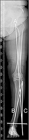
① Miniaci 방법: 수술 후 교정선이 대퇴골 두의 중심에서 술자가 원하는 체중 부하 위치로 경골 고평부를 지나면서 족관절까지 가도록 선을 그린다(Line A). 근위 경골 절골술의 경첩점에서 족관절의 중심까지 선을 그리고(Line B), 경첩점에서 Line A가 교차하는 선을 그린다(Line C). Line B와 Line C가 이루는 각도가 Miniaci 방법으로 계획된 교정각이다(Fig. 3).55)
② Dugdale 방법: 대퇴골 두의 중심에서 경골 고평부의 62.5% 지점까지 선을 그린다(Line A). 다음으로 족관절의 중심에서 경골고평부의 62.5%까지 선을 그린다(Line B). Line A와 Line B가 이루는 각도가 Dugdale 방법으로 계획된 교정각이다(Fig. 4).56)
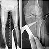
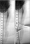
③ 체중 부하 단속 촬영법(weight bearing scanography)을 이용한 방법: 체중 부하 단속 촬영을 한 후, 이를 실제크기로 출력한다. 절골술을 시행할 부분을 자른 후 62%–65% 사이로 체중 부하 목표점이 지나도록 선을 그리고 회전시킨다. 절골술 부분의 크기를 자를 이용하여 직접 측정을 하여 교정 정도를 계획할 수 있다(Fig. 5).58)
④ 수술 중 하지 정렬을 확인하는 방법: 수술을 하는 도중에 수술 전 계획에 맞게 하지 정렬 교정이 되는지를 확인하는 것이 필요하다. 수술 중 하지 정렬을 확인하기 위해 cable이나 rod를 이용하는 방법이 고전적으로 사용되고 있으나 정확도 및 재현성에 문제점이 있다. 최근 사용되고 있는 항법 장치 시스템(navigation system)은 수술 중 실시간으로 해부학적 위치에 대한 정보를 제공하여 원하고자 하는 교정각을 보다 정확하게 얻을 수 있고, 오차 범위 및 이상치를 줄이며 교정의 정밀도를 향상시킬 수 있는 장점이 있다.585960) 하지만 임상적인 결과에 대해서는 추가적인 연구 및 장기 추시가 필요하다.
⑤ 최근 소개되고 있는 방법들: 최근 의료영상저장전송시스템(picture archiving and communication system, PACS) software나 Photoshop을 이용하여 Miniaci 방법으로 교정각을 계산하는 방법들이 소개되고 있다. PACS software나 Photoshop이 널리 보급되어 쉽게 접근이 가능하고, 앞의 방법들과는 달리 실제 크기의 출력물이 필요하지 않다는 장점이 있다.616263)

(4) 시상면에서 술 전 교정 계획

후방 경골 경사가 증가하면 굴곡 구축이 발생하고 경골의 전방 전위 및 전방 십자인대의 인장력을 증가시켜 후방 십자인대 결손시 나타나는 경골의 후방 아탈구가 감소하게 된다. 반대로 후방 경골 경사가 감소하면 과신전이 발생하고 후방 전위 및 후방 십자인대의 인장력을 증가시켜 전방 십자인대 결손 시 나타나는 경골의 전방 아탈구가 감소하게 된다.6465) 즉 후방 경골 경사를 조정하여 굴곡 구축 및 과신전과 같은 슬관절 변형을 교정할 수 있다는 것을 고려해야 하겠다. 하지만 후방 경골 경사가 10도 이상 증가하게 되면 전방 십자인대에 가해지는 힘이 3배 이상 증가하므로 주의해야 한다.66)
개방형 근위 경골 절골술의 경우 후방 경골 경사가 3도 정도 증가하는 경향을 보인다. 이러한 상황을 피하기 위해 절골술 부위의 전 후 간격을 2:3 정도로 벌려주는 것이 좋다.6768) 하지만 만성 후방 십자인대 손상이 있는 경우 전 후 간격을 비슷하게 하는 것이 더 좋은 결과를 기대할 수 있다. 이와는 반대로 폐쇄형 근위 경골 절골술에서는 후방 경골 경사가 3도 정도 감소하는 경향이 있고,69) 만성 십자인대 손상이 있는 경우 더 좋은 결과를 기대할 수 있겠다.

3. 원위 대퇴골 내반 절골술

1) 환자의 선택

원위 대퇴골 내반 절골술의 주된 적응증은 슬관절의 외측 구획에만 국한된 골관절염이 외반 변형과 동반된 경우이다.707172) 또 다른 적응증으로는 만성 내측 측부인대 부전으로 인한 인대 불안정성으로 외반 견인을 감소하기 위한 경우가 있고,46) 외반 변형으로 인한 슬개 대퇴 부정정렬을 교정하기 위해 시행하는 경우가 있다.73) 그 외에도 구루병, 신성 이영양증, 소아 마비, 기타 외상이나 근육 신경 질환에 의한 외반 변형 등에서 고려해볼 수 있다.2274) 원위 대퇴골 내반 절골술의 1차 적응증은 퇴행성 관절염과 외반변형이 동반되어 심한 통증을 호소하는 경우지만 최근에는 인대재건술, 연골 이식이나 반월연골판 동종 이식 등의 수술에서 수술 후 최상의 결과를 얻기 위해 함께 시행되기도 한다.
환자는 나이가 60세 이하이고, 술 전 운동범위가 90도 이상의 굴곡이 가능하고, 굴곡 구축이 15도 이하일 때, 슬관절의 불안정성이 없는 상태일 때 결과가 좋다.76) 통증의 원인이 슬개대퇴 관절에 있는 경우, 외측 구획뿐 아니라 내측 구획에도 관절염이 있는 경우, 내측 구획 반월연골판에 손상이 있는 경우, 슬관절에 감염성 질환이 있는 경우, 수술 후 재활에 협조가 불가능할 경우에는 수술을 시행하면 좋은 결과를 기대하기 어렵다.7677)

2) 수술 전 계획

(1) 방사선 검사

술 전 방사선적 평가를 위해 여러 촬영이 필요하다. 먼저 기본적인 슬관절의 진성 전후면과 측면 촬영을 시행하며, 슬관절 양측 구획의 퇴행성 변화를 평가하기 위해 슬관절을 45도 굴곡하여 체중 부하한 상태에서 후방에서 전방으로 촬영하는 Rogenberg view와 sky line view를 찍어야 한다. 또 슬관절을 완전히 신전한 상태에서 하지 정렬 평가를 위한 양측 장하지 체중 부하 전후방 촬영을 반드시 시행해야 한다. 만약 인대 불안정성이 의심되면 스트레스 촬영을 시행할 수 있고, 교정각 결정 시 인대 이완을 고려해야 한다.

(2) 원위 대퇴골 내반 절골술의 목표

원위 대퇴골 절골술의 교정각을 결정하는 방법에 대해 아직 일치된 의견은 없다. 역학적 대퇴 경골각이 1–3도 혹은 해부학적 대퇴경골각이 6–10도로 역학적 축이 슬관절의 내측을 지날 것을 권장하거나 하지의 역학적 축이 슬관절의 중심을 지나는 것이 좋다는 보고도 있다.227778798081) 원위 대퇴골 절골술이 생역학적으로 신전 상태를 교정하는 수술이기 때문에, 굴곡 상태에서 증상호전을 기대하기 어렵다. 90도 굴곡상태에서는 대퇴골 후방과에 부하가 걸리고, 이 부분은 대퇴 원위 절골술로 교정되는 부분이 아니므로 환자에게 이에 대한 설명을 미리 하는 것이 좋다.

(3) 술 전 교정계획

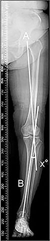
원위 대퇴골 절골술의 교정각은 양측 장하지 체중 부하 전후방 촬영 사진을 통해 결정할 수 있다. 대퇴골 두의 중심 A에서 족관절의 중심 B로 역학적 축을 그린다(Line A–B). 족관절의 중심 B에서 술 후 원하는 역학적 축이 경골 과간 융기 바로 내측 지점을 지나도록 Line B–C를 그린다. 이 때 Line A–B와 Line B–C의 길이는 동일해야 한다. 대퇴골 과상부 외측인 절골술의 경첩점 D에서 그린 Line A–D와 Line C–D가 이루는 각도가 교정각α가 된다. 교정각 α를 원위 대퇴골에 적용하였을 때 내측 피질골 위치에서 두 선이 이루는 사이 간격이 절골술에서 제거되는 쐐기 기저부의 길이가 된다(Fig. 6).2277)

4. 이중 절골술

관상면에서 하지의 축을 교정할 때 단일 절골술만 시행하는 경우, 과도한 관절선 경사를 만들어 관절면에 전단 부하를 증가시킬 수 있다. 또한 교정각도가 15도 이상으로 크고 경골 및 대퇴골에 모두 변형이 있는 경우 단일 절골술만으로는 좋은 결과를 기대하기 어렵다.82) 이러한 경우 고려할 수 있는 이중 절골술은 근위 경골 절골술과 원위 대퇴골 절골술을 모두 시행하는 것으로 Benjamin83)이 처음으로 제안하였다.
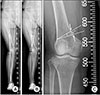
수술 전 교정 계획을 위해 체중 부하 단속 촬영이 필요하다. 체중 부하 단속 촬영 사진을 실제크기로 출력하고 대퇴골과 경골의 변형을 고려하여 절골술을 대퇴골과 경골에서 각각 개방형으로 할 것인지 혹은 폐쇄형으로 할 것인지를 결정한다.84) 절골술을 시행할 부분을 잘라 대퇴골과 경골의 변형이 교정되고 하지 정렬이 회복되도록 회전하여 교정 정도를 결정할 수 있다(Fig. 7). 또 다른 방법으로는 osteotomy analysis and simulation software (OASIS; Mayo Medical Ventures, Rochester, MN, USA)를 이용하여 수술전 계획을 세우는 방법도 소개되고 있다.85)

결론

인공관절 치환술은 진행된 슬관절의 퇴행성 관절염에서 환자의 증상을 호전시키고 술 후 빠른 통증 감소와 짧은 재활 기간 등의 장점이 있는 우수한 수술 방법이다. 하지만 모든 환자를 대상으로 시행하기에는 무리가 있고 특히 젊고 활동적이고 슬관절의 기능적 요구가 많은 환자인 경우 더욱 그러하다. 하지 정렬 이상을 동반한 단일 구획 관절염의 치료에 근위 경골 절골술과 원위 대퇴골 절골술이 좋은 결과를 보여주고 있다.35778687) 또 단일 절골술만으로 만족할 만한 결과를 얻기 힘든 경우 이중 절골술을 고려해볼 수도 있다.82) 물론 이러한 절골술을 시행할 때, 적절한 환자의 선택과 철저한 수술 전 계획이 수술의 성공을 결정하는 중요한 요소라고 할 수 있겠다

Figures and Tables

Figure 1

(A) Normal axis alignment. (B) Varus axis alignment. (C) Valgus axis alignment.

jkoa-53-283-g001
Figure 2

Normal physiologic axes and angles of the lower extremities: The physiologic axes and angles have an abnormal value in the malalignment of the lower extremities. CCDA, centrum column diaphyseal angle; aMPFA, anatomical medial proximal femoral angle; mLPFA, mechanical lateral proximal femoral angle; aLDFA, anatomical lateral distal femoral angle; mLDFA, mechanical lateral distal femoral angle; JLCA, joint line congruency angle; mMPTA, mechanical medial proximal tibial angle; mLDTA, mechanical lateral distal tibial angle; aPDFA, anatomical posterior distal femoral angle; aPPTA, anatomical posterior proximal tibial angle; aADTA, anatomical anterior distal tibial angle.

jkoa-53-283-g002
Figure 3

Bilateral weight bearing anteroposterior whole lower limb x-ray in full extension for planning the Miniaci method. Line A represents the planned weight bearing line for the postoperative correction extending from the center of the hip to about 60% to 70% of the tibial plateau width past the ankle. Line B connects the osteotomy hinge point with the center of the ankle. Line C connects the osteotomy hinge point with the arc intersection of Line A. The angle formed by Lines B and C is the planned correction angle.

jkoa-53-283-g003
Figure 4

Bilateral weight bearing anteroposterior whole lower limb x-ray in full extension for planning the Dugdale method. Line A is drawn from the center of the femoral head to 62.5% of the tibial width. Line B is drawn from the center of the tibiotalar joint to the 62.5% coordinate. The angle formed by these two lines is the correction angle.

jkoa-53-283-g004
Figure 5

Weight bearing scanography. A template was cut through the osteotomy site, and the tibia was rotated until the weight-bearing line passed through the 62% to 65% coordinate.

jkoa-53-283-g005
Figure 6

Bilateral weight bearing anteroposterior whole lower limb x-ray in full extension to plan the medial close-wedge distal femoral osteotomy. (A) The present mechanical axis is drawn from A, which is the center of the femoral head, to B, which is the center of the ankle joint. Line B–C is of equal length as Line A–B and passes the knee, just medial to the medial eminence, representing the desired postoperative mechanical axis. (B) The hinge point of the osteotomy D is marked just proximal to the upper border of the lateral condyle and 0.5–1.0 cm within the lateral cortex. The angle of correction (α) is defined by Line A–D between the present femoral head center and the hinge point and Line C–D connecting the new femoral head center position and the hinge point. (C) Correction angle α is projected at the distal femur using two oblique, down-sloping lines of equal length converging at the hinge point. The distance measured between those two lines at the level of the medial cortex represents the osteotomy wedge base length, which is to be removed during surgery.

jkoa-53-283-g006
Figure 7

Double level osteotomy. A template was cut through each osteotomy site, and the femur and tibia were rotated until the weight-bearing line passed through the 62% to 65% coordinate (Courtesy of Professor Dr. Seung-Beom Han, Department of Orthopaedic Surgery, Korea University Anam Hospital, Korea University College of Medicine, Seoul, Korea)

jkoa-53-283-g007

Notes

CONFLICTS OF INTEREST The authors have nothing to disclose.

References

1. Nakagawa Y, Mukai S, Yabumoto H, Tarumi E, Nakamura T. Cartilage degeneration and alignment in severe varus knee osteoarthritis. Cartilage. 2015; 6:208–215.
crossref
2. Aglietti P, Rinonapoli E, Stringa G, Taviani A. Tibial osteotomy for the varus osteoarthritic knee. Clin Orthop Relat Res. 1983; 176:239–251.
crossref
3. Coventry MB. Proximal tibial varus osteotomy for osteoarthritis of the lateral compartment of the knee. J Bone Joint Surg Am. 1987; 69:32–38.
crossref
4. Insall JN, Joseph DM, Msika C. High tibial osteotomy for varus gonarthrosis. A long-term follow-up study. J Bone Joint Surg Am. 1984; 66:1040–1048.
crossref
5. McDermott AG, Finklestein JA, Farine I, Boynton EL, MacIntosh DL, Gross A. Distal femoral varus osteotomy for valgus deformity of the knee. J Bone Joint Surg Am. 1988; 70:110–116.
crossref
6. Morrey BF. Upper tibial osteotomy for secondary osteoarthritis of the knee. J Bone Joint Surg Br. 1989; 71:554–559.
crossref
7. Morrey BF, Edgerton BC. Distal femoral osteotomy for lateral gonarthrosis. Instr Course Lect. 1992; 41:77–85.
8. Rinonapoli E, Mancini GB, Corvaglia A, Musiello S. Tibial osteotomy for varus gonarthrosis. A 10- to 21-year followup study. Clin Orthop Relat Res. 1998; 353:185–193.
9. Bonasia DE, Dettoni F, Sito G, et al. Medial opening wedge high tibial osteotomy for medial compartment overload/arthritis in the varus knee: prognostic factors. Am J Sports Med. 2014; 42:690–698.
10. Coventry MB, Ilstrup DM, Wallrichs SL. Proximal tibial osteotomy. A critical long-term study of eighty-seven cases. J Bone Joint Surg Am. 1993; 75:196–201.
crossref
11. Hunter DJ, Sharma L, Skaife T. Alignment and osteoarthritis of the knee. J Bone Joint Surg Am. 2009; 91:Suppl 1. 85–89.
crossref
12. Amis AA. Biomechanics of high tibial osteotomy. Knee Surg Sports Traumatol Arthrosc. 2013; 21:197–205.
crossref
13. Leonardi F, Rivera F, Zorzan A, Ali SM. Bilateral double osteotomy in severe torsional malalignment syndrome: 16 years follow-up. J Orthop Traumatol. 2014; 15:131–136.
14. Yoo JD, Kim NK. Distal femoral varization osteotomy. J Korean Orthop Assoc. 2014; 49:118–125.
crossref
15. Hsu RW, Himeno S, Coventry MB, Chao EY. Normal axial alignment of the lower extremity and load-bearing distribution at the knee. Clin Orthop Relat Res. 1990; 255:215–227.
crossref
16. Moreland JR, Bassett LW, Hanker GJ. Radiographic analysis of the axial alignment of the lower extremity. J Bone Joint Surg Am. 1987; 69:745–749.
crossref
17. Tetsworth K, Paley D. Malalignment and degenerative arthropathy. Orthop Clin North Am. 1994; 25:367–377.
crossref
18. Fujisawa Y, Masuhara K, Shiomi S. The effect of high tibial osteotomy on osteoarthritis of the knee. An arthroscopic study of 54 knee joints. Orthop Clin North Am. 1979; 10:585–608.
19. Moore J, Mychaltchouk L, Lavoie F. Applicability of a modified angular correction measurement method for open-wedge high tibial osteotomy. Knee Surg Sports Traumatol Arthrosc. 2017; 25:846–852.
crossref
20. Pape D, Rupp S. Preoperative planning for high tibial osteotomies. Oper Tech Orthop. 2007; 17:2–11.
crossref
21. Brown GA, Amendola A. Radiographic evaluation and preoperative planning for high tibial osteotomies. Oper Tech Sports Med. 2012; 20:93–102.
crossref
22. Brinkman JM, Lobenhoffer P, Agneskirchner JD, Staubli AE, Wymenga AB, van Heerwaarden RJ. Osteotomies around the knee: patient selection, stability of fixation and bone healing in high tibial osteotomies. J Bone Joint Surg Br. 2008; 90:1548–1557.
23. Flecher X, Parratte S, Aubaniac JM, Argenson JN. A 12-28-year followup study of closing wedge high tibial osteotomy. Clin Orthop Relat Res. 2006; 452:91–96.
crossref
24. Khoshbin A, Sheth U, Ogilvie-Harris D, et al. The effect of patient, provider and surgical factors on survivorship of high tibial osteotomy to total knee arthroplasty: a population-based study. Knee Surg Sports Traumatol Arthrosc. 2017; 25:887–894.
crossref
25. Naudie D, Bourne RB, Rorabeck CH, Bourne TJ. The Install Award. Survivorship of the high tibial valgus osteotomy. A 10- to -22-year followup study. Clin Orthop Relat Res. 1999; 367:18–27.
26. Trieb K, Grohs J, Hanslik-Schnabel B, Stulnig T, Panotopoulos J, Wanivenhaus A. Age predicts outcome of high-tibial osteotomy. Knee Surg Sports Traumatol Arthrosc. 2006; 14:149–152.
crossref
27. Goshima K, Sawaguchi T, Sakagoshi D, Shigemoto K, Hatsuchi Y, Akahane M. Age does not affect the clinical and radiological outcomes after open-wedge high tibial osteotomy. Knee Surg Sports Traumatol Arthrosc. 2017; 25:918–923.
crossref
28. Odenbring S, Egund N, Knutson K, Lindstrand A, Larsen ST. Revision after osteotomy for gonarthrosis. A 10-19-year follow-up of 314 cases. Acta Orthop Scand. 1990; 61:128–130.
crossref
29. van Raaij T, Reijman M, Brouwer RW, Jakma TS, Verhaar JN. Survival of closing-wedge high tibial osteotomy: good outcome in men with low-grade osteoarthritis after 10-16 years. Acta Orthop. 2008; 79:230–234.
30. Shin YS, Lee DH, Lee SH, Kim MJ, Han SB. Basic principles and current trends of medial opening-wedge high tibial osteotomy. J Korean Orthop Assoc. 2014; 49:85–94.
crossref
31. Nwachukwu BU, McCormick FM, Schairer WW, Frank RM, Provencher MT, Roche MW. Unicompartmental knee arthroplasty versus high tibial osteotomy: United States practice patterns for the surgical treatment of unicompartmental arthritis. J Arthroplasty. 2014; 29:1586–1589.
crossref
32. Fu D, Li G, Chen K, Zhao Y, Hua Y, Cai Z. Comparison of high tibial osteotomy and unicompartmental knee arthroplasty in the treatment of unicompartmental osteoarthritis: a meta-analysis. J Arthroplasty. 2013; 28:759–765.
33. W-Dahl A, Toksvig-Larsen S, Lindstrand A. Ten-year results of physical activity after high tibial osteotomy in patients with knee osteoarthritis. Knee Surg Sports Traumatol Arthrosc. 2017; 25:902–909.
crossref
34. W-Dahl A, Robertsson O, Lidgren L. Surgery for knee osteoarthritis in younger patients. Acta Orthop. 2010; 81:161–164.
crossref
35. Börjesson M, Weidenhielm L, Mattsson E, Olsson E. Gait and clinical measurements in patients with knee osteoarthritis after surgery: a prospective 5-year follow-up study. Knee. 2005; 12:121–127.
crossref
36. Stukenborg-Colsman C, Wirth CJ, Lazovic D, Wefer A. High tibial osteotomy versus unicompartmental joint replacement in unicompartmental knee joint osteoarthritis: 7-10-year follow-up prospective randomised study. Knee. 2001; 8:187–194.
37. Bonnin M, Chambat P. Current status of valgus angle, tibial head closing wedge osteotomy in media gonarthrosis. Orthopade. 2004; 33:135–142.
38. Kumagai K, Akamatsu Y, Kobayashi H, Kusayama Y, Koshino T, Saito T. Factors affecting cartilage repair after medial opening-wedge high tibial osteotomy. Knee Surg Sports Traumatol Arthrosc. 2017; 25:779–784.
crossref
39. Pape D, Seil R, Adam F, Rupp S, Kohn D, Lobenhoffer P. Imaging and preoperative planning of osteotomy of tibial head osteotomy. Orthopade. 2004; 33:122–134.
40. OAhlbäck S. Osteoarthrosis of the knee. A radiographic investigation. Acta Radiol Diagn (Stockh). 1968; Suppl 277. 7–72.
41. Murphy SB. Tibial osteotomy for genu varum. Indications, preoperative planning, and technique. Orthop Clin North Am. 1994; 25:477–482.
42. Berman AT, Bosacco SJ, Kirshner S, Avolio A Jr. Factors influencing long-term results in high tibial osteotomy. Clin Orthop Relat Res. 1991; 272:192–198.
crossref
43. Akizuki S, Shibakawa A, Takizawa T, Yamazaki I, Horiuchi H. The long-term outcome of high tibial osteotomy: a ten- to 20-year follow-up. J Bone Joint Surg Br. 2008; 90:592–596.
44. Nha KW, Kim HJ, Ahn HS, Lee DH. Change in posterior tibial slope after open-wedge and closed-wedge high tibial osteotomy: a meta-analysis. Am J Sports Med. 2016; 44:3006–3013.
45. Naudie DD, Amendola A, Fowler PJ. Opening wedge high tibial osteotomy for symptomatic hyperextension-varus thrust. Am J Sports Med. 2004; 32:60–70.
crossref
46. Phisitkul P, Wolf BR, Amendola A. Role of high tibial and distal femoral osteotomies in the treatment of lateral-posterolateral and medial instabilities of the knee. Sports Med Arthrosc. 2006; 14:96–104.
crossref
47. Arthur A, LaPrade RF, Agel J. Proximal tibial opening wedge osteotomy as the initial treatment for chronic posterolateral corner deficiency in the varus knee: a prospective clinical study. Am J Sports Med. 2007; 35:1844–1850.
48. Giagounidis EM, Sell S. High tibial osteotomy: factors influencing the duration of satisfactory function. Arch Orthop Trauma Surg. 1999; 119:445–449.
crossref
49. Lee YS, Lee BK, Lee SH, Park HG, Jun DS, Moon DH. Effect of foot rotation on the mechanical axis and correlation between knee and whole leg radiographs. Knee Surg Sports Traumatol Arthrosc. 2013; 21:2542–2547.
crossref
50. Lee YS, Lee BK, Kwon JH, et al. Serial assessment of weight-bearing lower extremity alignment radiographs after open-wedge high tibial osteotomy. Arthroscopy. 2014; 30:319–325.
51. Sancheti P, Patil K, Gugale S, Shyam A. How can preoperative planning prevent occurrence of a painful total knee replacement?. In : Hirschmann MT, Becker R, editors. The unhappy total knee replacement. Cham: Springer;2015. p. 59–68.
52. Phillips CL, Silver DA, Schranz PJ, Mandalia V. The measurement of patellar height: a review of the methods of imaging. J Bone Joint Surg Br. 2010; 92:1045–1053.
53. Coventry MB. Upper tibial osteotomy for osteoarthritis. J Bone Joint Surg Am. 1985; 67:1136–1140.
crossref
54. Hernigou P, Medevielle D, Debeyre J, Goutallier D. Proximal tibial osteotomy for osteoarthritis with varus deformity. A ten to thirteen-year follow-up study. J Bone Joint Surg Am. 1987; 69:332–354.
55. Miniaci A, Ballmer FT, Ballmer PM, Jakob RP. Proximal tibial osteotomy. A new fixation device. Clin Orthop Relat Res. 1989; 246:250–259.
56. Dugdale TW, Noyes FR, Styer D. Preoperative planning for high tibial osteotomy. The effect of lateral tibiofemoral separation and tibiofemoral length. Clin Orthop Relat Res. 1992; 274:248–264.
57. Jakob RP, Jacobi M. Closing wedge osteotomy of the tibial head in treatment of single compartment arthrosis. Orthopade. 2004; 33:143–152.
58. Lee DH, Han SB, Oh KJ, et al. The weight-bearing scanogram technique provides better coronal limb alignment than the navigation technique in open high tibial osteotomy. Knee. 2014; 21:451–455.
crossref
59. Akamatsu Y, Mitsugi N, Mochida Y, et al. Navigated opening wedge high tibial osteotomy improves intraoperative correction angle compared with conventional method. Knee Surg Sports Traumatol Arthrosc. 2012; 20:586–593.
crossref
60. Reising K, Strohm PC, Hauschild O, et al. Computer-assisted navigation for the intraoperative assessment of lower limb alignment in high tibial osteotomy can avoid outliers compared with the conventional technique. Knee Surg Sports Traumatol Arthrosc. 2013; 21:181–188.
crossref
61. Elson DW, Petheram TG, Dawson MJ. High reliability in digital planning of medial opening wedge high tibial osteotomy, using Miniaci's method. Knee Surg Sports Traumatol Arthrosc. 2015; 23:2041–2048.
crossref
62. Lee DH, Nha KW, Park SJ, Han SB. Preoperative and postoperative comparisons of navigation and radiologic limb alignment measurements after high tibial osteotomy. Arthroscopy. 2012; 28:1842–1850.
crossref
63. Yoon SD, Zhang G, Kim HJ, Lee BJ, Kyung HS. Comparison of cable method and miniaci method using picture archiving and communication system in preoperative planning for open wedge high tibial osteotomy. Knee Surg Relat Res. 2016; 28:283–288.
crossref
64. Giffin JR, Vogrin TM, Zantop T, Woo SL, Harner CD. Effects of increasing tibial slope on the biomechanics of the knee. Am J Sports Med. 2004; 32:376–382.
crossref
65. Dean CS, Liechti DJ, Chahla J, Moatshe G, LaPrade RF. Clinical outcomes of high tibial osteotomy for knee instability: a systematic review. Orthop J Sports Med. 2016; 4:2325967116633419.
66. Dejour H, Bonnin M. Tibial translation after anterior cruciate ligament rupture. Two radiological tests compared. J Bone Joint Surg Br. 1994; 76:745–749.
crossref
67. Chae DJ, Shetty GM, Lee DB, Choi HW, Han SB, Nha KW. Tibial slope and patellar height after opening wedge high tibia osteotomy using autologous tricortical iliac bone graft. Knee. 2008; 15:128–133.
crossref
68. Ogawa H, Matsumoto K, Ogawa T, Takeuchi K, Akiyama H. Effect of wedge insertion angle on posterior tibial slope in medial opening wedge high tibial osteotomy. Orthop J Sports Med. 2016; 4:2325967116630748.
crossref
69. Bae DK, Ko YW, Kim SJ, Baek JH, Song SJ. Computer-assisted navigation decreases the change in the tibial posterior slope angle after closed-wedge high tibial osteotomy. Knee Surg Sports Traumatol Arthrosc. 2016; 24:3433–3440.
crossref
70. Sharma L, Song J, Felson DT, Cahue S, Shamiyeh E, Dunlop DD. The role of knee alignment in disease progression and functional decline in knee osteoarthritis. JAMA. 2001; 286:188–195.
crossref
71. Stähelin T, Hardegger F, Ward JC. Supracondylar osteotomy of the femur with use of compression. Osteosynthesis with a malleable implant. J Bone Joint Surg Am. 2000; 82:712–722.
72. Chahla J, Mitchell JJ, Liechti DJ, et al. Opening- and closing-wedge distal femoral osteotomy: a systematic review of outcomes for isolated lateral compartment osteoarthritis. Orthop J Sports Med. 2016; 4:2325967116649901.
73. Hinterwimmer S, Rosenstiel N, Lenich A, Waldt S, Imhoff AB. Femoral osteotomy for patellofemoral instability. Unfallchirurg. 2012; 115:410–416.
74. Niklasch M, Dreher T, Döderlein L, et al. Superior functional outcome after femoral derotation osteotomy according to gait analysis in cerebral palsy. Gait posture. 2015; 41:52–56.
crossref
75. Kazi HA, Abdel-Rahman W, Brady PA, Cameron JC. Meniscal allograft with or without osteotomy: a 15-year follow-up study. Knee Surg Sports Traumatol Arthrosc. 2015; 23:303–309.
crossref
76. Wang JW, Hsu CC. Distal femoral varus osteotomy for osteoarthritis of the knee. J Bone Joint Surg Am. 2005; 87:127–133.
crossref
77. Thein R, Bronak S, Thein R, Haviv B. Distal femoral osteotomy for valgus arthritic knees. J Orthop Sci. 2012; 17:745–749.
crossref
78. Backstein D, Morag G, Hanna S, Safir O, Gross A. Long-term follow-up of distal femoral varus osteotomy of the knee. J Arthroplasty. 2007; 22:S2–S6.
crossref
79. Finkelstein JA, Gross AE, Davis A. Varus osteotomy of the distal part of the femur. A survivorship analysis. J Bone Joint Surg Am. 1996; 78:1348–1352.
crossref
80. Franco V, Cipolla M, Gerullo G, Gianni E, Puddu G. Open wedge osteotomy of the distal femur in the valgus knee. Orthopade. 2004; 33:185–192.
81. Mathews J, Cobb AG, Richardson S, Bentley G. Distal femoral osteotomy for lateral compartment osteoarthritis of the knee. Orthopedics. 1998; 21:437–440.
crossref
82. Paley D, Pfeil J. Principles of deformity correction around the knee. Orthopade. 2000; 29:18–38.
crossref
83. Benjamin A. Double osteotomy for the painful knee in rheumatoid arthritis and osteoarthritis. J Bone Joint Surg Br. 1969; 51:694–699.
crossref
84. Archbold P, Verdonk P, Servien E. Surgical technique for a double osteotomy. In : Neyret P, Demey G, editors. Surgery of the Knee. London: Springer;2014. p. 175–180.
85. Babis GC, An KN, Chao EY, Rand JA, Sim FH. Double level osteotomy of the knee: a method to retain joint-line obliquity. Clinical results. J Bone Joint Surg Am. 2002; 84:1380–1388.
86. Magyar G, Toksvig-Larsen S, Alkstedt J, et al. Frequent complications in distal femoral osteotomy: a retrospective 8 year multicenter follow-up. Chir Organi Mov. 1999; 84:19–26.
87. Terry GC, Cimino PM. Distal femoral osteotomy for valgus deformity of the knee. Orthopedics. 1992; 15:1283–1289. discussion 1289-90.
crossref
TOOLS
ORCID iDs

Oog Jin Shon
https://orcid.org/0000-0002-9123-5694

Similar articles