Journal List > World J Mens Health > v.36(3) > 1100483

Traish and Johansen: Impact of Testosterone Deficiency and Testosterone Therapy on Lower Urinary Tract Symptoms in Men with Metabolic Syndrome

Abstract

Lower urinary tract function is modulated by neural, vascular and urethral and bladder structural elements. The pathophysiological mechanisms of lower urinary tract symptoms (LUTS) encompass prostate enlargement, alterations in urethra histological structure bladder fibrosis and alterations in pelvic neuronal and vascular networks, The complex pathophysiological relationship between testosterone (T) deficiency (TD) and the constellations LUTS, and metabolic dysfunction manifested in the metabolic syndrome (Met S) remains poorly understood. TD has emerged as one the potential targets by which Met S may contribute to the onset and development as well as worsening of LUTS. Because it has been recognized that treatment of men with Met S with T therapy ameliorates Met S components, it is postulated that T therapy may represent a therapeutic target in improving LUTS. Furthermore, the effect of TD on the prostate remains unclear, and often debatable. It is believed that T exclusively promotes prostate growth, however recent evidence has strongly contradicted this belief. The true relationship between benign prostatic hyperplasia, TD, and LUTS remains elusive and further research will be required to clarify the role of T in both benign prostatic hypertrophy (BPH) and LUTS as a whole. Although there is conflicting evidence about the benefits of T therapy in men with BPH and LUTS, the current body of literature supports the safety of using this therapy in men with enlarged prostate. As the population afflicted with obesity epidemic continues to age, the number of men suffering from Met S and LUTS together is expected to increase.

INTRODUCTION

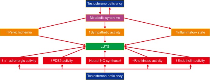
1. Impact of metabolic syndrome on lower urinary tract symptoms

Lower urinary tract symptoms (LUTS) are a set of bothersome voiding symptoms, including increased urinary frequency, urgency, nocturia, slow stream, postmicturition dribble, and are traditionally attributed to benign prostatic hypertrophy (BPH). Epidemiological studies have suggested that age is among the principal unmodifiable risk factor of LUTS [1]. Metabolic syndrome (Met S) is a complex pathology manifested through a host of interrelated factors, such as increased waist circumference (WC), elevated insulin resistance (IR), hyperglycemia, hypertension, visceral adiposity, dyslipidemia, endothelial dysfunction, and atherosclerotic disease concomitant with low-grade inflammation, as indicated by elevated non-specific inflammatory markers. The Met S components often exist concomitantly with a reduction in circulating serum testosterone (T) levels [2345678910111213]. Furthermore, Met S is thought to be associated with nephrolithiasis, BPH, LUTS, erectile dysfunction (ED), and infertility [14]. Met S and its related comorbidities are often associated with development and/or progression of BPH-LUTS [1].
Preclinical and clinical studies suggest that obesity, dyslipidemia, diabetes, components of Met S are critical determinants and are potentially modifiable risk factors in BPH-LUTS (Fig. 1). Testosterone deficiency (TD) is a predictor and contributor to Met S [23411]. Met S is associated with increased inflammatory state and increased sympathetic activity together with pelvic ischemia [14]. These pathophysiological states contribute to the worsening of LUTS. Furthermore, TD contributes to increased adrenergic activity and reduced nitric oxide synthase activity concomitant with increased endothelin and Rho kinase activity, further contributing to the worsening of LUTS.
Central obesity and WC, components of Met S, have been associated with increased risk for BPH and LUTS [1415161718192021222324252627282930]. However, central obesity may adversely impact LUTS independent of the prostate growth, as demonstrated by changes in severity of LUTS in men after prostatectomy [31323334]. An emerging body of evidence suggests that a relationship exists between LUTS severity and Met S [3233353637]. As reported by Kupelian et al [38], Met S was significantly associated with an elevated American Urological Association-symptom index score (multivariate odds ratio [OR]=1.68). After adjusting for age, men younger than 60 years with Met S were more likely to report intermittency, incomplete emptying, and nocturia [38]. We should point out that some studies [21] suggested that the presence of Met S was not significantly associated with moderate to severe LUTS (OR=1.13, p=0.53) and only altered serum triglycerides, an important component of Met S, and diabetes were associated with this risk. Yet, a positive association was noted between number of Met S components and LUTS/bladder outlet obstruction in this study [21]. It is possible that endocrine abnormalities attributed to central obesity and hypertriglyceridemia may play an important role in development or progression of the pathophysiology of lower urinary tract dysfunction [3839]. In particular, triglyceride and cholesterol levels seem to have a detrimental effect on prostatic function and inflammation, which may be associated with development and progression of LUTS/BPH [37]. Moreover, a meta-analysis, including 8 studies and 5,403 patients, demonstrated that Met S is associated with increased prostate size, supporting a potential role for metabolic dysfunction in progression of BPH, thus contributing to the pathophysiology of LUTS. It is likely that obesity, dyslipidemia and age represent significant risk factors in worsening LUTS [32]. Furthermore, an association between Met S and pelvic floor dysfunction exists independent of disease of the prostate [40].

2. Bidirectional relationship between testosterone deficiency and metabolic syndrome

As shown in Table 1, a considerable body of literature exists demonstrating a bidirectional link between TD and Met S [23441424344454647484950515253545556575859606162]. Several systematic reviews, crosssectional studies, and clinical trials have reported low levels of total T (TT) and sex-hormone binding globulin (SHBG) in men with Met S, irrespective of age [11446364]. The relationship between low TT and SHBG may be attributed to hyperinsulinemia in men with Met S. In addition, it was shown that serum estradiol (E2) levels are higher in men with Met S and are positively correlated with the number of Met S components [65]. Reduced TT levels predict the development of the Met S in men with normal body mass index (BMI) [41435666]. Additional evidence that low TT levels increase the risk of Met S is derived from experience with androgen deprivation therapy for prostate cancer [676869].
Laaksonen et al [41] evaluated 702 men who initially were without Met S or diabetes mellitus. After 11 years of follow-up, there were 147 incident cases of Met S (20.9%) and 57 incident cases of diabetes (8.1%). After adjustment for age, men with TT, free testosterone (FT) and SHBG levels in the lower T quartile had a several fold increased risk of developing Met S (OR=2.3, 95% confidence imterval [CI], 1.5–3.4; OR=1.7, 95% CI, 1.2–2.5; and OR=2.8, 95% CI, 1.9–4.1, respectively) and diabetes (OR=2.3, 95% CI, 1.3–4.1; OR=1.7, 95% CI, 0.9–3.0; and OR=4.3, 95% CI, 2.4–7.7, respectively). Furthermore, adjustment for potential confounders, such as cardiovascular disease, smoking, alcohol intake, and socioeconomic status did not alter the associations. The authors concluded that low TT and SHBG levels independently predict development of Met S and type 2 diabetes in middle-aged men. In addition, it was suggested that TD is an early marker for disturbances in both insulin and glucose metabolism, which may progress to Met S and diabetes. This may also contribute independently to the pathogenesis of such diseases. In a subsequent study, Laaksonen et al [42] reported that men with Met S at baseline had a 2.6-fold increased risk of developing TD (T levels<11 nM) at the 11-year follow-up independent of age, smoking, and other potential confounders. In addition, adjustment for BMI (OR=2.0, 95% CI, 1.1–3.8) or baseline TT levels (OR=1.9, 95% CI, 1.0–3.4) attenuated the association. The association of Met S with TD (FT<225 pmol/L) was similar. The adjusted decrease in T levels during the 11-year follow-up was also greater in men with than without Met S. Antonio et al [56] reported that in 1,651 men without Met S at baseline, 289 men developed incident Met S during the follow-up. Men with lower baseline TT levels were at higher risk for developing Met S (OR=1.72, p<0.001), even after adjustment for SHBG (OR=1.43, p<0.001), BMI (OR=1.44, p<0.001), or homeostasis model assessment of insulin resistance (HOMA-IR) (OR=1.64, p<0.001). E2 was not associated with development of Met S (OR=1.04, p<0.56). However, a lower E2/T ratio was associated with a lower risk of incident Met S (OR=0.38, p<0.001), even after adjustment for SHBG (OR=0.48, p<0.001), BMI (OR=0.60, p<0.001) or HOMA-IR (OR=0.41, p<0.001). It was concluded that lower T levels, but not E2, are linked with an increased risk of developing Met S, independent of SHBG, BMI, or IR. A lower E2/T ratio may be protective against developing Met S [56]. The authors suggested that low T but not E2 levels in men may be regarded as a biomarker or risk predictor for Met S, independently of SHBG, IR, and body composition. The importance of aromatase activity in Met S requires further investigation. These findings may have implications for the assessment of cardiometabolic risks in older and obese men [60]. The aforementioned studies strongly suggest that low TT levels independently predict Met S and the number of Met S components are inversely correlated with TT levels. Considering the Met S components individually, WC seems to be the strongest correlated factor with lower TT levels as demonstrated by multivariate analysis [11]. Therefore, it is reasonable to suggest that TD is an independent predictor of the Met S.

METABOLIC SYNDROME, TESTOSTERONE DEFICIENCY, AND PATHOPHYSIOLOGY OF LOWER URINARY TRACT SYMPTOMS

LUTS, which include frequency, urgency, incomplete voiding, and slow stream, are common in both men and women with advancing age. The most common cause of LUTS in men is prostate enlargement and progression of BPH. A number of studies have suggested that an inverse association exist between serum T levels and LUTS; however, the underlying biochemical and physiological mechanisms by which reduced T levels affect LUTS have yet to be fully delineated [70717273]. A significantly larger prostate volume was reported in men afflicted with Met S [1921747576], however it is unclear if this increase in prostate volume indeed exerts a clinical significance [32]. Met S which was associated with decreased improvement of postoperative symptoms and LUTS after prostate surgery, suggesting interplay of multiple metabolic risk factors contributing to this complex disorder [77].
It is important to note that in a study of 122 men Favilla et al [78] found no correlation between TT and FT and prostate size or volume. Thus, the increase in prostate volume in men with Met S may be, in part, attributed to intra-prostatic inflammation due to hyperinsulinemia and hypertriglyceridemia [3179]. In addition, reduced high-density lipoprotein (HDL), and increased triglyceride levels are significantly related to higher prostatic inflammation and increased secretion of interleukin (IL)-8, in response to oxidized low-density lipoprotein [2631]. Furthermore, increased WC in men with Met S was positively correlated with prostate volume, which may contribute to severity of LUTS [8081]. In contrast, a recent meta-analysis found that Met S components, such as alteration in WC and serum HDL levels were not associated with increased odds of having moderate to severe LUTS [21]. This meta-analysis suggested that serum triglycerides and diabetes may be more significantly associated with increased risk of LUTS and severity.
LUTS are frequently associated with BPH in men, concomitant with a progressive decline in serum T levels and a gradual increase in prostate volume, reflecting pathophysiology of BPH and LUTS. As summarized in Table 2 [16192125272829303137387980818283], Met S, visceral obesity, ED, and increased IR are conditions associated with progressive decline of T with age [84]. Schatzl et al [85] reported that TD was detected in 22.1% of 312 men with LUTS but had no impact on International Prostate Symptom Score (IPSS), maximum urinary flow rate (Qmax), prostate volume, or prostate specific antigen (PSA) level. A large population study suggested that circulating levels of sex hormones are not significant predictors of LUTS in men, after adjustment for age, and the pathophysiology of LUTS is complex and probably includes factors other than circulating sex steroid levels [86]. Furthermore, no consistent correlations between T or FT and LUTS were shown in cross-sectional study [87]. In one prospective study, the 20-year risk of LUTS in 185 men with a mean age of 58 years was examined. In fact, the authors reported a significant inverse association between 5-dihydrotestosterone and LUTS. It was also found that men with higher concentrations of bioavailable T (BT) had a 56% decreased risk of LUTS compared with men with TD [88]. Interestingly, Miwa et al [89] noted that LUTS were not associated with serum levels of TT or FT, but storage symptom scores of the IPSS could be affected by the serum dehydroepiandrosterone sulfate level in older men. Additionally, Tan et al [90] reported that the decrease in serum FT concentrations with a relative rise in serum E2 levels with advancing age may be an important factor in the development of BPH. These findings indicated that the relationships between sex hormone levels and the development or severity of LUTS are conflicting, and further studies in large populations of men with and without LUTS and with various severities of BPH are required to reach more definitive conclusions.
TD may alter the structures of the lower urinary tract, such as urethral and bladder epithelial cells. The role of Met S components including WC, HDL, triglycerides, and diabetes on the risk of having moderate-to-severe LUTS have been reported [38919293949596]. More specifically, decreasing T levels have been associated with altered release of adenosine triphosphate (ATP) and acetylcholine (ACh) responsiveness to stretch, micturition reflex, and functional factors like bladder capacity [919798]. Met S contributes to increased sympathetic nervous system activity and increased muscle tone of the prostate, resulting in more severe LUTS, even in the absence of prostate enlargement [21].

PATHOPHYSIOLOGICAL MECHANISMS OF LOWER URINARY TRACT SYMPTOMS

Animal model studies demonstrated that T elicits direct and indirect effects on neural tissue involved in bladder control [99100]. Bladder function is coordinated by a complex interplay of central and peripheral mechanisms [99]. At the local level within the bladder wall, the urothelium, a highly specialized transitional epithelium, acts as a distensible barrier between the underlying tissues and the urine and is also thought to play a major role in the sensation and transmission of information from the bladder to the nervous system [101]. Distension and stretch of the bladder during filling with urine causes the release of non-neuronal ATP from the urothelial cells, activating sensory nerves via purinergic P2X3 receptors and resulting in afferent signaling to higher central nervous system centers via the spinal cord to initiate bladder emptying. Thus, ATP release is crucial in initiating the micturition reflex and voiding [97]. The urothelium also releases other chemical mediators including ACh [102], which is thought to communicate with the underlying tissues, nerves, smooth muscle, myofibroblasts and interstitial cells, to regulate sensory mechanisms and bladder contraction [101]. Whilst the role of ACh is less well understood than that of ATP, urothelial ACh can stimulate the release of other mediators such as NO or ATP and may also stimulate muscarinic receptors on the detrusor smooth muscle (DSM) and nerves [102].
Maggi et al [103] reported that T treatment in castrated rats increased prostate weight, but had no effect on bladder weight, bladder capacity, or amplitude of bladder contractions. However, T treatment induced detrusor instability and markedly increased residual volume, indicating intravesical outflow obstruction. Pandita et al [104] also reported that T treatment for 2 weeks increased micturition pressure, bladder capacity, residual volume, and micturition volume. Other studies have suggested that T treatment improves bladder capacity, reverses the parameters of DSM contractility, and improves smooth muscle/collagen ratio [98105106]. Tek et al [106] reported that T treatment of orchiectomized rats inhibited bladder activity. Castration reduced bladder capacity, and T treatment reversed this change. A similar effect of T treatment increased bladder capacity in castrated rabbits [107]. The discrepancies in these findings may be attributed to differing T concentrations used in such experimentation. Moreover, it is possible that supraphysiologic T levels may affect the urethral outlet and may induce urinary retention which indirectly influence bladder function. Cheng and de Groat [108] reported that castration altered several storage and voiding functions of the LUT and these changes are attributed to effects on activity of both the urethra and the bladder. Some of the observed changes in LUT function were completely reversed by T treatment, however, others were not reversed, suggesting that TD alters the LUT by several overlapping physiological mechanisms. T has been reported to relax the pig urinary bladder neck [109], and rapidly inhibit the contractility of DSM preparations [110]. In rat DSM, inhibition of androgen receptor with flutamide does not abolish the relaxing effects of T on DSM, suggesting that T is acting via a non-genomic mechanism [110]. Hristov et al [111] examined the effects of T treatment on bladder smooth muscle excitability in the animal model and suggested that T at physiological levels decreases DSM cell excitability by directly activating BK channels via a non-genomic mechanism. The authors demonstrated that T at 100 nM increased the whole cell BK current in freshly isolated guinea pig DSM cells, suggesting a regulatory role of T in DSM cell excitability. Ito et al [112] have shown that significant increase in prostaglandin E2 (PGE2) release from stretched bladder epithelium after castration concomitant with significant increase in bladder IL-1β and cyclooxygenase type 2 (COX-2) expression. T treatment reversed the PGE2 increase, suggesting that castration induces inflammation in the rat bladder, which causes elevated PGE2 release from bladder epithelium and may contribute to the disruption of bladder storage function.
In a rabbit model of Met S, Vignozzi et al [26] demonstrated that in animals fed high-fat diet (HFD), the mRNA expression of several proinflammatory markers in the prostate (IL-8, IL-6, IL-1, and tumor necrosis factor-α), T lymphocyte (CD4, CD8, T-bet, GATA3, and ROR-γt), macrophage (toll-like receptor 2 [TLR2], TLR4, and STAMP2), neutrophil (lactoferrin), inflammation (COX-2 and receptor for advanced glycation end products), and fibrosis/myofibroblast activation (tumor growth factor [TGF]-β, SM22-α, α-SMA, RhoA, and Rho-associated kinase1 [ROCK1]/ROCK2) were significantly increased. T treatment normalized all the HFD-induced prostate alterations. These observations suggest that T treatment protects rabbit prostate from Met S-induced prostatic hypoxia, fibrosis, and inflammation, which can play a role toward the development/progression of BPH/LUTS. Juan et al [113] reported that castration resulted in decreased activity in the mitochondria specific enzyme, citrate synthase, the activity of which was greatest in the urethra and lowest in the corpora. Cholinergic nerve density indicator, choline acetyl-transferase activity was greatest in the bladder body and lowest in the urethra. Zhang et al [114] demonstrated that TD in the animal model induces bladder fibrosis and decreases bladder maximal volume and compliance. TD induced TGF-β mRNA and treatment of the castrated animals with anti-TGF-β antibodies abolished the effects of TD on induced bladder fibrosis and dysfunction. T treatment prevented the histological and functional abnormalities induced by TD in the bladder of the animal model. Changes in bladder capacity and compliance in this study were similar to the findings of Cayan et al [115] observed these changes in rats treated with hormone replacement therapy including T and E2 alone and in combination.

DOES TESTOSTERONE THERAPY IMPROVES LOWER URINARY TRACT SYMPTOMS?

1. Testosterone therapy ameliorates metabolic syndrome components

T therapy has been shown to improve hyperglycemia and IR and ameliorate some of the Met S components (Table 3) [116117118119120121122123124125126127128129]. However, conflicting evidence exists suggesting changes in weight loss and WC are not maintained [63]. Mårin et al [130] reported that T therapy improved insulin sensitivity in obese men and reduced central adiposity. Recently it has also been shown that T therapy improves insulin sensitivity in the HOMA-IR model. A significant reduction in IR in men with TD and type 2 diabetes was reported in men treated with intramuscular T therapy over a 3-month period [131]. Although this study reported reduction in glycated hemoglobin and fasting blood glucose, the effect of T therapy on these factors remains unclear [123131]. In one multicenter, double-blind, placebo-controlled trial, T treatment reduced hemoglobin A1c (HbA1c) and WC [132]. Heufelder et al [117] also demonstrated this in 32 hypogonadal (TT<12 nmol/L) men with Met S and newly diagnosed type 2 diabetes randomized to 52 weeks of treatment with diet and exercise with or without transdermal T (50 mg/d). Addition of T treatment to supervised diet and exercise resulted in greater therapeutic improvement of glycemic control and reversed the Met S in 81% vs. 31% of controls after 52 weeks. Hoyos et al [133] investigated the effects of T therapy in obese men with severe obstructive sleep apnoea in an 18-week randomized, placebo-controlled trial. Sixty-seven men were randomized to intramuscular T therapy or placebo. T therapy improved insulin sensitivity, reduced liver fat, and increased muscle mass, as well as decreased arterial stiffness, but did not differentially reduce overall weight or the Met S and this may be attributed to the short treatment duration.
Cai et al [134] have identified only five randomized clinical trials with a total of 351 subjects who were treated with T for an average of 6.5 months. A meta analysis of these clinical trials to determine the metabolic effects of T therapy in hypogonadal men with type 2 diabetes found that fasting plasma glucose levels decreased 1.1 mmol/L (95% CI, −1.88 to −0.31), fasting serum insulin levels decreased by 2.73 mIU/L (95% CI, −3.62 to −1.84), HbA1c decreased by 0.87% (95% CI, −1.32% to −0.42%). The authors concluded that T therapy can improve glycemic control and decrease triglyceride levels in men with TD and type 2 diabetes.
Interestingly, other trials have failed to show improvements in glucose metabolism. Gianatti et al [135] conducted a randomized, double-blind, parallel, placebo-controlled trial in 88 men with type 2 diabetes, aged 35 to 70 years with an HbA1c ≤8.5% (69 mmol/mol), and a TT level of ≤12.0 nmol/L (346 ng/dL). Participants were randomly assigned to 40 weeks of intramuscular T therapy (n=45) or matching placebo (n=43). T therapy did not improve IR despite a decrease in fat mass and an increase in lean mass. T therapy reduced subcutaneous but not visceral abdominal adipose tissue. T therapy did not improve glucose metabolism or visceral adiposity in obese men with moderately controlled type 2 diabetes and modest reductions in circulating T levels typical for men with type 2 diabetes.
Haider et al [136] reported data from a registry study on T therapy in men with obesity and diabetes. A total of 156 obese, diabetic men with TD, aged 61.17±6.18 years, fulfilled selection criteria. T treatment for up to 6 years decreased WC by 11.6 cm, weight declined by 17.5 kg (15.04%), fasting glucose declined from 7.06±1.74 to 5.59±0.94 mmol/L (127±31 to 101±17 mg/dL, p<0.0001) and HbA1c decreased from 8.08% to 6.14%, with a mean change of 1.93%. Systolic and diastolic blood pressure, lipid profiles including total cholesterol: HDL ratio, C-reactive protein, and liver enzymes all improved (p<0.0001). While this is not a randomized controlled clinical trial, it does suggest the potential benefits of T treatment on metabolic parameters when weight loss is achieved, and it suggests that long term treatment with T may contribute to weight loss.
It is widely recognized that T therapy reduces fat mass and increases lean body mass; however, until recently, most reports have not been associated with much weight loss [137]. Changes in body composition and weight loss are considered potential mechanisms by which T treatment improves insulin sensitivity and glucose control in patients with diabetes. Effects on inflammatory cytokines [138] and changes in oxidative metabolism [139] also have been reported to improve glucose metabolism. In a HFD induced model of Met S, Maneschi et al [8] demonstrated that Met S is associated with visceral adipose tissue ischemia, which contributes to altered cytokine production and endocrine signaling. They demonstrated that the insulin-regulated GLUT4 transporter expression was reduced by a HFD, but that this was reversed with T therapy administration. These findings suggest that adipose tissue signaling is modulated by T and that T therapy could potentially contribute to normalization of adipose function in subject suffering from Met S.

2. Testosterone therapy and lower urinary tract symptoms

It has been shown by a number of studies that T therapy improves LUTS (Table 4) [73126128140141142143144145]. According to the study by Favilla et al [78] in men with BPH a significant association exists between severity of LUTS and reduced serum TT level, but not other serum sex hormones. Importantly, this study demonstrated that serum T levels are not associated with prostate size and volume. In fact, it has been shown that there exists a negative relationship of FT and BT with IPSS on multivariate analysis [24]. This negative relationship provides a framework with which to approach the evidence regarding T Therapy in men with LUTS.
The way in which T is hypothesized to improve LUTS includes regulation of the expression of alpha1-adrenergic receptors, phosphodiesterase type 5 activity, Rho-kinase activation, endothelin activity, and neural nitric oxide synthase [146147]. As shown in an animal model, T therapy also improves LUTS and bladder function parameters, in part by increasing bladder capacity and compliance and by decreasing detrusor pressure at maximal flow [106]. In that animal model, the authors found that orchiectomy decreased the mean bladder capacity by 38.9% while T therapy actually increased bladder capacity by an average of 46.6%. Celayir [107] measured the baseline urodynamic and T levels in control and orchiectomized rabbits treated with or without T. Bladder capacity and compliance were increased on days 5 and 10 with T treatment but decreased thereafter and returned to the baseline levels on day 30, suggesting that T modulates bladder capacity and compliance.
Aside from storage factors, T therapy is also observed to decrease DSM cell excitability by direct activation of large conductance voltage and calcium activated K+ channels through a non-genomic mechanism [111]. Additionally, T therapy can induced bladder neck smooth muscle relaxation and inhibit contractility in the DSM [109110]. Such findings suggest that there are underlying T-mediated physiological and anatomical mechanisms in bladder function.
It has long been believed that the most common etiology for LUTS in older men is BPH [108]. An inverse associations between serum T and LUTS was reported by Trifiro et al [88]. T therapy was reported to improve lower urinary function by increasing bladder capacity and compliance and by decreasing detrusor pressure at maximal flow in men TD [140141].
Crawford et al [148] found that endogenous T levels did not correlate with LUTS or prostate size and these findings support the saturation theory [149]. This is consistent with data reported by Schatzl et al [85], in which T levels were shown to have no impact on LUTS status or prostate volume. On the other hand, preliminary evidence indicates that men with LUTS benefit from T treatment [116] and pilot studies have also shown that T therapy has a positive effect on LUTS men with TD [140]. In a recent review Baas and Köhler [150] suggested that T may be beneficial for BPH/LUTS, likely via increased expression and activity of nitric oxide synthase concomitant with smooth muscle relaxation. Concern remains that T therapy may worsen LUTS secondary to BPH, although few major adverse events have been reported with T treatment. We examined the effects of long-term T therapy in men with TD on urinary and erectile function and compared the findings with those of men in the same registry who remained untreated. More importantly we performed propensity matching analysis [151] to adjust for a number of variables to ensure that confounding factors such as age and obesity were considered. This was done to assess changes in urinary and sexual function as well as in quality of life (QoL). The currently reported data and those reported previously [73151] were derived from the same registry. We report that T therapy produced significant improvements in LUTS as assessed subjectively by IPSS and objectively by the significant reduction in post-voiding residual volume (PVR). Improvements in IPSS and PVR were progressive and sustained during the entire follow-up of approximately 8 years. Further analysis of IPSS showed a clear difference between the treated and untreated groups. Others also reported that T therapy significantly increased mean maximal bladder capacity and compliance (p=0.007 and p=0.032, respectively) while mean detrusor pressure at maximum urine flow significantly decreased from before to after treatment (p=0.017) [141].
Amano et al [142] treated 41 men with TD (mean baseline FT of 6.8±1.5 pg/mL) using a 1% T ointment. Prior to treatment total IPSS ranged from 0 to 25 (mean score, 8.5±6.5), suggesting mild to moderate LUTS symptoms. After three months of treatment, the mean FT increased markedly (6.82±1.51 to 8.24±2.84 pg/mL) and total IPSS significantly decreased (mean, 6.0±5.4, p=0.0086), Most importantly, all sub-scores of IPSS, including post-voiding, storage, and voiding symptoms and QoL check to see if abbreviated all improved with T therapy [142].
Debruyne et al [152] reported on 999 men with clinically diagnosed TD and 750 men (75%) initiated T therapy. There were no differences in PSA levels, total IPSS, or the IPSS obstructive sub-scale score in response to T therapy. Lower IPSS irritative sub-scale scores were reported in T treated compared to untreated men. Moderate to severe LUTS (IPSS≥8) were reported at baseline by 38.3% of untreated men and 41.1% in treated men. Adjusted and unadjusted longitudinal models showed modest, positive effects of T therapy on LUTS, with 7.1% lower total IPSS over time (p=0.004) and 7.9% reduction of irritative scores (p<0.001) in T treated compared with untreated men, but no change in IPSS obstructive scores (p=0.33). Shigehara et al [143] found that at 6 months, T therapy with 250 mg of T enanthate every 4 weeks was associated with decreased nocturia, improved sleep conditions, and improved QoL, which in and of itself has been shown to improve endogenous T production [153]. T treatment has also been shown to improve voiding function, urinary symptoms and changes in bladder wall thickness observed with androgen deficiency [140141145].

3. Testosterone therapy improves lower urinary tract symptoms in men with metabolic syndrome

Several studies have suggested a relationship between LUTS and Met S, and it is accepted that severe LUTS are more likely to develop in men with the Met S [96154155]. In a recent study of the epidemiological relationship between Met S and LUTS it was hypothesized that Met S was associated with over activity of the autonomic nervous system and that IR, a key element of Met S, might be responsible [154155]. Furthermore, the presence of Met S might mediate intraprostatic inflammation because of its association with an increased serum high sensitivity C-reactive protein concentration, which would link Met S to symptomatic BPH [96156].
Liu et al [157] investigated the effects of T therapy on LUTS in 632 diabetic men and reported that men with the lowest T levels (2.21±0.51 ng/dL) had the highest prevalence of nocturia, and it was more likely to be severe (≥3 times/night). Francomano et al [126] examined the effects of T therapy for 5 years on LUTS in 20 obese men with TD and Met S and compared the findings in 20 matched subjects who remained untreated. After 5 years of T therapy no significant changes were reported in IPSS, PVR, or Qmax. The authors suggested that the improvement in the components of the Met S may have played some role in maintaining unaltered urinary function. Furthermore, in a propensity matched subgroup analysis of 164 men with low T, it was shown that long term T therapy of as much as 10 years contributed to improved IPSS and Aging males' symptom scores, as well as decreased post-void residual volume [73].
Kohn et al [158] reported that marked differences exist in LUTS between obese and non-obese patients, including differences in voiding and storage symptoms. The increased obesity associated with TD and worsening LUTS may have a common pathophysiological mechanism. Obesity increases aromatase activity, leading to a state of TD, while in the meantime the prostate continues to enlarge, worsening LUTS [126]. Since T therapy results in a sustained and marked decrease in body weight, WC, and BMI [151], weight loss associated with T treatment may contribute to the observed improvement in LUTS. Our findings are also supported by a number of human and animal studies demonstrating a strong independent association between the components of Met S and BPH/LUTS as discussed above [153].

SUMMARY

Lower urinary tract function is modulated by neural, vascular and urethral and bladder structural elements. The pathophysiological mechanisms of LUTS encompass prostate enlargement, alterations in urethra histological structure bladder fibrosis and alterations in pelvic neuronal and vascular networks, thus contributing to lower urinary tract dysfunction [154155]. Androgen deficiency in the animal model is reported to produce marked alterations in the bladder wall structure as shown by decreased elastic fibers and increased collagen fiber density, a pathology that is reversible by T treatment [159160]. This observation has clinical implication as reported by Yassin et al [145161] in which T therapy was shown to reduce bladder wall thickness interruption of T therapy led to increase in bladder wall thickness. The relationship between TD, LUTS, and Met S is complex and remains poorly understood. Although a pathophysiological link between components of Met S and LUTS has been proposed [162163], our understanding of the complex interaction between these elements is limited. While a host of studies point to a link between Met S and LUTS (Table 2), other studies have suggested that no relationship exists between Met S and LUTS [164165166]. Thus, large, prospective studies are needed to ascertain such relationship. Cohen [167] has advanced the hypothesis that intra-abdominal pressure caused by visceral obesity plays a dynamic role in LUTS by damaging the one-way valves of the internal spermatic veins. T and TD have emerged as one potential avenue by which these two diseases may interact. The bidirectional relationship between TD and Met S, as we have discussed, certainly may represent a therapeutic target in future research, however, the current body of evidence is still developing. Furthermore, the effect of TD on the prostate is unclear. For many years it was believed that T exclusively produced prostate growth, however recent evidence has strongly contradicted this belief. The true relationship between the two factors remains elusive and further research will be required to clarify the role of T in both BPH and LUTS as a whole. The evidence for T therapy in men with LUTS is quite reassuring. Although there is conflicting evidence about the benefits of T therapy in men with BPH and LUTS, the current body of literature does support the safety of using this therapy in men with enlarged prostate. As the population afflicted by the obesity epidemic continues to age, the number of men suffering from Met S and LUTS together should be expected to increase. Given the association between Met S and TD, as well as the potential association with LUTS, the safety and efficacy of T therapy in these men will require further study.

ACKNOWLEDGEMENTS

This work was solely supported by the Department of Urology, Boston University School of Medicine.
And we wish to thank Dr. Farid Saad for his careful reading of the manuscript and for his thoughtful comments.

Notes

Disclosure: The authors have no potential conflicts of interest to disclose.

Author Contribution: Developed the framework for the manuscript and was responsible for drafting the manuscript: Traish AM. Contributed to the literature search and data acquisition: Johansen V. Assembled the figure and the tables for this manuscript: Johansen V. Responsible for the collating the references: Johansen V. Responsible for editing and reviewing of the final draft of the manuscript: all authors.

References

1. Vignozzi L, Gacci M, Maggi M. Lower urinary tract symptoms, benign prostatic hyperplasia and metabolic syndrome. Nat Rev Urol. 2016; 13:108–119. PMID: 26754190.
crossref
2. Traish AM, Guay A, Feeley R, Saad F. The dark side of testosterone deficiency: I. Metabolic syndrome and erectile dysfunction. J Androl. 2009; 30:10–22. PMID: 18641413.
crossref
3. Traish AM, Saad F, Guay A. The dark side of testosterone deficiency: II. Type 2 diabetes and insulin resistance. J Androl. 2009; 30:23–32. PMID: 18772488.
crossref
4. Traish AM, Saad F, Feeley RJ, Guay A. The dark side of testosterone deficiency: III. Cardiovascular disease. J Androl. 2009; 30:477–494. PMID: 19342698.
crossref
5. Grosman H, Fabre B, Lopez M, Scorticati C, Lopez Silva M, Mesch V, et al. Complex relationship between sex hormones, insulin resistance and leptin in men with and without prostatic disease. Aging Male. 2016; 19:40–45. PMID: 26526283.
crossref
6. Hsu B, Cumming RG, Naganathan V, Blyth FM, Le Couteur DG, Seibel MJ, et al. Associations between circulating reproductive hormones and SHBG and prevalent and incident metabolic syndrome in community-dwelling older men: the Concord Health and Ageing in Men Project. J Clin Endocrinol Metab. 2014; 99:E2686–E2691. PMID: 25259909.
crossref
7. Rao PM, Kelly DM, Jones TH. Testosterone and insulin resistance in the metabolic syndrome and T2DM in men. Nat Rev Endocrinol. 2013; 9:479–493. PMID: 23797822.
crossref
8. Maneschi E, Morelli A, Filippi S, Cellai I, Comeglio P, Mazzanti B, et al. Testosterone treatment improves metabolic syndrome-induced adipose tissue derangements. J Endocrinol. 2012; 215:347–362. PMID: 23045189.
crossref
9. Wang C, Jackson G, Jones TH, Matsumoto AM, Nehra A, Perelman MA, et al. Low testosterone associated with obesity and the metabolic syndrome contributes to sexual dysfunction and cardiovascular disease risk in men with type 2 diabetes. Diabetes Care. 2011; 34:1669–1675. PMID: 21709300.
crossref
10. Muraleedharan V, Jones TH. Testosterone and the metabolic syndrome. Ther Adv Endocrinol Metab. 2010; 1:207–223. PMID: 23148165.
11. Corona G, Monami M, Rastrelli G, Aversa A, Tishova Y, Saad F, et al. Testosterone and metabolic syndrome: a meta-analysis study. J Sex Med. 2011; 8:272–283. PMID: 20807333.
crossref
12. Zitzmann M. Testosterone deficiency, insulin resistance and the metabolic syndrome. Nat Rev Endocrinol. 2009; 5:673–681. PMID: 19859074.
crossref
13. Stanworth RD, Jones TH. Testosterone in obesity, metabolic syndrome and type 2 diabetes. Front Horm Res. 2009; 37:74–90. PMID: 19011290.
crossref
14. Gorbachinsky I, Akpinar H, Assimos DG. Metabolic syndrome and urologic diseases. Rev Urol. 2010; 12:e157–e180. PMID: 21234260.
15. Lee JH, Kwon H, Park YW. Association of lower urinary tract symptom/benign prostatic hyperplasia measures with international index of erectile function 5 in middle-aged policemen of Korea and the role of metabolic syndrome and testosterone in their relationship. Urology. 2013; 82:1008–1012. PMID: 24041675.
crossref
16. De Nunzio C, Cindolo L, Gacci M, Pellegrini F, Carini M, Lombardo R, et al. Metabolic syndrome and lower urinary tract symptoms in patients with benign prostatic enlargement: a possible link to storage symptoms. Urology. 2014; 84:1181–1187. PMID: 25443931.
crossref
17. Kseneva SI, Yurmazov ZA, Timofeev MS, Borodulina EV, Udut VV. Lower urinary tract symptoms in metabolic syndrome in young adults. Endocr Metab Immune Disord Drug Targets. 2018; 18:230–234. PMID: 29437022.
crossref
18. Ozer K, Horsanali MO, Gorgel SN, Horsanali BO, Ozbek E. Association between benign prostatic hyperplasia and neutrophil-lymphocyte ratio, an indicator of inflammation and metabolic syndrome. Urol Int. 2017; 98:466–471. PMID: 27464069.
crossref
19. Zhao S, Chen C, Chen Z, Xia M, Tang J, Shao S, et al. Relationship between metabolic syndrome and predictors for clinical benign prostatic hyperplasia progression and international prostate symptom score in patients with moderate to severe lower urinary tract symptoms. Urol J. 2016; 13:2717–2726. PMID: 27351328.
20. Rył A, Rotter I, Słojewski M, Jędrzychowska A, Marcinowska Z, Grabowska M, et al. Can metabolic disorders in aging men contribute to prostatic hyperplasia eligible for transurethral resection of the prostate (TURP)? Int J Environ Res Public Health. 2015; 12:3327–3342. PMID: 25809513.
21. Russo GI, Castelli T, Urzì D, Privitera S, Fragalà E, La Vignera S, et al. Connections between lower urinary tract symptoms related to benign prostatic enlargement and metabolic syndrome with its components: a systematic review and meta-analysis. Aging Male. 2015; 18:207–216. PMID: 26171768.
crossref
22. Gooren L. Androgen deficiency in the aging male: benefits and risks of androgen supplementation. J Steroid Biochem Mol Biol. 2003; 85:349–355. PMID: 12943721.
crossref
23. Pradidarcheep W. Lower urinary tract symptoms and its potential relation with late-onset hypogonadism. Aging Male. 2008; 11:51–55. PMID: 18570055.
crossref
24. Chang IH, Oh SY, Kim SC. A possible relationship between testosterone and lower urinary tract symptoms in men. J Urol. 2009; 182:215–220. PMID: 19447426.
crossref
25. Demir O, Akgul K, Akar Z, Cakmak O, Ozdemir I, Bolukbasi A, et al. Association between severity of lower urinary tract symptoms, erectile dysfunction and metabolic syndrome. Aging Male. 2009; 12:29–34. PMID: 19326294.
crossref
26. Vignozzi L, Cellai I, Santi R, Lombardelli L, Morelli A, Comeglio P, et al. Antiinflammatory effect of androgen receptor activation in human benign prostatic hyperplasia cells. J Endocrinol. 2012; 214:31–43. PMID: 22562653.
crossref
27. Park YW, Min SK, Lee JH. Relationship between lower urinary tract symptoms/benign prostatic hyperplasia and metabolic syndrome in Korean men. World J Mens Health. 2012; 30:183–188. PMID: 23596610.
crossref
28. Kwon H, Kang HC, Lee JH. Relationship between predictors of the risk of clinical progression of benign prostatic hyperplasia and metabolic syndrome in men with moderate to severe lower urinary tract symptoms. Urology. 2013; 81:1325–1329. PMID: 23602796.
crossref
29. Kristal AR, Arnold KB, Schenk JM, Neuhouser ML, Weiss N, Goodman P, et al. Race/ethnicity, obesity, health related behaviors and the risk of symptomatic benign prostatic hyperplasia: results from the prostate cancer prevention trial. J Urol. 2007; 177:1395–1400. PMID: 17382740.
crossref
30. Dahle SE, Chokkalingam AP, Gao YT, Deng J, Stanczyk FZ, Hsing AW. Body size and serum levels of insulin and leptin in relation to the risk of benign prostatic hyperplasia. J Urol. 2002; 168:599–604. PMID: 12131317.
crossref
31. Gacci M, Vignozzi L, Sebastianelli A, Salvi M, Giannessi C, De Nunzio C, et al. Metabolic syndrome and lower urinary tract symptoms: the role of inflammation. Prostate Cancer Prostatic Dis. 2013; 16:101–106. PMID: 23165431.
crossref
32. Gacci M, Corona G, Vignozzi L, Salvi M, Serni S, De Nunzio C, et al. Metabolic syndrome and benign prostatic enlargement: a systematic review and meta-analysis. BJU Int. 2015; 115:24–31.
crossref
33. Gacci M, Corona G, Sebastianelli A, Serni S, De Nunzio C, Maggi M, et al. Male lower urinary tract symptoms and cardiovascular events: a systematic review and meta-analysis. Eur Urol. 2016; 70:788–796. PMID: 27451136.
crossref
34. Gacci M, Sebastianelli A, Salvi M, De Nunzio C, Tubaro A, Gravas S, et al. The impact of central obesity on storage luts and urinary incontinence after prostatic surgery. Curr Urol Rep. 2016; 17:61. PMID: 27432378.
crossref
35. Kasturi S, Russell S, McVary KT. Metabolic syndrome and lower urinary tract symptoms secondary to benign prostatic hyperplasia. Curr Urol Rep. 2006; 7:288–292. PMID: 16930500.
crossref
36. Gratzke C, Bachmann A, Descazeaud A, Drake MJ, Madersbacher S, Mamoulakis C, et al. EAU Guidelines on the assessment of non-neurogenic male lower urinary tract symptoms including benign prostatic obstruction. Eur Urol. 2015; 67:1099–1109. PMID: 25613154.
crossref
37. Vignozzi L, Gacci M, Cellai I, Santi R, Corona G, Morelli A, et al. Fat boosts, while androgen receptor activation counteracts, BPH-associated prostate inflammation. Prostate. 2013; 73:789–800. PMID: 23620238.
crossref
38. Kupelian V, McVary KT, Kaplan SA, Hall SA, Link CL, Aiyer LP, et al. Association of lower urinary tract symptoms and the metabolic syndrome: results from the Boston Area Community Health Survey. J Urol. 2009; 182:616–624. PMID: 19539955.
crossref
39. Besiroglu H, Dursun M, Otunctemur A, Ozbek E. The association between triglyceride high density lipoprotein cholesterol ratio and benign prostate hyperplasia in non-diabetic patients:a cross-sectional study. Aging Male. 2017; 20:198–204. PMID: 28332908.
crossref
40. Kim YH, Kim JJ, Kim SM, Choi Y, Jeon MJ. Association between metabolic syndrome and pelvic floor dysfunction in middle-aged to older Korean women. Am J Obstet Gynecol. 2011; 205:71.e1–71.e8. PMID: 21481837.
crossref
41. Laaksonen DE, Niskanen L, Punnonen K, Nyyssönen K, Tuomainen TP, Valkonen VP, et al. Testosterone and sex hormone-binding globulin predict the metabolic syndrome and diabetes in middle-aged men. Diabetes Care. 2004; 27:1036–1041. PMID: 15111517.
crossref
42. Laaksonen DE, Niskanen L, Punnonen K, Nyyssönen K, Tuomainen TP, Valkonen VP, et al. The metabolic syndrome and smoking in relation to hypogonadism in middle-aged men: a prospective cohort study. J Clin Endocrinol Metab. 2005; 90:712–719. PMID: 15536158.
crossref
43. Kupelian V, Page ST, Araujo AB, Travison TG, Bremner WJ, McKinlay JB. Low sex hormone-binding globulin, total testosterone, and symptomatic androgen deficiency are associated with development of the metabolic syndrome in nonobese men. J Clin Endocrinol Metab. 2006; 91:843–850. PMID: 16394089.
crossref
44. Rodriguez A, Muller DC, Metter EJ, Maggio M, Harman SM, Blackman MR, et al. Aging, androgens, and the metabolic syndrome in a longitudinal study of aging. J Clin Endocrinol Metab. 2007; 92:3568–3572. PMID: 17595254.
crossref
45. Akishita M, Fukai S, Hashimoto M, Kameyama Y, Nomura K, Nakamura T, et al. Association of low testosterone with metabolic syndrome and its components in middle-aged Japanese men. Hypertens Res. 2010; 33:587–591. PMID: 20339372.
crossref
46. Katabami T, Kato H, Asahina T, Hinohara S, Shin T, Kawata T, et al. Serum free testosterone and metabolic syndrome in Japanese men. Endocr J. 2010; 57:533–539. PMID: 20371984.
crossref
47. Liao CH, Huang CY, Li HY, Yu HJ, Chiang HS, Liu CK. Testosterone and sex hormone-binding globulin have significant association with metabolic syndrome in Taiwanese men. Aging Male. 2012; 15:1–6. PMID: 22091971.
crossref
48. Chin KY, Ima-Nirwana S, Mohamed IN, Aminuddin A, Ngah WZ. Total testosterone and sex hormone-binding globulin are significantly associated with metabolic syndrome in middle-aged and elderly men. Exp Clin Endocrinol Diabetes. 2013; 121:407–412. PMID: 23765753.
crossref
49. Chrysohoou C, Panagiotakos D, Pitsavos C, Siasos G, Oikonomou E, Varlas J, et al. Low total testosterone levels are associated with the metabolic syndrome in elderly men: the role of body weight, lipids, insulin resistance, and inflammation; the Ikaria study. Rev Diabet Stud. 2013; 10:27–38. PMID: 24172696.
crossref
50. Haring R, Völzke H, Spielhagen C, Nauck M, Wallaschofski H. The role of sex hormone-binding globulin and testosterone in the risk of incident metabolic syndrome. Eur J Prev Cardiol. 2013; 20:1061–1068. PMID: 22711969.
crossref
51. Kweon SS, Shin MH, Nam HS, Jeong SK, Park KS, Choi JS, et al. Sex differences in the associations of testosterone and sex hormone-binding globulin with metabolic syndrome in middle-aged and elderly Koreans: the Namwon study. Circ J. 2013; 77:734–740. PMID: 23182833.
52. Tsujimura A, Miyagawa Y, Takezawa K, Okuda H, Fukuhara S, Kiuchi H, et al. Is low testosterone concentration a risk factor for metabolic syndrome in healthy middle-aged men? Urology. 2013; 82:814–819. PMID: 24074976.
crossref
53. Zhang J, Huang X, Liao M, Gao Y, Tan A, Yang X, et al. Both total testosterone and sex hormone-binding globulin are independent risk factors for metabolic syndrome: results from Fangchenggang Area Male Health and Examination Survey in China. Diabetes Metab Res Rev. 2013; 29:391–397. PMID: 23463406.
crossref
54. García-Cruz E, Leibar-Tamayo A, Romero-Otero J, Asiaín I, Carrión A, Castañeda R, et al. Marked testosterone deficiency-related symptoms may be associated to higher metabolic risk in men with low testosterone levels. J Sex Med. 2014; 11:2292–2301. PMID: 24975551.
55. Grosman H, Rosales M, Fabre B, Nolazco C, Mazza O, Berg G, et al. Association between testosterone levels and the metabolic syndrome in adult men. Aging Male. 2014; 17:161–165. PMID: 24828370.
crossref
56. Antonio L, Wu FC, O'Neill TW, Pye SR, Carter EL, Finn JD, et al. Associations between sex steroids and the development of metabolic syndrome: a longitudinal study in European men. J Clin Endocrinol Metab. 2015; 100:1396–1404. PMID: 25636052.
crossref
57. Naifar M, Rekik N, Messedi M, Chaabouni K, Lahiani A, Turki M, et al. Male hypogonadism and metabolic syndrome. Andrologia. 2015; 47:579–586. PMID: 25040289.
crossref
58. Wickramatilake CM, Mohideen MR, Pathirana C. Association of metabolic syndrome with testosterone and inflammation in men. Ann Endocrinol (Paris). 2015; 76:260–263. PMID: 26142486.
crossref
59. Yang YH, Zhao MJ, Zhou SJ, Lu WH, Liang XW, Xiong CL, et al. Is serum sex hormone-binding globulin a dominant risk factor for metabolic syndrome? Asian J Androl. 2015; 17:991–995. PMID: 25851658.
crossref
60. Blaya R, Thomaz LD, Guilhermano F, Paludo Ade O, Rhoden L, Halmenschlager G, et al. Total testosterone levels are correlated to metabolic syndrome components. Aging Male. 2016; 19:85–89. PMID: 26961662.
crossref
61. Moon H, Choi I, Kim S, Ko H, Shin J, Lee K, et al. Cross-sectional association between testosterone, sex hormone-binding globulin and metabolic syndrome: the healthy twin study. Clin Endocrinol (Oxf). 2017; 87:523–531. PMID: 28581026.
crossref
62. Laouali N, Brailly-Tabard S, Helmer C, Ancelin ML, Tzourio C, Singh-Manoux A, et al. Testosterone and all-cause mortality in older men: the role of metabolic syndrome. J Endocr Soc. 2018; 2:322–335. PMID: 29577108.
crossref
63. Cunningham GR. Testosterone and metabolic syndrome. Asian J Androl. 2015; 17:192–196. PMID: 25652634.
crossref
64. Corona G, Vignozzi L, Sforza A, Mannucci E, Maggi M. Obesity and late-onset hypogonadism. Mol Cell Endocrinol. 2015; 418:120–133. PMID: 26143633.
crossref
65. Maggio M, Lauretani F, Ceda GP, Bandinelli S, Basaria S, Paolisso G, et al. Estradiol and metabolic syndrome in older italian men: The InCHIANTI Study. J Androl. 2010; 31:155–162. PMID: 19059904.
crossref
66. Braga-Basaria M, Dobs AS, Muller DC, Carducci MA, John M, Egan J, et al. Metabolic syndrome in men with prostate cancer undergoing long-term androgen-deprivation therapy. J Clin Oncol. 2006; 24:3979–3983. PMID: 16921050.
crossref
67. Brand JS, Rovers MM, Yeap BB, Schneider HJ, Tuomainen TP, Haring R, et al. Testosterone, sex hormone-binding globulin and the metabolic syndrome in men: an individual participant data meta-analysis of observational studies. PLoS One. 2014; 9:e100409. PMID: 25019163.
crossref
68. Saylor PJ, Smith MR. Metabolic complications of androgen deprivation therapy for prostate cancer. J Urol. 2013; 189:S34–S42. PMID: 23234628.
crossref
69. Mitsuzuka K, Arai Y. Metabolic changes in patients with prostate cancer during androgen deprivation therapy. Int J Urol. 2018; 25:45–53. PMID: 29052905.
crossref
70. Yucel C, Keskin MZ, Peskircioglu CL. The effect of transdermal testosterone administration on lower urinary tract symptoms and erectile dysfunction: a prospective, randomized, placebo-controlled trial. Curr Urol. 2017; 11:4–8. PMID: 29463970.
crossref
71. Shigehara K, Izumi K, Mizokami A, Namiki M. Testosterone deficiency and nocturia: a review. World J Mens Health. 2017; 35:14–21. PMID: 28459143.
crossref
72. Shim JS, Kim JH, Yoon YS, Choi H, Park JY, Bae JH. Serum testosterone levels are negatively correlated with international prostate symptom score and transitional prostate volume. Low Urin Tract Symptoms. 2018; 10:143–147. PMID: 29664237.
crossref
73. Haider KS, Haider A, Doros G, Traish A. Long-term testosterone therapy improves urinary and sexual function, and quality of life in men with hypogonadism: results from a propensity matched subgroup of a controlled registry study. J Urol. 2018; 199:257–265. PMID: 28728990.
crossref
74. De Nunzio C, Brassetti A, Proietti F, Deroma M, Esperto F, Tubaro A. Metabolic syndrome and smoking are associated with an increased risk of nocturia in male patients with benign prostatic enlargement. Prostate Cancer Prostatic Dis. 2017; [Epub]. DOI: 10.1038/s41391-017-0003-z.
crossref
75. Russo GI, Regis F, Spatafora P, Frizzi J, Urzì D, Cimino S, et al. Association between metabolic syndrome and intravesical prostatic protrusion in patients with benign prostatic enlargement and lower urinary tract symptoms (MIPS Study). BJU Int. 2018; 121:799–804. PMID: 28872764.
crossref
76. Wang JY, Fu YY, Kang DY. The association between metabolic syndrome and characteristics of benign prostatic hyperplasia: a systematic review and meta-analysis. Medicine (Baltimore). 2016; 95:e3243. PMID: 27175628.
77. Kwon T, Park S, Park S, Moon KH. Metabolic syndrome is predictive of lower urinary tract symptom improvement after holmium laser enucleation of the prostate for benign prostatic obstruction. Int Urol Nephrol. 2017; 49:1105–1110. PMID: 28361474.
crossref
78. Favilla V, Cimino S, Castelli T, Madonia M, Barbagallo I, Morgia G. Relationship between lower urinary tract symptoms and serum levels of sex hormones in men with symptomatic benign prostatic hyperplasia. BJU Int. 2010; 106:1700–1703. PMID: 20575982.
crossref
79. Fu Y, Zhou Z, Yang B, Zhang K, He L, Zhang X. The relationship between the clinical progression of benign prostatic hyperplasia and metabolic syndrome: a prospective study. Urol Int. 2016; 97:330–335. PMID: 27509202.
crossref
80. Lee S, Min HG, Choi SH, Kim YJ, Oh SW, Kim YJ, et al. Central obesity as a risk factor for prostatic hyperplasia. Obesity (Silver Spring). 2006; 14:172–179. PMID: 16493136.
crossref
81. Muller RL, Gerber L, Moreira DM, Andriole G Jr, Hamilton RJ, Fleshner N, et al. Obesity is associated with increased prostate growth and attenuated prostate volume reduction by dutasteride. Eur Urol. 2013; 63:1115–1121. PMID: 23541458.
crossref
82. Pashootan P, Ploussard G, Cocaul A, de Gouvello A, Desgrandchamps F. Association between metabolic syndrome and severity of lower urinary tract symptoms (LUTS): an observational study in a 4666 European men cohort. BJU Int. 2015; 116:124–130. PMID: 25229124.
crossref
83. Rył A, Rotter I, Miazgowski T, Słojewski M, Dołęgowska B, Lubkowska A, et al. Metabolic syndrome and benign prostatic hyperplasia: association or coincidence? Diabetol Metab Syndr. 2015; 7:94. PMID: 26516352.
crossref
84. Shigehara K, Namiki M. Late-onset hypogonadism syndrome and lower urinary tract symptoms. Korean J Urol. 2011; 52:657–663. PMID: 22087358.
crossref
85. Schatzl G, Brössner C, Schmid S, Kugler W, Roehrich M, Treu T, et al. Endocrine status in elderly men with lower urinary tract symptoms: correlation of age, hormonal status, and lower urinary tract function. The Prostate Study Group of the Austrian Society of Urology. Urology. 2000; 55:397–402. PMID: 10699620.
86. Litman HJ, Bhasin S, O'Leary MP, Link CL, McKinlay JB. An investigation of the relationship between sex-steroid levels and urological symptoms: results from the Boston Area Community Health survey. BJU Int. 2007; 100:321–326. PMID: 17506868.
crossref
87. Rohrmann S, Nelson WG, Rifai N, Kanarek N, Basaria S, Tsilidis KK, et al. Serum sex steroid hormones and lower urinary tract symptoms in Third National Health and Nutrition Examination Survey (NHANES III). Urology. 2007; 69:708–713. PMID: 17445656.
crossref
88. Trifiro MD, Parsons JK, Palazzi-Churas K, Bergstrom J, Lakin C, Barrett-Connor E. Serum sex hormones and the 20-year risk of lower urinary tract symptoms in community-dwelling older men. BJU Int. 2010; 105:1554–1559. PMID: 20002438.
crossref
89. Miwa Y, Kaneda T, Yokoyama O. Association between lower urinary tract symptoms and serum levels of sex hormones in men. Urology. 2008; 72:552–555. PMID: 18597822.
crossref
90. Tan MO, Karabiyik I, Uygur MC, Diker Y, Erol D. Serum concentrations of sex hormones in men with severe lower urinary tract symptoms and benign prostatic hyperplasia. Int Urol Nephrol. 2003; 35:357–363. PMID: 15160539.
crossref
91. Bravo G, Massa H, Rose'Meyer R, Chess-Williams R, McDermott C, Sellers DJ. Effect of short-term androgen deficiency on bladder contractility and urothelial mediator release. Naunyn Schmiedebergs Arch Pharmacol. 2017; 390:547–556. PMID: 28190243.
crossref
92. Vanella L, Russo GI, Cimino S, Fragalà E, Favilla V, Li Volti G, et al. Correlation between lipid profile and heme oxygenase system in patients with benign prostatic hyperplasia. Urology. 2014; 83:1444.e7–1444.e13.
crossref
93. Park HK, Lee HW, Lee KS, Byun SS, Jeong SJ, Hong SK, et al. Relationship between lower urinary tract symptoms and metabolic syndrome in a community-based elderly population. Urology. 2008; 72:556–560. PMID: 18597823.
crossref
94. Wang CC, Chancellor MB, Lin JM, Hsieh JH, Yu HJ. Type 2 diabetes but not metabolic syndrome is associated with an increased risk of lower urinary tract symptoms and erectile dysfunction in men aged <45 years. BJU Int. 2010; 105:1136–1140. PMID: 19804425.
95. Gao Y, Wang M, Zhang H, Tan A, Yang X, Qin X, et al. Are metabolic syndrome and its components associated with lower urinary tract symptoms? Results from a Chinese male population survey. Urology. 2012; 79:194–201. PMID: 21924462.
crossref
96. Rohrmann S, Smit E, Giovannucci E, Platz EA. Association between markers of the metabolic syndrome and lower urinary tract symptoms in the Third National Health and Nutrition Examination Survey (NHANES III). Int J Obes (Lond). 2005; 29:310–316. PMID: 15672112.
crossref
97. Burnstock G. Therapeutic potential of purinergic signalling for diseases of the urinary tract. BJU Int. 2011; 107:192–204. PMID: 21208364.
crossref
98. Tanidir Y, Ercan F, Tarcan T. Exogenous testosterone and estrogen affect bladder tissue contractility and histomorphology differently in rat ovariectomy model. J Sex Med. 2011; 8:1626–1637. PMID: 21366882.
crossref
99. Fowler CJ. Editorial comment on: Influence of nerve transsections and combined bladder filling on intravesical electrostimulation-induced bladder contraction in the rat. Eur Urol. 2009; 56:533. PMID: 18472210.
crossref
100. Keast JR, Saunders RJ. Testosterone has potent, selective effects on the morphology of pelvic autonomic neurons which control the bladder, lower bowel and internal reproductive organs of the male rat. Neuroscience. 1998; 85:543–556. PMID: 9622251.
crossref
101. Birder L, Andersson KE. Urothelial signaling. Physiol Rev. 2013; 93:653–680. PMID: 23589830.
crossref
102. Hanna-Mitchell AT, Beckel JM, Barbadora S, Kanai AJ, de Groat WC, Birder LA. Non-neuronal acetylcholine and urinary bladder urothelium. Life Sci. 2007; 80:2298–2302. PMID: 17363007.
crossref
103. Maggi CA, Manzini S, Giuliani S, Meli A. Infravesical outflow obstruction in rats: a comparison of two models. Gen Pharmacol. 1989; 20:345–349. PMID: 2744400.
crossref
104. Pandita RK, Persson K, Hedlund P, Andersson KE. Testosterone-induced prostatic growth in the rat causes bladder overactivity unrelated to detrusor hypertrophy. Prostate. 1998; 35:102–108. PMID: 9568673.
crossref
105. Abdel-Hamid AA, Ali EM. Effect of testosterone therapy on the urinary bladder in experimental hypogonadism of rats. J Mol Histol. 2015; 46:263–272. PMID: 25805595.
crossref
106. Tek M, Balli E, Cimen B, Efesoy O, Oğuz I, Cayan S. The effect of testosterone replacement therapy on bladder functions and histology in orchiectomized mature male rats. Urology. 2010; 75:886–890. PMID: 19815256.
crossref
107. Celayir S. Effects of different sex hormones on male rabbit urodynamics: an experimental study. Horm Res. 2003; 60:215–220. PMID: 14614225.
crossref
108. Cheng CL, de Groat WC. Effect of orchiectomy and testosterone replacement on lower urinary tract function in anesthetized rats. Am J Physiol Renal Physiol. 2016; 311:F864–F870. PMID: 27534993.
crossref
109. Fernandes VS, Barahona MV, Recio P, Martínez-Sáenz A, Ribeiro AS, Contreras C, et al. Mechanisms involved in testosterone-induced relaxation to the pig urinary bladder neck. Steroids. 2012; 77:394–402. PMID: 22285851.
crossref
110. Hall R, Andrews PL, Hoyle CH. Effects of testosterone on neuromuscular transmission in rat isolated urinary bladder. Eur J Pharmacol. 2002; 449:301–309. PMID: 12167473.
crossref
111. Hristov KL, Parajuli SP, Provence A, Petkov GV. Testosterone decreases urinary bladder smooth muscle excitability via novel signaling mechanism involving direct activation of the BK channels. Am J Physiol Renal Physiol. 2016; 311:F1253–F1259. PMID: 27605581.
crossref
112. Ito H, Wang D, Zha X, Inamura S, Seki M, Taga M, et al. Castration increases PGE2 release from the bladder epithelium in male rats. Life Sci. 2018; 193:252–256. PMID: 29080694.
113. Juan YS, Onal B, Broadaway S, Cosgrove J, Leggett RE, Whitbeck C, et al. Effect of castration on male rabbit lower urinary tract tissue enzymes. Mol Cell Biochem. 2007; 301:227–233. PMID: 17294138.
crossref
114. Zhang Y, Chen J, Hu L, Chen Z. Androgen deprivation induces bladder histological abnormalities and dysfunction via TGF-β in orchiectomized mature rats. Tohoku J Exp Med. 2012; 226:121–128. PMID: 22277325.
115. Cayan F, Tek M, Balli E, Oztuna S, Karazindiyanoğlu S, Cayan S. The effect of testosterone alone and testosterone + estradiol therapy on bladder functions and smooth muscle/collagen content in surgically menopause induced rats. Maturitas. 2008; 60:248–252. PMID: 18774243.
116. Saad F, Gooren LJ, Haider A, Yassin A. A dose-response study of testosterone on sexual dysfunction and features of the metabolic syndrome using testosterone gel and parenteral testosterone undecanoate. J Androl. 2008; 29:102–105. PMID: 17916569.
crossref
117. Heufelder AE, Saad F, Bunck MC, Gooren L. Fifty-two-week treatment with diet and exercise plus transdermal testosterone reverses the metabolic syndrome and improves glycemic control in men with newly diagnosed type 2 diabetes and subnormal plasma testosterone. J Androl. 2009; 30:726–733. PMID: 19578132.
crossref
118. Aversa A, Bruzziches R, Francomano D, Rosano G, Isidori AM, Lenzi A, et al. Effects of testosterone undecanoate on cardiovascular risk factors and atherosclerosis in middle-aged men with late-onset hypogonadism and metabolic syndrome: results from a 24-month, randomized, double-blind, placebo-controlled study. J Sex Med. 2010; 7:3495–3503. PMID: 20646185.
crossref
119. Haider A, Gooren LJ, Padungtod P, Saad F. Beneficial effects of 2 years of administration of parenteral testosterone undecanoate on the metabolic syndrome and on non-alcoholic liver steatosis and C-reactive protein. Horm Mol Biol Clin Investig. 2010; 1:27–33.
crossref
120. Kalinchenko SY, Tishova YA, Mskhalaya GJ, Gooren LJ, Giltay EJ, Saad F. Effects of testosterone supplementation on markers of the metabolic syndrome and inflammation in hypogonadal men with the metabolic syndrome: the double-blinded placebo-controlled Moscow study. Clin Endocrinol (Oxf). 2010; 73:602–612. PMID: 20718771.
crossref
121. Bhattacharya RK, Khera M, Blick G, Kushner H, Nguyen D, Miner MM. Effect of 12 months of testosterone replacement therapy on metabolic syndrome components in hypogonadal men: data from the Testim Registry in the US (TRiUS). BMC Endocr Disord. 2011; 11:18. PMID: 22044661.
crossref
122. Jeong SM, Ham BK, Park MG, Oh MM, Yoon DK, Kim JJ, et al. Effect of testosterone replacement treatment in testosterone deficiency syndrome patients with metabolic syndrome. Korean J Urol. 2011; 52:566–571. PMID: 21927705.
crossref
123. Jones TH, Arver S, Behre HM, Buvat J, Meuleman E, Moncada I, et al. Testosterone replacement in hypogonadal men with type 2 diabetes and/or metabolic syndrome (the TIMES2 study). Diabetes Care. 2011; 34:828–837. PMID: 21386088.
crossref
124. Hoyos CM, Yee BJ, Phillips CL, Machan EA, Grunstein RR, Liu PY. Body compositional and cardiometabolic effects of testosterone therapy in obese men with severe obstructive sleep apnoea: a randomised placebo-controlled trial. Eur J Endocrinol. 2012; 167:531–541. PMID: 22848006.
crossref
125. Stanworth RD, Akhtar S, Channer KS, Jones TH. The role of androgen receptor CAG repeat polymorphism and other factors which affect the clinical response to testosterone replacement in metabolic syndrome and type 2 diabetes: TIMES2 sub-study. Eur J Endocrinol. 2013; 170:193–200. PMID: 24165020.
crossref
126. Francomano D, Ilacqua A, Bruzziches R, Lenzi A, Aversa A. Effects of 5-year treatment with testosterone undecanoate on lower urinary tract symptoms in obese men with hypogonadism and metabolic syndrome. Urology. 2014; 83:167–173. PMID: 24139347.
crossref
127. Traish AM, Haider A, Doros G, Saad F. Long-term testosterone therapy in hypogonadal men ameliorates elements of the metabolic syndrome: an observational, long-term registry study. Int J Clin Pract. 2014; 68:314–329. PMID: 24127736.
crossref
128. Yassin DJ, El Douaihy Y, Yassin AA, Kashanian J, Shabsigh R, Hammerer PG. Lower urinary tract symptoms improve with testosterone replacement therapy in men with late-onset hypogonadism: 5-year prospective, observational and longitudinal registry study. World J Urol. 2014; 32:1049–1054. PMID: 24135918.
crossref
129. Shigehara K, Konaka H, Nohara T, Izumi K, Kitagawa Y, Kadono Y, et al. Effects of testosterone replacement therapy on metabolic syndrome among Japanese hypogonadal men: A subanalysis of a prospective randomised controlled trial (EARTH study). Andrologia. 2018; 50:[Epub]. DOI: 10.1111/and.12815.
crossref
130. Mårin P, Holmäng S, Gustafsson C, Jönsson L, Kvist H, Elander A, et al. Androgen treatment of abdominally obese men. Obes Res. 1993; 1:245–251. PMID: 16350577.
crossref
131. Kapoor D, Goodwin E, Channer KS, Jones TH. Testosterone replacement therapy improves insulin resistance, glycaemic control, visceral adiposity and hypercholesterolaemia in hypogonadal men with type 2 diabetes. Eur J Endocrinol. 2006; 154:899–906. PMID: 16728551.
crossref
132. Hackett G, Cole N, Bhartia M, Kennedy D, Raju J, Wilkinson P. Testosterone replacement therapy improves metabolic parameters in hypogonadal men with type 2 diabetes but not in men with coexisting depression: the BLAST study. J Sex Med. 2014; 11:840–856. PMID: 24308723.
crossref
133. Hoyos CM, Yee BJ, Phillips CL, Machan EA, Grunstein RR, Liu PY. Body compositional and cardiometabolic effects of testosterone therapy in obese men with severe obstructive sleep apnea: a randomized placebo-controlled trial. Eur J Endocrinol. 2015; 173:X3. PMID: 26516206.
crossref
134. Cai X, Tian Y, Wu T, Cao CX, Li H, Wang KJ. Metabolic effects of testosterone replacement therapy on hypogonadal men with type 2 diabetes mellitus: a systematic review and meta-analysis of randomized controlled trials. Asian J Androl. 2014; 16:146–152. PMID: 24369149.
crossref
135. Gianatti EJ, Dupuis P, Hoermann R, Strauss BJ, Wentworth JM, Zajac JD, et al. Effect of testosterone treatment on glucose metabolism in men with type 2 diabetes: a randomized controlled trial. Diabetes Care. 2014; 37:2098–2107. PMID: 24804695.
crossref
136. Haider A, Yassin A, Doros G, Saad F. Effects of long-term testosterone therapy on patients with “diabesity”: results of observational studies of pooled analyses in obese hypogonadal men with type 2 diabetes. Int J Endocrinol. 2014; [Epub]. DOI: 10.1155/2014/683515.
crossref
137. Traish AM. Testosterone and weight loss: the evidence. Curr Opin Endocrinol Diabetes Obes. 2014; 21:313–322. PMID: 25105998.
138. Høst C, Gormsen LC, Hougaard DM, Christiansen JS, Pedersen SB, Gravholt CH. Acute and short-term chronic testosterone fluctuation effects on glucose homeostasis, insulin sensitivity, and adiponectin: a randomized, double-blind, placebo-controlled, crossover study. J Clin Endocrinol Metab. 2014; 99:E1088–E1096. PMID: 24606070.
crossref
139. Pitteloud N, Mootha VK, Dwyer AA, Hardin M, Lee H, Eriksson KF, et al. Relationship between testosterone levels, insulin sensitivity, and mitochondrial function in men. Diabetes Care. 2005; 28:1636–1642. PMID: 15983313.
crossref
140. Kalinchenko S, Vishnevskiy EL, Koval AN, Mskhalaya GJ, Saad F. Beneficial effects of testosterone administration on symptoms of the lower urinary tract in men with late-onset hypogonadism: a pilot study. Aging Male. 2008; 11:57–61. PMID: 18570056.
crossref
141. Karazindiyanoğlu S, Cayan S. The effect of testosterone therapy on lower urinary tract symptoms/bladder and sexual functions in men with symptomatic late-onset hypogonadism. Aging Male. 2008; 11:146–149. PMID: 18821291.
142. Amano T, Imao T, Takemae K, Iwamoto T, Nakanome M. Testosterone replacement therapy by testosterone ointment relieves lower urinary tract symptoms in late onset hypogonadism patients. Aging Male. 2010; 13:242–246. PMID: 20795793.
crossref
143. Shigehara K, Konaka H, Koh E, Izumi K, Kitagawa Y, Mizokami A, et al. Effects of testosterone replacement therapy on nocturia and quality of life in men with hypogonadism: a subanalysis of a previous prospective randomized controlled study in Japan. Aging Male. 2015; 18:169–174. PMID: 26075538.
crossref
144. Permpongkosol S, Khupulsup K, Leelaphiwat S, Pavavattananusorn S, Thongpradit S, Petchthong T. Effects of 8-year treatment of long-acting testosterone undecanoate on metabolic parameters, urinary symptoms, bone mineral density, and sexual function in men with late-onset hypogonadism. J Sex Med. 2016; 13:1199–1211. PMID: 27436076.
crossref
145. Yassin A, Nettleship JE, Talib RA, Almehmadi Y, Doros G. Effects of testosterone replacement therapy withdrawal and re-treatment in hypogonadal elderly men upon obesity, voiding function and prostate safety parameters. Aging Male. 2016; 19:64–69. PMID: 26742589.
crossref
146. Koritsiadis G, Stravodimos K, Mitropoulos D, Doumanis G, Fokitis I, Koritsiadis S, et al. Androgens and bladder outlet obstruction: a correlation with pressure-flow variables in a preliminary study. BJU Int. 2008; 101:1542–1546. PMID: 18294304.
crossref
147. Traish AM, Park K, Dhir V, Kim NN, Moreland RB, Goldstein I. Effects of castration and androgen replacement on erectile function in a rabbit model. Endocrinology. 1999; 140:1861–1868. PMID: 10098525.
148. Crawford ED, Poage W, Nyhuis A, Price DA, Dowsett SA, Muram D. Effects of testosterone level on lower urinary tract symptoms. Am J Mens Health. 2016; 10:440–442. PMID: 26077271.
crossref
149. Morgentaler A, Traish AM. Shifting the paradigm of testosterone and prostate cancer: the saturation model and the limits of androgen-dependent growth. Eur Urol. 2009; 55:310–320. PMID: 18838208.
crossref
150. Baas W, Köhler TS. Testosterone replacement therapy and voiding dysfunction. Transl Androl Urol. 2016; 5:890–897. PMID: 28078221.
crossref
151. Traish AM, Haider A, Haider KS, Doros G, Saad F. Long-term testosterone therapy improves cardiometabolic function and reduces risk of cardiovascular disease in men with hypogonadism: a real-life observational registry study setting comparing treated and untreated (control) groups. J Cardiovasc Pharmacol Ther. 2017; 22:414–433. PMID: 28421834.
152. Debruyne FM, Behre HM, Roehrborn CG, Maggi M, Wu FC, Schröder FH, et al. Testosterone treatment is not associated with increased risk of prostate cancer or worsening of lower urinary tract symptoms: prostate health outcomes in the Registry of Hypogonadism in Men. BJU Int. 2017; 119:216–224. PMID: 27409523.
crossref
153. Kim JW, Oh MM, Yoon CY, Bae JH, Kim JJ, Moon du G. Nocturnal polyuria and decreased serum testosterone: is there an association in men with lower urinary tract symptoms? Int J Urol. 2014; 21:518–523. PMID: 24286364.
crossref
154. Björntorp P, Rosmond R. The metabolic syndrome: a neuroendocrine disorder? Br J Nutr. 2000; 83(Suppl 1):S49–S57. PMID: 10889792.
155. Rosmond R, Dallman MF, Björntorp P. Stress-related cortisol secretion in men: relationships with abdominal obesity and endocrine, metabolic and hemodynamic abnormalities. J Clin Endocrinol Metab. 1998; 83:1853–1859. PMID: 9626108.
crossref
156. Teoh H, Verma S. C-reactive protein, metabolic syndrome, and end organ damage. Metabolism. 2007; 56:1620–1622. PMID: 17998011.
crossref
157. Liu HY, Chung MS, Wang HJ, Liu RT, Chuang YC. Nocturia indicates a poor health status and increases mortality in male patients with type 2 diabetes mellitus. Int Urol Nephrol. 2016; 48:1209–1214. PMID: 27156073.
crossref
158. Kohn TP, Mata DA, Ramasamy R, Lipshultz LI. Effects of testosterone replacement therapy on lower urinary tract symptoms: a systematic review and meta-analysis. Eur Urol. 2016; 69:1083–1090. PMID: 26874809.
crossref
159. Lorenzetti F, Pintarelli VL, Seraphim DC, Dambros M. Low testosterone levels induce apoptosis via active 3-caspase dependent signaling in the bladder wall of male rats. Aging Male. 2012; 15:216–219. PMID: 22967305.
crossref
160. Gallo CB, Miranda AF, Felix-Patricio B, Ramos CF, Cardoso LE, Costa WS, et al. Effects of castration and hormone replacement in the urinary bladder of rats: structural, ultrastructural, and biochemical analysis. J Androl. 2012; 33:684–690. PMID: 22052775.
crossref
161. Yassin AA, El-Sakka AI, Saad F, Gooren LJ. Lower urinary-tract symptoms and testosterone in elderly men. World J Urol. 2008; 26:359–364. PMID: 18594831.
crossref
162. Morelli A, Comeglio P, Filippi S, Sarchielli E, Cellai I, Vignozzi L, et al. Testosterone and farnesoid X receptor agonist INT-747 counteract high fat diet-induced bladder alterations in a rabbit model of metabolic syndrome. J Steroid Biochem Mol Biol. 2012; 132:80–92. PMID: 22406511.
crossref
163. Kirby MG, Wagg A, Cardozo L, Chapple C, Castro-Diaz D, de Ridder D, et al. Overactive bladder: is there a link to the metabolic syndrome in men? Neurourol Urodyn. 2010; 29:1360–1364. PMID: 20589717.
crossref
164. Temml C, Obermayr R, Marszalek M, Rauchenwald M, Madersbacher S, Ponholzer A. Are lower urinary tract symptoms influenced by metabolic syndrome? Urology. 2009; 73:544–548. PMID: 19118873.
crossref
165. Yeh HC, Liu CC, Lee YC, Wu WJ, Li WM, Li CC, et al. Associations of the lower urinary tract symptoms with the lifestyle, prostate volume, and metabolic syndrome in the elderly males. Aging Male. 2012; 15:166–172. PMID: 22452270.
crossref
166. Ohgaki K, Hikima N, Horiuchi K, Kondo Y. Association between metabolic syndrome and male lower urinary tract symptoms in Japanese subjects using three sets of criteria for metabolic syndrome and International Prostate Symptom Score. Urology. 2011; 77:1432–1438. PMID: 21316081.
crossref
167. Cohen PG. Abdominal obesity and intra-abdominal pressure: a new paradigm for the pathogenesis of the hypogonadal-obesity-BPH-LUTS connection. Horm Mol Biol Clin Investig. 2012; 11:317–320.
crossref
Fig. 1

Potential mechanisms underlying the interrelationship between testosterone deficiency, metabolic syndrome, and lower urinary tract symptoms (LUTS). PDE5: phosphodiesterase 5, NO: nitric oxide.

wjmh-36-199-g001
Table 1

The complex relationship between TD (hypogonadism) and Met S

wjmh-36-199-i001
Study Nature of study Major finding Comment
Laaksonen et al (2004) [41] An epidemiological study of 702 middle-aged men participating in a population-based cohort study and had neither diabetes nor Met S and followed for 11 years. Low TT and SHBG levels independently predict development of Met S and T2DM in middle-aged men. Men with TT, FT, and SHBG levels in the lower quartile had severalfold increased risk of developing the Met S. Low TT and SHBG levels independently predict development of Met S and T2DM in middle-aged men.
Laaksonen et al (2005) [42] A prospective cohort study of 651 men of which 114 exhibited Met S at baseline were assessed for the development of TD at the 11-year follow-up. Met S was associated with 2.6-fold increased risk of developing TD, an association mildly attenuated by BMI and baseline TT. Met S predisposes to development of TD in middle-aged men.
Kupelian et al (2006) [43] A prospective cohort study of 950 men without Met S at baseline assessing the relationship between sex hormone levels and clinical TD, defined by T levels and clinical presentation and Met S. Among men with normal BMI low TT and SHBG levels are predictive of incident Met S. Low T and SHBG are associated with increased risk of developing Met S over time.
Rodriguez et al (2007) [44] A cohort of 618 men from the Baltimore Longitudinal Study of Aging were investigated for an association between androgens, SHBG levels, and components of Met S. TT and SHBG levels are inversely related to the development of Met S over a period of 5.8 years follow-up. The prevalence of the Met S increased with age, and this was associated with lower T levels. Lower TT and SHBG predicted a higher incidence of the Met S.
Akishita et al (2010) [45] A cross-sectional study of 194 men aged 30–64 years investigating hormone levels and Met S. TT is significantly related to the Met S components, including: obesity, hypertension, dyslipidemia, IR, and adiponectin. Reduced T levels are associated with increased incidence of Met S.
Katabami et al (2010) [46] A cross-sectional study of 274 men (average age of 46 years) were evaluated by anthropometric parameters, indices of glucose and lipid metabolism during general health checks. Approximately 25.5% were diagnosed as having Met S, while the frequency of TD was 8.0%. Men with Met S had significantly lower FT levels compared with men without Met S (11.7 vs . 14.7 pg/mL, p<0.0001). The number of Met S components increased as FT decreased. Met S is associated with TD and the lower the T levels the greater the number of Met S components. Met S and IR decrease serum T, and the reduction in T levels in turn cause further obesity and IR, consequently initiating a vicious cycle.
Liao et al (2012) [47] A cross-sectional study of 237 men, aged 20–88 years, investigating the relationship between lipids, glucose, insulin, TT, E2, SHBG, and DHEA-S levels and anthropometric measurements. Men with Met S exhibited lower levels of TT, BT, FT, SHBG, and DHEA-S compared to men without Met S. Met S is associated with TD. A significant trend was observed between decreasing levels of TT, BT, FT, and SHBG and increasing numbers of Met S components.
Chin et al (2013) [48] A cross-sectional study of 332 men aged 40 or older assessing levels of T, SHBG, glucose, and lipids and correlating with medical records. Men with Met S had significantly reduced levels of T and SHBG. Triglycerides were the only component of Met S which were inversely associated with T and SHBG. Patients with Met S had reduced T and SHBG suggesting that Met S is associated with TD. T and SHBG are potential intervention targets for the prevention of Met S in men.
Chrysohoou et al (2013) [49] A cross-sectional study included 467 patients, 220 men and 247 women, with a mean age of 75 which investigated Met S components and TT levels. Fifty-two percent of men and 64% of women exhibited Met S. With each 10 ng/dL increase in TT was associated with 3% lower odds of Met S in men. TT was inversely associated with WC, hs-CRP, insulin, and HDL levels. TD is associated with Met S and increased WC. Lipids, BMI, inflammation, and IR levels may explain this relationship, suggesting a potential interrelationship between T and Met S.
Haring et al (2013) [50] A cross-sectional study of 1,906 men, aged 20–79 years examined the associations between TT, SHBG, and FT and Met S. A significant inverse relationship of TT (OR per SD decrease: 1.28), and FT (OR per SD decrease: 1.29), but not SHBG (OR per SD decrease: 1.13) with prevalent Met S. Decline in T levels is the main driver of the association between sex hormones and Met S.
Kweon et al (2013) [51] A cross-sectional study of 9,424 men aged 45–74 years (mean age, 63.7 years) investigated the relationship between Met S and TT, SHBG. TT levels were inversely associated with Met S and SHBG levels were negatively associated with Met S. In men, TD is strongly associated with Met S. Higher TT levels were associated with a reduced prevalence of Met S. Higher SHBG levels were associated with decreased prevalence of Met S.
Tsujimura et al (2013) [52] A cross-sectional study of 1,150 men, 30 years or older, were tested for TT levels and Met S components. Approximately 8% of the men were classified as having Met S at baseline. Increased TT levels were independently associated with a decreased risk of Met S, even after adjusted for age. TD is associated with Met S and increased T levels is inversely related to Met S.
Zhang et al (2013) [53] A cross-sectional study 2,361 assessing TT, FT, BT, SHBG, and Met S. Lower TT, BT, FT, and SHBG levels were found in men with Met S compared to those without Met S. Men with Met S are likely to exhibit signs and symptoms of TD. TT and SHBG are independent risk factors for Met S.
García-Cruz et al (2014) [54] A cross-sectional study of men aged ≥45 years (mean, 61.2 years) with low TT or FT and data on Met S components including anthropometrics. Met S prevalence was 69.6%. Men with moderate to severe AMS scores had a significantly higher prevalence of Met S (75.3% vs . 57.9%, p<0.001). Men with TD have increased rates of Met S.
Grosman et al (2014) [55] A cross-sectional study of 660 men, aged 45–70 years, selected from a population screening for Pca were evaluated for T levels and components of Met S. Increased prevalence of Met S with decreasing levels of T (p<0.001) independent of age. Decline in T levels increases the prevalence of Met S independently of age. TD is a determinant for developing Met S.
Hsu et al (2014) [6] A prospective cohort study of 1,705 men, ages 70 years and older, from the Concord Health and Ageing in Men Project study were evaluated at baseline and at a 2-year follow-up; TT, DHT, E2, estrone, SHBG, LH, and FSH were measured. At baseline, there was a significant association between Met S and TT, SHBG, DHT, and FT. However, in the longitudinal analysis, SHBG was the only factor significantly associated with incidence of Met S (p=0.04) in 2 years of follow-up. Low T, DHT, SHBG, and FT were associated cross-sectionally with Met S.
SHBG remained significant after multivariate adjustment, suggesting that androgens (T and DHT) may be biomarkers rather than causally related to incident Met S.
Antonio et al (2015) [56] An epidemiological study comprising 3,369 community-dwelling men aged 40–79 years in which T and E2 levels were measured and related to development of Met S. Lower T levels are linked with an increased risk of developing Met S, independent of SHBG, BMI, or IR. Low T levels may be regarded as a biomarker or risk predictor for Met S, independently of SHBG, IR, and body composition.
Naifar et al (2015) [57] An investigation of Met S in 120 men including 40 men with TD and 80 healthy controls. Met S components were assessed. In men with TD, WC, BMI, and blood pressure were significantly higher when compared with healthy controls. TD was an independent risk factor for Met S (p<0.001). Low T levels are important predictors of development of Met S.
Wickramatilake et al (2015) [58] A cross-sectional study of 309 men, ages 30–70 years old, including men with Met S and men without Met S. Plasma glucose, serum lipids, TT, and hs-CRP were assessed in all men. Independent significant (all p<0.05) predictors of Met S included age, BMI, obesity, dyslipidemia, LDL, low TT (OR, 0.76) and elevated hs-CRP (OR, 1.56). Low T and high hs-CRP levels are independent predictors of Met S.
Although a host of factors appear to predict Met S; however, low T, obesity, and BMI are strong predictors.
Yang et al (2015) [59] A multi-center, cross-sectional study examined the association between TT, SHBG, and the risk of Met S in 3,332 men. Lower TT and SHBG levels were found in men with Met S (p <0.001). The association with Met S and SHBG levels persisted through multivariate adjustment. An association exists between reduced T and SHBG levels and Met S. TT and SHBG levels, but not serum FT, are inversely associated with the prevalence of Met S and SHBG is an independent and dominant risk factor for Met S.
Blaya et al (2016) [60] A cross-sectional study of 143 nondiabetic men >40 years which assessed the association between TT and Met S. TT levels were inversely associated with Met S. Low TT levels were associated with Met S and abdominal obesity may be the strongest component to correlate Met S with T levels.
Moon et al (2017) [61] A cross-sectional study of 1,098 men including 139 monozygotic twins, which evaluated the association of Met S, SHBG, and T. With every 1-SD increase in TT, FT and SHBG, the risk of Met S decreased by 31%, 29%, and 48%, respectively. A complex relationship between T levels and Met S components exist and may involve a host of biochemical factors. T and SHBG were inversely associated with Met S.
Laouali et al (2018) [62] A prospective cohort study involving 444 men which stratified sex hormone levels and presence of Met S and related it to all-cause mortality Among men who suffered mortality there was a significant interaction between T levels and Met S across 12 years of follow-up. In men with Met S, lower T levels predicted increased mortality. Met S may be a modifier of the association between TD and overall mortality.

TD: testosterone deficiency, Met S: metabolic syndrome, T: testosterone, SHBG: sex-hormone binding globulin, TT: total testosterone, E2: estradiol, DHEA-S: dehydroepiandrosterone sulfate, FT: free testosterone, BT: bioavailable testosterone, PCa: prostate cancer, DHT: dihydrotestosterone, LH: luteinizing hormone, FSH: follicle stimulating hormone, hs-CRP: high-sensitivity C-reactive protein, T2DM: type 2 diabetes mellitus, BMI: body mass index, IR: insulin resistance, WC: waist circumference, HDL: high-density lipoprotein, OR: odds ratio, SD: standard deviation, AMS: aging males' symptom, LDL: low-density lipoprotein.

Table 2

The complex relationship between TD, Met S, and LUTS

wjmh-36-199-i002
Study Nature of study Major finding Comment
Dahle et al (2002) [30] A cross-sectional study of 302 men with newly diagnosed BPH. Men were assessed for BMI, WHR, insulin, and leptin levels. Elevated WHR ratio and serum insulin levels were significantly associated with increased risk for BPH. The effect of insulin levels on risk of BPH was largest among men with lower WHR. Abdominal obesity and increasing serum insulin are associated with a higher risk of BPH. Insulin levels increase with presence of IR which could potentially lead to increased growth of the prostate.
Lee et al (2006) [80] A cross-sectional study of 146 men age >40 years, without overt Met S, TRUS was performed. Mean prostate volume was significantly greater among obese men compared with normal healthy men. There was a positive correlation with BMI when adjusting for age, and central obesity and was positively correlated with presence of BPH when adjusting for multiple confounders. Central obesity is an independent from other Met S components and affects prostatic growth and development of BPH.
Kristal et al (2007) [29] A retrospective review including 5,667 men in the Prostate Cancer Prevention Trial. The relationship between incident symptomatic BPH to risk factors related to obesity were analyzed. There were 34.4 incident cases of BPH per 1,000 person-years. Factors associated with incident BPH were age, ethnicity and increasing WHR. Ethnicity and particularly abdominal obesity are associated with increased BPH risk. Abdominal obesity was assessed separately from Met S but incident of BPH increases with WHR.
Demir et al (2009) [25] A cross-sectional study of 190 patients with LUTS, with and without Met S, were assessed by IIEF and IPSS, as well as fasting blood glucose, triglycerides, HDL, and TT levels. The prevalence of Met S was significantly higher among patients with severe LUTS (26% vs. 46%, p=0.009). Severe LUTS was significantly correlated with WC>102 cm, BP≥130/85 mmHg, and fasting blood glucose >110 mg/dL (all p<0.05). Met S may play a key role in the pathogenesis of LUTS. There was significant correlation between severe LUTS and WC, suggesting an association with Met S.
Kupelian et al (2009) [38] An epidemiological study of 1,899 men assessed by AUA symptom index and correlated with Met S components. Men with mild to severe LUTS demonstrated significantly increased risk of Met S, particularly those with voiding symptom scores greater than 5. These associations were limited to men younger than 60 years. An association between urological symptoms and Met S suggests a common underlying factor between LUTS and comorbidities impacting the urinary tract.
Park et al (2012) [27] A cross-sectional study of 1,224 male police officers aged 50–59 years who had participated in a health examination. The number of Met S components was associated with the BPH positive ratio. However, there was no significant association between Met S and IPSS, Qmax, or PVR. No association was found between Met S and IPSS or Qmax or PVR in men in their 50s.
Gacci et al (2013) [31] A retrospective study of 271 men who were treated for LUTS with prostatectomy were investigated for Met S. Met S is associated with prostate volume, prostatic AP diameter and intra-prostatic IS. Met S may be considered as a new determinant of prostate inflammation and BPH progression. Met S is associated with prostate volume, prostatic AP diameter and intra-prostatic IS. This study suggested the existence of an association among Met S features, prostate enlargement (in particular AP diameter) and prostate inflammation and may impact LUTS severity.
Kwon et al (2013) [28] A cross-sectional study of 778 men with IPSS >7 were assessed for the presence of Met S and predictors of BPH progression. The % of patients with ≥1 predictors for BPH progression, total prostate volume ≥ 31 mL, and PVR ≥39 mL increased significantly (p=0.003, p=0.001, and p=0.007, respectively) with increasing number of Met S components. After adjustment for age and TT levels, Met S was significantly associated with the presence of ≥1 predictor for progression of BPH (OR, 1.42). An association exists between Met S components and factors that contribute to LUTS. Met S is associated with predictors of risk of clinical progression of BPH in men in their 50s with moderate to severe LUTS.
Muller et al (2013) [81] A subgroup analysis of 8,122 men from the REDUCE trial for PCa risk reduction examined whether obesity enhances prostate growth measured by TRUS. Among men receiving placebo, prostate growth was significantly greater at 2-year and 4-year followup in obese men compared with men of normal weight. Dutasteride significantly reduced prostate volume, however in obese men this reduction in volume was significantly attenuated. Obesity enhanced PV growth and attenuated PV reduction by dutasteride. Obesity is associated with increased prostate growth throughout 4 years of follow-up.
Vignozzi et al (2013) [37] A multi-center cohort study of 244 patients undergoing prostatectomy for BPH examined the association with Met S components. Inflammatory infiltrate increased with decreasing TT level. A biochemical link between Met S components and the presence of inflammation of the prostate associated with BPH is postulated. T-via its conversion into DHT-may have unexpected beneficial effects on prostate health.
De Nunzio et al (2014) [16] A cross-sectional study of 431 patients (mean age of 67 years) with 23.8% exhibited Met S were assessed for LUTS. Patients with Met S had significantly higher IPSS storage sub-scores compared to non-Met S subjects (4 vs. 3, p=0.002). This relationship was preserved after multivariate adjustment (OR, 1.78, 95% CI, 1.045–3.042). Met S is associated with an increased risk of storage symptoms in patients with BPH. Met S and its metabolic components may be considered as potential factors involved in LUTS-BPH pathogenesis.
Pashootan et al (2015) [82] A cross-sectional study of 4,666 men >55 years of assessed the relationship between LUTS and Met S. Met S was reported in 51.5% of the patients and 47% were treated for LUTS. There was a significant link between Met S syndrome and treated LUTS (p<0.001). A significant relationship between LUTS linked to BPH and Met S, regarding frequency and severity.
Russo et al (2015) [21] A meta-analysis of 19 studies enrolling 18,746 patients in which 30% had Met S. A pooled analysis of IPSS was performed. There was no relationship between presence of Met S and moderate to severe LUTS. However, elevated serum triglycerides and diabetes were associated with increased risk of LUTS. A complex relationship exists between Met S and LUTS suggesting not only metabolic disease influences prostatic symptoms, but specific inflammatory elements also contribute to this pathology.
Rył et al (2015) [83] A cross-sectional study of 128 men with BPH and 141 men without BPH were assessed for fasting glucose, insulin, lipid levels, and sex steroid levels (including TT, FT, E2, SHBG, and DHEA-S). Among men with BPH there was a higher prevalence of Met S (58% vs. 41%, p=0.007). WC was inversely correlated with TT, FT, and SHBG. There appears to be an association between Met S and incidence of BPH.
Fu et al (2016) [79] A prospective cohort study of 525 men with LUTS followed for 3 years. Men with Met S had a significantly higher IPSS, lower Qmax, and higher PVR. The clinical progression rate of BPH symptoms was also higher in men with Met S compared to those without. Hypertension and T2DM as individual components of Met S were positively correlated with worsening IPSS. Met S may not only influence the initiation of BPH and LUTS as a disease process but may contribute independently to worsening symptoms and disease progression.
Zhao et al (2016) [19] A cross-sectional study of 530 men with moderate to severe IPSS were assessed for predictors of BPH progression, LUTS, and Met S components. A significant positive association between the number of Met S components and the percentage of subjects exhibiting ≥1 predictor for clinical BPH progression, including elevated PVR, prostate volume, and decreased Qmax (all p<0.05). Furthermore, after adjustment for baseline T level, each Met S component was independently associated with increased risk for severe LUTS (IPSS≥19, p<0.001). There appears to be an association between Met S and incidence of BPH and increased risk of severe LUTS.
Russo et al (2018) [75] A multicenter cross-sectional study comprised of 224 consecutive men aged >50 years presenting with LUTS suggestive of BPH investigated the relationship between BPH and Met S. Patients with Met S have a significant increase in IPP (p<0.01), TPV (p<0.01), and TZV (p=0.02). On age-adjusted logistic regression analysis, Met S was significantly associated with IPP≥10 mm (OR, 34.0; p<0.01), TZV≥20 mL (OR, 4.40; p<0.01), and TPV ≥40 mL (OR, 5.89; p=0.03). An association exist between Met S and BPH, demonstrating a relationship with IPP.

TD: testosterone deficiency, Met S: metabolic syndrome, LUTS: lower urinary tract symptoms, BPH: benign prostatic hypertrophy, BMI: body mass index, WHR: waist-to-hip ratio, TRUS: transrectal ultrasound, IIEF: international index of erectile function, IPSS: International Prostate Symptom Score, HDL: high-density lipoprotein, TT: total testosterone, AUA: American Urological Association, PCa: prostate cancer, FT: free testosterone, E2: estradiol, SHBG: sex-hormone binding globulin, DHEA-S: dehydroepiandrosterone sulfate, WC: waist circumference, BP: blood pressure, Qmax: maximum urinary flow rate, PVR: post-voiding residual volume, AP: anteriorposterior, IS: inflammatory score, OR: odds ratio, CI: confidence interval, T2DM: type 2 diabetes mellitus, T: testosterone, IPP: intravesical prostatic protrusion, TPV: total prostate volume, TZV: transitional zone volume, IR: insulin resistance, PV: prostate volume, DHT: dihydrotestosterone.

Table 3

T therapy ameliorates components of Met S

wjmh-36-199-i003
Study Nature of study Major finding Comment
Saad et al (2008) [116] This was a 9-month observational study of men with hypogonadism who were treated with either IM with TU 1,000 mg every 12 weeks (mean age, 60 years) or T gel 50 mg daily (mean age, 61 years). T therapy resulted in reduction WC concomitant with changes in total cholesterol, LDL, and HDL. Furthermore, in men treated with TU, SHBG levels increased, while T gel was associated with a reduction in SHBG levels. T therapy improves Met S components.
Heufelder et al (2009) [117] Randomized control trial of 32 hypogonadal men with Met S and newly diagnosed T2DM were treated with diet and exercise alone (n=16) or in combination with transdermal T 50 mg (n=16). Patients were followed for 52 weeks for assessment of Met S components, TD, and diabetes. T therapy significantly improved fasting glucose, HbA1c, triglycerides insulin sensitivity and reduced hs-CRP levels and WC. All patients on T therapy achieved goal HbA1c of <7.0%, and 87.5% achieved HbA1c<6.5%. T therapy improves WC, HbA1c, glucose, and triglyceride levels and reduces hs-CRP, suggesting improvement in Met S.
Aversa et al (2010) [118] Fifty patients with Met S were randomized to receive IM TU (1,000 mg every 12 weeks) or placebo gel daily for 24 months. Outcomes included HOMA-IR, carotid intima-media thickness, and hs-CRP. At 24 months, there was an absolute reduction in the prevalence of Met S of 65% in subjects randomized to TU. The primary changes in Met S components occurred in WC, visceral fat mass, and HOMA-IR (all p<0.0001). T therapy ameliorates Met S components and reduces IR and reduces WC and fat mass.
Haider et al (2010) [119] An observational study comprised of 122 men with TD (mean age, 59.6 years) were treated with IM TU for 24 months and assessed for elements of Met S and anthropometric parameters. A decrease over the study period was observed in BMI and WC. At baseline 38.5% of subjects met criteria for Met S, while only 9.0% met these criteria by study end. T therapy ameliorates Met S and reduces BMI and WC.
Kalinchenko et al (2010) [120] A randomized, placebo-controlled, phase 3 trial in which 184 men with Met S and TD were evaluated. Subjects were randomized to receive either IM TU (1,000 mg) or placebo at baseline, 6 weeks, and 18 weeks. Follow-up was for 30 weeks. Outcomes included BMI, WC, WHR, insulin, leptin, lipids, CRP, and several inflammatory markers. T therapy was associated with significant decreases in weight, BMI, WC, leptin, and insulin. There were no significant changes in lipid profile. Additionally, decreased levels of IL-1β, TNF-α, and CRP were observed. T therapy reduces weight, BMI, and WC and improves insulin sensitivity and attenuates inflammatory cytokines.
Bhattacharya et al (2011) [121] In this observational study, 849 men from the Testim® Registry in the United States who were prescribed 1% testosterone gel were assessed for hormonal milieu and Met S components. The 37% of patients had Met S at baseline. Among patients with Met S there were significantly lower TT and SHBG levels (p<0.0001, p=0.01, respectively). Patients within the lowest quartile of TT (<206 ng/ dL) had a significantly increased risk of Met S compared to the highest quartile (≥331 ng/dL) (OR=2.66, 95% CI, 1.60–4.43). After 12 months of T therapy, Met S patients demonstrated improved WC, fasting glucose, and BP. Lipid measures were unaffected in both groups. Hypogonadal patients with Met S had on average lower baseline TT levels and a higher number of comorbidities. Because T therapy resulted in improved Met S parameters, the authors suggest there may be a role for measuring T levels in men with Met S.
Jeong et al (2011) [122] This was a retrospective review of 200 men diagnosed with TD and undergoing T therapy. Patients were divided into two groups, 71 men with Met S and 129 without Met S. Among men with Met S, T therapy was associated with improved fasting glucose and WC. Among men without Met S, T therapy was associated with improved WC, BMI, total cholesterol, LDL, and fasting glucose. In both groups of men, T therapy was associated with improved AMS and IIEF scores. T therapy improves fasting glucose and reduces WC and BMI concomitant with reduction in LDL and total cholesterol.
Jones et al (2011) [123] 220 hypogonadal men with T2DM and/ or Met S were enrolled in a randomized, double-blind, placebo-controlled study to assess the efficacy and safety of 2% transdermal T gel over 12 months. The primary outcome was change in HOMA-IR, while secondary outcomes included body composition, glycemic control, lipid levels, and sexual function. No medication changes were allowed in months 0–6, while they were allowed in months 7–12. T therapy reduced HOMA-IR measures by 15.2% at 6 months and 16.4% at 12 months (p=0.018, 0.006, respectively). Among men with T2DM, T therapy resulted in significantly better glycemic control as measured by HbA1c (−0.446%, p=0.035). There were no differences between groups in terms of serious adverse events. T therapy improves insulin sensitivity and reduces HOMA-IR.
Hoyos et al (2012) [124] This study was an 18-week randomized, double-blind, placebo-controlled trial of 67 men. Men in the experimental group were given 1,000 mg of IM TU and placed on a low-calorie diet. Experimental group had increased insulin sensitivity (−1.14 units, p<0.05), reduced liver fat educed liver fat (0.09 Hounsfield attenuation ratio, p=0.03) and increased muscle mass (1.6 kg, p=0.0009) compared to placebo. Additionally, testosterone decreased arterial stiffness in the experimental group by 3.2%. Although across the 18 weeks, components of Met S were ameliorated, the overall prevalence of Met S remained the same, regardless of T therapy.
Stanworth et al (2013) [125] 139 men with hypogonadism and T2DM, 73 of whom received 2% transdermal T gel, were assessed in a subgroup analysis from a larger randomized, placebo-controlled trial (TIMES2 study). The main outcome measure was the regression coefficient of androgen receptor CAG polymorphism from linear regression models for multiple covariates. The presence of androgen receptor CAG polymorphism as associated with changes in fasting insulin, triglycerides, and diastolic blood pressure in response to T therapy. There were further trends to association with HOMA-IR and PSA levels, but these were non-significant. The authors posit that the presence of androgen receptor CAG polymorphism may modulate the response to T therapy in men with regards to several, but not all, Met S and diabetes related variables.
Francomano et al (2014) [126] 20 hypogonadal men with Met S (mean age, 58 years) were treated with IM TU every 12 weeks for 5 years. Twenty matched controls were also identified, and primary endpoints were variations in hormonal and metabolic parameters. Among men who received T therapy, significant reductions in WC (p<0.0001), weight (−15 kg, p<0.0001), and HbA1c (−1.6%, p<0.0001) were observed. Additionally, HOMA-IR, lipid profile, and BP were all improved significantly compared to controls. T therapy improves body composition and reduces weight and improves hyperglycemia and insulin sensitivity and dyslipidemia.
Traish et al (2014) [127] This was a retrospective study of 255 men (mean age, 58 years) with below normal TT levels (mean, 9.93 nmol/L) including at least mild TD symptoms assessed by AMS score. All subjects received IM TU 1,000 mg at baseline, 6 weeks, and every 3 months thereafter for up to 5 years. T therapy resulted in changes in total cholesterol (−93.46 mg/dL), LDL (−53.95 mg/dL), triglycerides (−86.38 mg/dL), and HDL (+2.68 mg/dL). There were additionally reductions in systolic and diastolic BP, HbA1c, CRP level, and liver function enzymes. T therapy ameliorates Met S components in men with TD.
Yassin et al (2014) [128] This was an observational study in which 261 men (mean age, 59.5 years) with hypogonadism and ED were treated with IM TU 1,000 mg at baseline, 6 weeks, and subsequently every 3 months. Primary outcome measures included Met S and obesity parameters, lipids, glucose and HbA1c, BP, and health-related QoL. T therapy was associated with significant improvement in body weight, WC, BMI, and cholesterol. Additionally, fasting glucose, HbA1c, and BP were significantly reduced over the 5-year follow-up period. T therapy also improved erectile function and health-related QoL. T therapy improves WC, BMI and lipid profiles and reduces glucose and HbA1c in men with TD.
Shigehara et al (2018) [129] A randomized controlled study including 65 hypogonadal men with Met S. The T therapy group (n=32) was administered 250 mg of TE as an intramuscular injection every 4 week for 1 year. Met S components including lipid levels and HbA1c were evaluated. T therapy produced significant improvements in WC, body fat percentage, and triglycerides at 12-month follow-up (all p<0.05), all of which were significantly different from changes in the control group. T therapy in men with TD improves WC and body composition.

T: testosterone, Met S: metabolic syndrome, IM: intramuscular, TU: testosterone undecanoate, T2DM: type 2 diabetes mellitus, TD: testosterone deficiency, HOMA-IR: homeostasis model assessment of insulin resistance, hs-CRP: high-sensitivity C-reactive protein, BMI: body mass index, WC: waist circumference, WHR: waist-to-hip ratio, TT: total testosterone, AMS: aging males' symptom, ED: erectile dysfunction, HbA1c: hemoglobin A1c, BP: blood pressure, QoL: quality of life, TE: testosterone enanthate, LDL: low-density lipoprotein, HDL: high-density lipoprotein, SHBG: sex-hormone binding globulin, IL: interleukin, TNF: tumor necrosis factor, OR: odds ratio, CI: confidence interval, IIEF: international index of erectile function, PSA: prostate specific antigen.

Table 4

T therapy improves LUTS in men with TD and Met S

wjmh-36-199-i003
Study Nature of study Major finding Comment
Saad et al (2008) [116] This was a 9-month observational study of men with hypogonadism who were treated with either IM with TU 1,000 mg every 12 weeks (mean age, 60 years) or T gel 50 mg daily (mean age, 61 years). T therapy resulted in reduction WC concomitant with changes in total cholesterol, LDL, and HDL. Furthermore, in men treated with TU, SHBG levels increased, while T gel was associated with a reduction in SHBG levels. T therapy improves Met S components.
Heufelder et al (2009) [117] Randomized control trial of 32 hypogonadal men with Met S and newly diagnosed T2DM were treated with diet and exercise alone (n=16) or in combination with transdermal T 50 mg (n=16). Patients were followed for 52 weeks for assessment of Met S components, TD, and diabetes. T therapy significantly improved fasting glucose, HbA1c, triglycerides insulin sensitivity and reduced hs-CRP levels and WC. All patients on T therapy achieved goal HbA1c of <7.0%, and 87.5% achieved HbA1c<6.5%. T therapy improves WC, HbA1c, glucose, and triglyceride levels and reduces hs-CRP, suggesting improvement in Met S.
Aversa et al (2010) [118] Fifty patients with Met S were randomized to receive IM TU (1,000 mg every 12 weeks) or placebo gel daily for 24 months. Outcomes included HOMA-IR, carotid intima-media thickness, and hs-CRP. At 24 months, there was an absolute reduction in the prevalence of Met S of 65% in subjects randomized to TU. The primary changes in Met S components occurred in WC, visceral fat mass, and HOMA-IR (all p<0.0001). T therapy ameliorates Met S components and reduces IR and reduces WC and fat mass.
Haider et al (2010) [119] An observational study comprised of 122 men with TD (mean age, 59.6 years) were treated with IM TU for 24 months and assessed for elements of Met S and anthropometric parameters. A decrease over the study period was observed in BMI and WC. At baseline 38.5% of subjects met criteria for Met S, while only 9.0% met these criteria by study end. T therapy ameliorates Met S and reduces BMI and WC.
Kalinchenko et al (2010) [120] A randomized, placebo-controlled, phase 3 trial in which 184 men with Met S and TD were evaluated. Subjects were randomized to receive either IM TU (1,000 mg) or placebo at baseline, 6 weeks, and 18 weeks. Follow-up was for 30 weeks. Outcomes included BMI, WC, WHR, insulin, leptin, lipids, CRP, and several inflammatory markers. T therapy was associated with significant decreases in weight, BMI, WC, leptin, and insulin. There were no significant changes in lipid profile. Additionally, decreased levels of IL-1β, TNF-α, and CRP were observed. T therapy reduces weight, BMI, and WC and improves insulin sensitivity and attenuates inflammatory cytokines.
Bhattacharya et al (2011) [121] In this observational study, 849 men from the Testim® Registry in the United States who were prescribed 1% testosterone gel were assessed for hormonal milieu and Met S components. The 37% of patients had Met S at baseline. Among patients with Met S there were significantly lower TT and SHBG levels (p<0.0001, p=0.01, respectively). Patients within the lowest quartile of TT (<206 ng/ dL) had a significantly increased risk of Met S compared to the highest quartile (≥331 ng/dL) (OR=2.66, 95% CI, 1.60–4.43). After 12 months of T therapy, Met S patients demonstrated improved WC, fasting glucose, and BP. Lipid measures were unaffected in both groups. Hypogonadal patients with Met S had on average lower baseline TT levels and a higher number of comorbidities. Because T therapy resulted in improved Met S parameters, the authors suggest there may be a role for measuring T levels in men with Met S.
Jeong et al (2011) [122] This was a retrospective review of 200 men diagnosed with TD and undergoing T therapy. Patients were divided into two groups, 71 men with Met S and 129 without Met S. Among men with Met S, T therapy was associated with improved fasting glucose and WC. Among men without Met S, T therapy was associated with improved WC, BMI, total cholesterol, LDL, and fasting glucose. In both groups of men, T therapy was associated with improved AMS and IIEF scores. T therapy improves fasting glucose and reduces WC and BMI concomitant with reduction in LDL and total cholesterol.
Jones et al (2011) [123] 220 hypogonadal men with T2DM and/ or Met S were enrolled in a randomized, double-blind, placebo-controlled study to assess the efficacy and safety of 2% transdermal T gel over 12 months. The primary outcome was change in HOMA-IR, while secondary outcomes included body composition, glycemic control, lipid levels, and sexual function. No medication changes were allowed in months 0–6, while they were allowed in months 7–12. T therapy reduced HOMA-IR measures by 15.2% at 6 months and 16.4% at 12 months (p=0.018, 0.006, respectively). Among men with T2DM, T therapy resulted in significantly better glycemic control as measured by HbA1c (−0.446%, p=0.035). There were no differences between groups in terms of serious adverse events. T therapy improves insulin sensitivity and reduces HOMA-IR.
Hoyos et al (2012) [124] This study was an 18-week randomized, double-blind, placebo-controlled trial of 67 men. Men in the experimental group were given 1,000 mg of IM TU and placed on a low-calorie diet. Experimental group had increased insulin sensitivity (−1.14 units, p<0.05), reduced liver fat educed liver fat (0.09 Hounsfield attenuation ratio, p=0.03) and increased muscle mass (1.6 kg, p=0.0009) compared to placebo. Additionally, testosterone decreased arterial stiffness in the experimental group by 3.2%. Although across the 18 weeks, components of Met S were ameliorated, the overall prevalence of Met S remained the same, regardless of T therapy.
Stanworth et al (2013) [125] 139 men with hypogonadism and T2DM, 73 of whom received 2% transdermal T gel, were assessed in a subgroup analysis from a larger randomized, placebo-controlled trial (TIMES2 study). The main outcome measure was the regression coefficient of androgen receptor CAG polymorphism from linear regression models for multiple covariates. The presence of androgen receptor CAG polymorphism as associated with changes in fasting insulin, triglycerides, and diastolic blood pressure in response to T therapy. There were further trends to association with HOMA-IR and PSA levels, but these were non-significant. The authors posit that the presence of androgen receptor CAG polymorphism may modulate the response to T therapy in men with regards to several, but not all, Met S and diabetes related variables.
Francomano et al (2014) [126] 20 hypogonadal men with Met S (mean age, 58 years) were treated with IM TU every 12 weeks for 5 years. Twenty matched controls were also identified, and primary endpoints were variations in hormonal and metabolic parameters. Among men who received T therapy, significant reductions in WC (p<0.0001), weight (−15 kg, p<0.0001), and HbA1c (−1.6%, p<0.0001) were observed. Additionally, HOMA-IR, lipid profile, and BP were all improved significantly compared to controls. T therapy improves body composition and reduces weight and improves hyperglycemia and insulin sensitivity and dyslipidemia.
Traish et al (2014) [127] This was a retrospective study of 255 men (mean age, 58 years) with below normal TT levels (mean, 9.93 nmol/L) including at least mild TD symptoms assessed by AMS score. All subjects received IM TU 1,000 mg at baseline, 6 weeks, and every 3 months thereafter for up to 5 years. T therapy resulted in changes in total cholesterol (−93.46 mg/dL), LDL (−53.95 mg/dL), triglycerides (−86.38 mg/dL), and HDL (+2.68 mg/dL). There were additionally reductions in systolic and diastolic BP, HbA1c, CRP level, and liver function enzymes. T therapy ameliorates Met S components in men with TD.
Yassin et al (2014) [128] This was an observational study in which 261 men (mean age, 59.5 years) with hypogonadism and ED were treated with IM TU 1,000 mg at baseline, 6 weeks, and subsequently every 3 months. Primary outcome measures included Met S and obesity parameters, lipids, glucose and HbA1c, BP, and health-related QoL. T therapy was associated with significant improvement in body weight, WC, BMI, and cholesterol. Additionally, fasting glucose, HbA1c, and BP were significantly reduced over the 5-year follow-up period. T therapy also improved erectile function and health-related QoL. T therapy improves WC, BMI and lipid profiles and reduces glucose and HbA1c in men with TD.
Shigehara et al (2018) [129] A randomized controlled study including 65 hypogonadal men with Met S. The T therapy group (n=32) was administered 250 mg of TE as an intramuscular injection every 4 week for 1 year. Met S components including lipid levels and HbA1c were evaluated. T therapy produced significant improvements in WC, body fat percentage, and triglycerides at 12-month follow-up (all p<0.05), all of which were significantly different from changes in the control group. T therapy in men with TD improves WC and body composition.

T: testosterone, LUTS: lower urinary tract symptoms, TD: testosterone deficiency, Met S: metabolic syndrome, IM: intramuscular, TU: testosterone undecanoate, GL: Glowmin, IPSS: International Prostate Symptom Score, AMS: aging males' symptom, IIEF: international index of erectile function, Qmax: maximum urinary flow rate, FT: free testosterone, PVR: post-voiding residual volume, WC: waist circumference, LDL: low-density lipoprotein, HbA1c: hemoglobin A1c, CRP: C-reactive protein.

TOOLS
Similar articles