

 PDF
PDF ePub
ePub Citation
 Print
Print


INTRODUCTION
Maxillofacial cellulitis, abscess, and fracture are commonly treated by maxillofacial surgeons. These conditions are frequently accompanied by swelling of the oral and surrounding facial tissues. Since maxillofacial surgery is typically performed near the upper respiratory tract, secondary swelling due to surgery could block this region. This in turn causes dyspnea, which could lead to the patient's death in the absence of appropriate treatment. Therefore, airway patency should be thoroughly monitored following maxillofacial surgery. Additionally, several other factors could cause complications in the lower respiratory tract, and maxillofacial surgeons should be aware of these.
Atelectasis is a common postoperative complication that causes decreased or absent gas exchange [1]. This leads to partial or total collapse or closure of the lung [12]. Atelectasis is usually caused by three mechanisms: airway obstruction, altered alveolar surface tension, and compression of lung tissue [123]. Obstruction, the most common etiological factor, is the blockage of lung units (bronchioles) or a major bronchus by excess mucus, blood stasis, or an aspirated foreign body [123]. Gas within the alveoli distal to the obstruction becomes trapped and is absorbed by pulmonary blood flowing through that area, resulting in alveolar collapse [123]. Another mechanism is poor surfactant spreading during inspiration, causing the surface tension to be at its highest, which tends to collapse smaller alveoli [13]. Compression atelectasis is caused by supine positioning, and the use of paralytic agents during general anesthesia. These factors increase the pleural pressure and decrease the lung volume [13].
Maxillofacial surgery occurs in proximity to the airway, and requires nasal intubation. It also frequently involves intraoral pus discharge, which increases the risk of obstructive atelectasis. The purpose of this report is to describe the case of a patient who underwent maxillofacial surgery for an abscess, and developed atelectasis due to blockage of the bronchus by the intraoral pus discharge. This report also reviews the diagnosis and treatment of similar cases of atelectasis arising after maxillofacial surgery.
CASE
A 58-year-old male patient, who gave a medical history of hypertension and diabetes, reported to the emergency department complaining of a painful swelling in the right submandibular and submental areas.
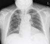
The patient presented with a mouth opening of 10 mm, a reddish face, and tenderness on palpation over the swelling present in right submental area which also extended into the submandibular area. Moderate pus discharge was observed at the #37 extraction site. Severe caries was observed on #46. His preoperative status was normal and without pulmonary or cardiovascular problems (Fig. 1).
The patient underwent facial and intraoral incision and drainage (I&D) under general anesthesia. Nasotracheal intubation using a fiberscope was performed without any incident. The I&D was performed without any surgical complications in the right submental, submandibular, buccal, pterygomandibular, and left sublingual spaces. The operative time was 2 hours. The SPO2 of the patient immediately after the operation was stable (97% to 99%).
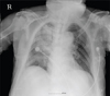
The patient was transferred to the post-anesthesia recovery unit without removing the endotracheal tube after the surgery. 30 minutes later, the patient became hypoxic with the SPO2 decreasing to 82%. During the next 20 minutes, the patient continued to maintain the SPO2 of 89 to 91%, with O2 flow rate of 15 L/min using a connector. The patient was transferred to the intensive care unit (ICU) for ventilator care. However, the patient's SPO2 was still unstable in the ICU. Portable chest X-ray imaging, at 1 hour after surgery, confirmed the collapse of the right upper lung, as well as lobar pneumonia of right lower lobe (Fig. 2).
In flexible fiberoptic bronchoscopy (FFB), there was thick pus on the right main stem bronchus and the passage was cleared through suction and lavage using a suction catheter. The patient's SPO2 improved to 97% after suctioning.
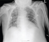
Follow-up chest radiograph, taken 6 hours after FFB, confirmed improvement of the right lobe atelectasis (Fig. 3). On the 4th day post-surgery, the patient was extubated and moved to the general ward. The patient's condition continued to improve, and he was discharged, 13 days post-surgery.
DISCUSSION
Atelectasis is diagnosed based on clinical and radiographic findings [1]. Clinical findings of atelectasis include dyspnea, tachypnea, asymmetric chest movement, and reduced breath sounds in the affected lung field [13]. Persistent wheezing raises the suspicion of atelectasis [3]. Hypoxia is a direct result of atelectasis, and thus, measurement of SPO2 with pulse oximetry is the best approach to diagnosing atelectasis, given its low cost, minimal invasiveness, and immediate results [4]. Monitoring oxygen saturation trends using pulse oximetry can raise the suspicion for atelectasis when hypoxia is present [45].
Radiographic findings of atelectasis include increased opacity in the affected lung field and loss of volume [1]. Further radiographic findings such as, changes in the mediastinum and diaphragm, facilitate diagnosis [5]. As the lung volume decreases with atelectasis, the mediastinum shifts towards the affected field on the radiograph [5]. Furthermore, a unilateral elevated diaphragm increases the suspicion for atelectasis [5]. Moreover, ultrasonography can distinguish between opacity due to fluid accumulation and collapsed lung [5].
Treatment options include incentive spirometry, chest physiotherapy, bronchodilators, fiberoptic bronchoscopy, DNase (deoxyribonuclease), positive-expiratory pressure, and surfactant therapy [6]. Chest physiotherapy is the traditional first-line therapy for atelectasis [6]. If the symptoms do not improve within 24 hours after initiating chest physiotherapy, obstructive atelectasis secondary to obstruction should be suspected and fiberoptic bronchoscopy is recommended for lavage after the obstruction is confirmed [12]. Using fiberoptic bronchoscopy, without first attempting chest physiotherapy, is not likely to improve the symptoms in patients with atelectasis, and caution is required since it can increase intracranial pressure [7]. Furthermore, fiberoptic bronchoscopy requires a skilled and experienced technician [8]. In this case, the patient was admitted for swelling and was transferred out of the surgery room without extubation, to prevent obstructive upper respiratory failure.
It may be better to attempt postural drainage and chest percussion along with bronchodilators in case of atelectasis symptoms after the surgery. However, in this case, although mechanical respiration was provided by the ventilator, the patient presented symptoms of hypoxemia, which required immediate management. Hypoxemia secondary to obstruction of the tracheobronchial tree was suspected and FFB was performed. Although FFB is usually used to confirm the existence of obstruction, or for suction and lavage when patients do not show improvement within 24 hours, its immediate use should be considered in patients who present with clear evidence of atelectasis. In this case, the intubation was leading to easier access.
Lastly, obstructive atelectasis increases the risk for infection, regardless of the cause of the obstruction. Hence, broad spectrum antibiotics should be prescribed if the patient presents with symptoms such as fever, night sweats, and leukocytosis [1]. If the suspected cause of obstruction is pus, such as in our case, the likelihood of infection is very high and aggressive postoperative antibiotic therapy is required.
Pulmonary atelectasis is a common postoperative complication [9]. Postoperative atelectasis occurs in 90% of major thoracic surgeries and in 20–60% of abdominal surgeries [9]. Postoperative atelectasis was observed in 27% of maxillofacial surgery patients [9]. Atelectasis can occur at any time between before surgery to days after surgery, and untreated atelectasis can lead to complications, such as pneumonia, acute pulmonary injury, and reintubation and extubation failure [4].
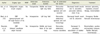
Reports of atelectasis after maxillofacial surgery are summarized in Table 1. Aziz et al. [1] reported cases of atelectasis after orthognathic surgery for 3rd degree malocclusion. The atelectasis was secondary to obstruction, possibly caused by oral secretion or epistaxis caused by nasotracheal intubation. Vikari et al. [10] reported obstructive atelectasis caused by bleeding from an open fracture and nasotracheal intubation. Furthermore, they reported that mechanical ventilation contributed to the development of atelectasis by pushing the residual blood into the bronchus. The report of Skouteris et al. [2] described acute bronchial obstruction and intraoperative atelectasis secondary to thick mucus plugging during routine nasotracheal intubation, prior to the correction of bilateral, severely displaced mandibular fracture. The patient's thick mucus secretion is suspected to have been due to a habit of heavy smoking. Aframian-Farnad et al. [9] reported that obstruction of the airway and accumulation of secretions were the major causes. Risk factors in patients with mandibulo-maxillary fixation (MMF) include upper airway obstruction and difficulty in clearing pharyngeal secretions.
Most cases of atelectasis reported in association with maxillofacial surgery are obstructive. Risk factors include nasotracheal intubation epistaxis, oral bleeding, and heavy smoking-induced thick tracheal mucus secretion. In our case, the persistent drainage of pus caused obstruction that resulted in atelectasis. No bleeding was present in our case, and oral contents could not be directly visualized due to the patient's restricted mouth opening. Therefore, it was difficult to predict that pus could cause obstructive atelectasis when the patient was intubated. Furthermore, patients with abscess can suffer from upper respiratory failure due to swelling requiring immediate intubation. However, in such cases, intubation can push the pus into the bronchus. Hence, patients with persistent pus drainage should be thoroughly suctioned prior to intubation, and patients with risk factors should be continually followed up with SPO2, breath sounds, and chest X-ray evaluations.
 XML Download