

 PDF
PDF ePub
ePub Citation
 Print
Print


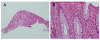
Figures and Tables
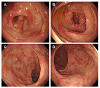
Fig. 1
Colonoscopic finding of a patient with eosinophilic colitis showing non-specific inflammatory changes, mucosal edema, patchy eythema, and loss of vascularity. (A) Cecum. (B) Proximal ascending colon. (C) Upper rectum. (D) Lower rectum.
References
1. Fenoglio LM, Benedetti V, Rossi C, et al. Eosinophilic gastroenteritis with ascites: a case report and review of the literature. Dig Dis Sci. 2003; 48:1013–1020.
2. Rothenberg ME. Eosinophilic gastrointestinal disorders (EGID). J Allergy Clin Immunol. 2004; 113:11–28. quiz 29.

3. Gaertner WB, Macdonald JE, Kwaan MR, et al. Eosinophilic colitis: university of Minnesota experience and literature review. Gastroenterol Res Pract. 2011; 2011:857508.
4. Talley NJ, Shorter RG, Phillips SF, Zinsmeister AR. Eosinophilic gastroenteritis: a clinicopathological study of patients with disease of the mucosa, muscle layer, and subserosal tissues. Gut. 1990; 31:54–58.
5. Khan S, Orenstein SR. Eosinophilic gastroenteritis. Gastroenterol Clin North Am. 2008; 37:333–348. v
6. Klein NC, Hargrove RL, Sleisenger MH, Jeffries GH. Eosinophilic gastroenteritis. Medicine (Baltimore). 1970; 49:299–319.
7. Cello JP. Eosinophilic gastroenteritis--a complex disease entity. Am J Med. 1979; 67:1097–1104.
8. Chen MJ, Chu CH, Lin SC, Shih SC, Wang TE. Eosinophilic gastroenteritis: clinical experience with 15 patients. World J Gastroenterol. 2003; 9:2813–2816.
9. Sundaram S, Sunku B, Nelson SP, et al. Adherent white plaques: an endoscopic finding in eosinophilic esophagitis. J Pediatr Gastroenterol Nutr. 2004; 38:208–212.
10. Straumann A, Simon HU. The physiological and pathophysiological roles of eosinophils in the gastrointestinal tract. Allergy. 2004; 59:15–25.
11. Rubio CA. A method for the detection of eosinophilic granulocytes in colonoscopic biopsies from IBD patients. Pathol Res Pract. 2003; 199:145–150.
 XML Download