

 PDF
PDF ePub
ePub Citation
 Print
Print


INTRODUCTION
Endoscopic Gall Bladder Drainage (EGBD) has been used to treat patients with acute cholecystitis or to relieve malignant biliary obstruction in patients who are poor candidates for surgery due to underlying comorbidities.1 An endoscopic connection is created between the gastric - or duodenal - and the gallbladder lumen to achieve drainage of the gall bladder (GB) and biliary tract. EGBD was first described in 2007, and since, its approach has evolved from the placement of naso-gallbladder drain to plastic biliary stents, and then to self-expanding metallic stents (SEMS).2 Lately, lumen apposing metallic stent (LAMS), which utilizes dumbbell-shaped flanges on both ends to appose the two visceral walls, has been made available. Initially, it was placed utilizing the “Cold technique”, which included endoscopic ultrasound (EUS)-guided needle puncture followed by guidewire placement and dilation of the tract. A recently approved second generation LAMS allows the endoscopist to perform anastomosis without the need for a guidewire or tract dilation, with the electrocautery tip mounted delivery system, which is also called the “Hot technique”. Here, we report a case of SEMS placement using LAMS after stent migration during EGBD via the “Hot Technique” and propose the routine use of guidewire in patients undergoing the procedure.
CASE REPORT
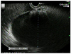
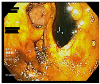
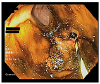
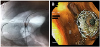
An 88-year-old female patient presented with obstructive jaundice. The patient underwent EUS examination and a large periampullary mass without duodenal obstruction was found. She was scheduled for EUS-guided biliary drainage via the rendezvous technique. EUS examination with linear echoendoscope revealed a distended gallbladder abutting the gastric wall (Fig. 1). Therefore, an EGBD was performed to decompress the GB and biliary system. An appropriate puncture site without intervening the blood vessels was identified. Under EUS guidance, the GB wall was punctured from antrum with an electrocautery tip, followed by deployment of a 10×10 mm AXIOS (Boston Scientific, Marlborough, MA, USA) stent with the proximal end in the GB lumen and with the distal end in the gastric lumen. A copious amount of dark, black bile was observed flowing through the stent (Fig. 2). On further inspection, the distal flange of stent was noted to be retracted in the gastric wall due to GB contraction (Fig. 2). Attempts to reposition the stent with cold biopsy and raptor forceps failed. Due to anatomical position and difficult access, we were unable to advance a guidewire through the stent. A double channel upper endoscope (GIF-2TH180, Olympus, Tokyo, Japan) was then passed into the stomach. The tip of a 5 Fr biliary catheter was modified into a hockey stick shape and advanced through the biopsy channel of the endoscope. A 0.035-inch jag guidewire (Boston Scientific, Marlborough, MA, USA) was passed through the biliary catheter and stent under endoscopic and fluoroscopic guidance (Fig. 3). Multiple loops of the guidewire were coiled inside the GB lumen through LAMS. A 10×40 mm fully covered biliary SEMS (Boston Scientific, Marlborough, MA, USA) was then deployed over the guidewire with distal flange inside the stomach (Fig. 4). Our patient tolerated the procedure well, and follow-up acute abdomen series showed no evidence of free air with continuous improvement of her liver enzymes. She was discharged in stable condition two days after the procedure. Due to involvement of the superior mesenteric artery and poor functional status, she then decided to undergo palliative measures and was discharged to home hospice care.
DISCUSSION
According to literature review, similar cases describing the placement of fully covered SEMS within the LAMS have been reported previously. A case series reported as many as 4 out of 13 patients needing a second stent placement during EGBD.34 However, such cases have been reported with the use of the ‘Cold LAMS’ technique, which already involves the placement of a guidewire before stent deployment.34 Technical failure has been attributed to thickened GB wall, resulting in stent migration. There have been no significant differences in the outcomes and adverse events of patients with EGBD when compared with percutaneous GBD; however, favorable trend towards endoscopic intervention has been observed.567 A study reported that about 10.7% (8/75) of patients experienced adverse events, including recurrent cholecystitis (3), stent migration (2), and Bouveret syndrome (1), with all these complications managed conservatively.8 EGBD has also been shown to be better tolerated with low risk of adverse events as compared to percutaneous GBD.7 Catheter-related problems are minimal in EGBD; however, the risk of perforation, bile peritonitis, bleeding, and stent migration have been reported.79 In our patient, the proposed mechanism of displacement is sudden retraction of the GB wall after decompression with stomach distention as a potential contributor. Based on our literature review, cases of spontaneous migration of LAMS on repeat endoscopy have been reported, but cases of immediate migration have not been reported. We propose that there might be a component of selective reporting here since patients with immediate migration require urgent surgical treatment for the prevention of peritonitis and perforation, which might preclude their study inclusion. We propose that a guidewire should routinely be used for patients undergoing EGBD with ‘Hot LAMS’ technique, which can be passed through the delivery system before stent deployment. Placement of a guidewire in GB lumen before stent deployment would likely allow for a safe and reliable placement of another stent in case of displacement or migration of the first stent. A careful endoscopic examination should be performed to confirm the correct placement of LAMS. If the stent is deployed without any complications, the guidewire can then be removed through the stent, or if needed, it can be used to place another stent in case of malalignment or displacement of the first stent. Hence, we believe that routine use of a guidewire for the placement of “Hot LAMS” might as well “Save the day” for patients who otherwise might experience complications.
 XML Download