

 PDF
PDF ePub
ePub Citation
 Print
Print


Abstract
Purpose
To assess the venographic findings of central venous abnormalities before exchanging dysfunctional tunneled hemodialysis catheters and the outcome of endovascular salvage techniques.
Materials and Methods
A total of 110 episodes of tunneled hemodialysis catheter dysfunction in 78 patients undergoing catheter-directed hemodialysis treatment from January 2011 to December 2015 were retrospectively evaluated. Venography was performed before catheter exchange, and the following procedures were conducted according to the venographic findings: balloon disruption of a fibrin sheath, angioplasty for central vein stenosis, or stent insertion. Technical success was defined as at least one successful session of hemodialysis with the exchanged catheter. Patients were followed until the study endpoints or the last hospital visit.
Results
Venography showed abnormalities in patients with 67 of the 110 exchanged catheters, including central vein stenosis (n = 27), fibrin sheath formation (n = 17), and thrombus formation (n = 12). Technical success was confirmed in all cases. The estimated 30-day catheter patency for all assessable catheters was 61.7%. Nine catheters were removed during the follow-up period because of suspected catheter-related infections.
Go to : 
REFERENCES
1.Lee H., Manns B., Taub K., Ghali WA., Dean S., Johnson D, et al. Cost analysis of ongoing care of patients with end-stage renal disease: the impact of dialysis modality and dialysis access. Am J Kidney Dis. 2002. 40:611–622.

2.Lacson E Jr., Wang W., Hakim RM., Teng M., Lazarus JM. Associates of mortality and hospitalization in hemodialysis: potentially actionable laboratory variables and vascular access. Am J Kidney Dis. 2009. 53:79–90.
3.Allon M., Daugirdas J., Depner TA., Greene T., Ornt D., Schwab SJ. Effect of change in vascular access on patient mortality in hemodialysis patients. Am J Kidney Dis. 2006. 47:469–477.
4.Robinson BM., Akizawa T., Jager KJ., Kerr PG., Saran R., Pisoni RL. Factors affecting outcomes in patients reaching end-stage kidney disease worldwide: differences in access to renal replacement therapy, modality use, and haemodialysis practices. Lancet. 2016. 388:294–306.
5.Little MA., O'Riordan A., Lucey B., Farrell M., Lee M., Conlon PJ, et al. A prospective study of complications associated with cuffed, tunnelled haemodialysis catheters. Nephrol Dial Transplant. 2001. 16:2194–2200.
6.Shingarev R., Barker-Finkel J., Allon M. Natural history of tunneled dialysis catheters placed for hemodialysis initiation. J Vasc Interv Radiol. 2013. 24:1289–1294.
7.Sampathkumar K., Ramakrishnan M., Sah AK., Sooraj Y., Ma-haldhar A., Ajeshkumar R. Tunneled central venous catheters: experience from a single center. Indian J Nephrol. 2011. 21:107–111.
8.Quarello F., Forneris G., Borca M., Pozzato M. Do central venous catheters have advantages over arteriovenous fistulas or grafts? J Nephrol. 2006. 19:265–279.
9.Sacks D., McClenny TE., Cardella JF., Lewis CA. Society of interventional radiology clinical practice guidelines. J Vasc Interv Radiol. 2003. 14:S199–S202.
10.Leblanc M., Bosc JY., Paganini EP., Canaud B. Central venous dialysis catheter dysfunction. Adv Ren Replace Ther. 1997. 4:377–389.
11.Gray RJ., Levitin A., Buck D., Brown LC., Sparling YH., Jablonski KA, et al. Percutaneous fibrin sheath stripping versus transcatheter urokinase infusion for malfunctioning well-positioned tunneled central venous dialysis catheters: a prospective, randomized trial. J Vasc Interv Radiol. 2000. 11:1121–1129.
12.Schwab SJ., Buller GL., McCann RL., Bollinger RR., Stickel DL. Prospective evaluation of a Dacron cuffed hemodialysis catheter for prolonged use. Am J Kidney Dis. 1988. 11:166–169.
13.Macrae JM., Loh G., Djurdjev O., Shalansky S., Werb R., Levin A, et al. Short and long alteplase dwells in dysfunctional hemodialysis catheters. Hemodial Int. 2005. 9:189–195.
14.Garofalo RS., Zaleski GX., Lorenz JM., Funaki B., Rosenblum JD., Leef JA. Exchange of poorly functioning tunneled permanent hemodialysis catheters. AJR Am J Roentgenol. 1999. 173:155–158.
15.Merport M., Murphy TP., Egglin TK., Dubel GJ. Fibrin sheath stripping versus catheter exchange for the treatment of failed tunneled hemodialysis catheters: randomized clinical trial. J Vasc Interv Radiol. 2000. 11:1115–1120.
16.Angle JF., Shilling AT., Schenk WG., Bissonette EA., Stadtlander KS., Hagspiel KD, et al. Utility of percutaneous intervention in the management of tunneled hemodialysis catheters. Cardiovasc Intervent Radiol. 2003. 26:9–18.
17.Crain MR., Mewissen MW., Ostrowski GJ., Paz-Fumagalli R., Beres RA., Wertz RA. Fibrin sleeve stripping for salvage of failing hemodialysis catheters: technique and initial results. Radiology. 1996. 198:41–44.
18.Haskal ZJ., Leen VH., Thomas-Hawkins C., Shlansky-Goldberg RD., Baum RA., Soulen MC. Transvenous removal of fibrin sheaths from tunneled hemodialysis catheters. J Vasc Interv Radiol. 1996. 7:513–517.
19.Janne d'Othée B., Tham JC., Sheiman RG. Restoration of patency in failing tunneled hemodialysis catheters: a comparison of catheter exchange, exchange and balloon disruption of the fibrin sheath, and femoral stripping. J Vasc Interv Radiol. 2006. 17:1011–1015.
20.Oliver MJ., Mendelssohn DC., Quinn RR., Richardson EP., Rajan DK., Pugash RA, et al. Catheter patency and function after catheter sheath disruption: a pilot study. Clin J Am Soc Nephrol. 2007. 2:1201–1206.
21.Shanaah A., Brier M., Dwyer A. Fibrin sheath and its relation to subsequent events after tunneled dialysis catheter exchange. Semin Dial. 2013. 26:733–737.
22.Alomari AI., Falk A. The natural history of tunneled hemodialysis catheters removed or exchanged: a single-institution experience. J Vasc Interv Radiol. 2007. 18:227–235.
23.Weber E., Liberek T., Wołyniec W., Gruszecki M., Rutkowski B. Survival of tunneled hemodialysis catheters after percutaneous placement. Acta Biochim Pol. 2016. 63:139–143.
24.Ewing F., Patel D., Petherick A., Winney R., McBride K. Radiological placement of the AshSplit haemodialysis catheter: a prospective analysis of outcome and complications. Nephrol Dial Transplant. 2002. 17:614–619.
25.Ervo S., Cavatorta F., Zollo A. Implantation of permanent jugular catheters in patients on regular dialysis treatment: ten years' experience. J Vasc Access. 2001. 2:68–72.
Go to :
 | Fig. 1Normal venography findings in 75-year-old male. The patient was managed with simple catheter exchange over the wire without additional intervention. |
 | Fig. 2A 68-year-old female. A. Venography reveals central vein stenosis at junction of superior vena cava-right atrium. B. The patient was managed with catheter exchange over the wire with adjustment of catheter tip position, avoiding the site of central vein stenosis. |
 | Fig. 3A 51-year-old female. A. Venography reveals fibrin sheath limiting contrast flow along the catheter insertion pathway. B, C. Balloon angioplasty was performed to rupture the fibrin sheath using a 14 mm × 4 cm Atlas percutaneous transluminal angioplasty dilatation catheter (Bard peripheral Vascular). Note the waist formation at the balloon during partial inflation (arrow). D. Repeated venography after the intervention no longer visualizes contrast flow limitation. |
 | Fig. 4A 45-year-old female. A. Venography reveals central vein stenosis at SVC level. B. Balloon angioplasty was performed at the level of stenotic central vein using a 12 mm × 8 cm Mustang balloon (Boston Scientific Corporation). C. Repeated venography after the intervention reveals improved stenosis at SVC level. SVC = superior vena cava |
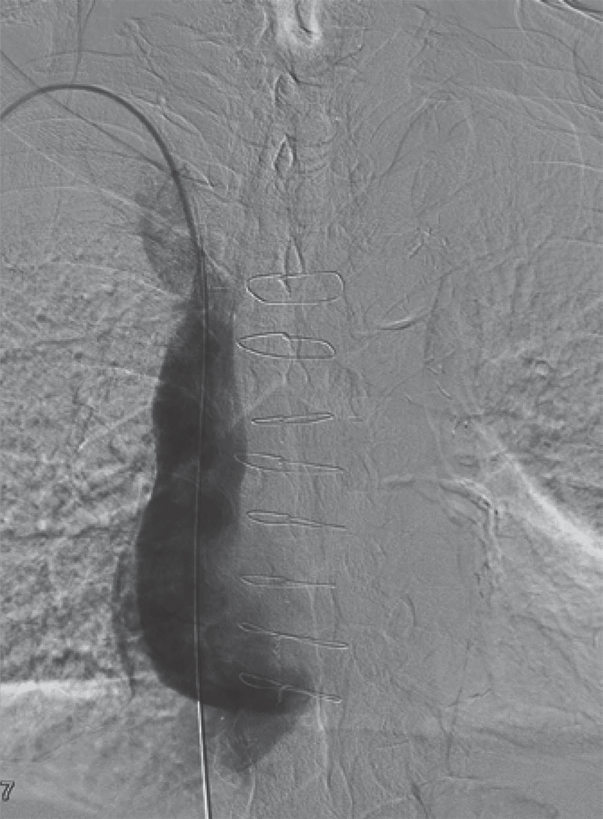
 | Fig. 5A 81-year-old female. A. Venography reveals complex findings of central vein stenosis at SVC level (arrow) with fibrin sheath along catheter insertion pathway (arrowheads). B. Stenosis of SVC remained after several sessions of balloon angioplasty (not seen), and a 20 mm x 6 cm Palmaz Genesis peripheral stent (Cordis) was placed in the SVC despite the fact the patient had cardiac pacemaker installed. C. Repeated venography after the intervention reveals improved stenosis and disappearance of fibrin sheath. D. New catheter was inserted over the wire and lasted for 47 days. SVC = superior vena cava |
Table 1.
Venography Findings
 XML Download