

 PDF
PDF ePub
ePub Citation
 Print
Print


Abstract
Purpose
We compared the outcomes of percutaneous transthoracic needle aspiration biopsy (PCNA) of lung masses in cases with and without prior positron emission tomography/computed tomography (PET/CT) information, and investigated the factors associated with false-negative pathological results.
Materials and Methods
From a total of 291 patients, 161 underwent PCNA without prior PET/CT imaging, while 130 underwent PET/CT before PCNA. Clinical characteristics, procedural variables, pathological results, and diagnostic success rates were compared between the 2 groups. Among patients with initial negative (non-specific benign) PCNA results, the radiological findings of these groups were compared to evaluate the predictors of false-negative lesions.
Results:
No significant difference was found in the clinical characteristics, procedural characteristics, and pathological results of the 2 groups, nor was the diagnostic rate significantly different between them (p = 0.818). Among patients with initial negative PCNA results, radiological characteristics were similar in both the groups. In multivariate analysis, the presence of necrosis (p = 0.005) and ground-glass opacity (GGO) (p = 0.011) were the significant characteristics that indicated an increased probability of initial false-negative results in PCNA.
Go to : 
REFERENCES
1.National Lung Screening Trial Research Team. Church TR., Black WC., Aberle DR., Berg CD., Clingan KL, et al. Results of initial low-dose computed tomographic screening for lung cancer. N Engl J Med. 2013. 368:1980–1991.

2.Hoffman JM., Gambhir SS. Molecular imaging: the vision and opportunity for radiology in the future. Radiology. 2007. 244:39–47.
3.Bomanji JB., Costa DC., Ell PJ. Clinical role of positron emission tomography in oncology. Lancet Oncol. 2001. 2:157–164.
4.Cornelis F., Silk M., Schoder H., Takaki H., Durack JC., Erinjeri JP, et al. Performance of intra-procedural 18-fluorodeoxy-glucose PET/CT-guided biopsies for lesions suspected of malignancy but poorly visualized with other modalities. Eur J Nucl Med Mol Imaging. 2014. 41:2265–2272.
5.Klaeser B., Mueller MD., Schmid RA., Guevara C., Krause T., Wiskirchen J. PET-CT-guided interventions in the management of FDG-positive lesions in patients suffering from solid malignancies: initial experiences. Eur Radiol. 2009. 19:1780–1785.
6.Guralnik L., Rozenberg R., Frenkel A., Israel O., Keidar Z. Metabolic PET/CT-guided lung lesion biopsies: impact on diagnostic accuracy and rate of sampling error. J Nucl Med. 2015. 56:518–522.
7.Purandare NC., Kulkarni AV., Kulkarni SS., Roy D., Agrawal A., Shah S, et al. 18F-FDG PET/CT-directed biopsy: does it offer incremental benefit? Nucl Med Commun. 2013. 34:203–210.
8.Stattaus J., Kuehl H., Ladd S., Schroeder T., Antoch G., Baba HA, et al. CT-guided biopsy of small liver lesions: visibility, artifacts, and corresponding diagnostic accuracy. Cardiovasc Intervent Radiol. 2007. 30:928–935.
9.Gelbman BD., Cham MD., Kim W., Libby DM., Smith JP., Port JL, et al. Radiographic and clinical characterization of false negative results from CT-guided needle biopsies of lung nodules. J Thorac Oncol. 2012. 7:815–820.
10.Hiraki T., Mimura H., Gobara H., Iguchi T., Fujiwara H., Sakurai J, et al. CT fluoroscopy-guided biopsy of 1,000 pulmonary lesions performed with 20-gauge coaxial cutting needles: diagnostic yield and risk factors for diagnostic failure. Chest. 2009. 136:1612–1617.
11.Hansell DM., Bankier AA., MacMahon H., McLoud TC., Müller NL., Remy J. Fleischner Society: glossary of terms for thoracic imaging. Radiology. 2008. 246:697–722.
12.de Geus-Oei LF., van der Heijden HF., Visser EP., Hermsen R., van Hoorn BA., Timmer-Bonte JN, et al. Chemotherapy response evaluation with 18F-FDG PET in patients with nonsmall cell lung cancer. J Nucl Med. 2007. 48:1592–1598.
13.Hicks RJ., Kalff V., MacManus MP., Ware RE., Hogg A., McKenzie AF, et al. (18)F-FDG PET provides high-impact and powerful prognostic stratification in staging newly diagnosed nonsmall cell lung cancer. J Nucl Med. 2001. 42:1596–1604.
14.Takeuchi S., Khiewvan B., Fox PS., Swisher SG., Rohren EM., Bassett RL Jr, et al. Impact of initial PET/CT staging in terms of clinical stage, management plan, and prognosis in 592 patients with nonsmall-cell lung cancer. Eur J Nucl Med Mol Imaging. 2014. 41:906–914.
15.Truong MT., Viswanathan C., Erasmus JJ. Positron emission tomography/computed tomography in lung cancer staging, prognosis, and assessment of therapeutic response. J Thorac Imaging. 2011. 26:132–146.
16.Cerci JJ., Pereira Neto CC., Krauzer C., Sakamoto DG., Vitola JV. The impact of coaxial core biopsy guided by FDG PET/CT in oncological patients. Eur J Nucl Med Mol Imaging. 2013. 40:98–103.
17.Klaeser B., Wiskirchen J., Wartenberg J., Weitzel T., Schmid RA., Mueller MD, et al. PET/CT-guided biopsies of metabolically active bone lesions: applications and clinical impact. Eur J Nucl Med Mol Imaging. 2010. 37:2027–2036.
18.Kim JI., Park CM., Kim H., Lee JH., Goo JM. Non-specific benign pathological results on transthoracic core-needle biopsy: how to differentiate false-negatives? Eur Radiol. 2017. 27:3888–3895.
19.Minot DM., Gilman EA., Aubry MC., Voss JS., Van Epps SG., Tuve DJ, et al. An investigation into false-negative transthoracic fine needle aspiration and core biopsy specimens. Diagn Cy-topathol. 2014. 42:1063–1068.
20.Tsukada H., Satou T., Iwashima A., Souma T. Diagnostic accuracy of CT-guided automated needle biopsy of lung nodules. AJR Am J Roentgenol. 2000. 175:239–243.
21.Yeow KM., Tsay PK., Cheung YC., Lui KW., Pan KT., Chou AS. Factors affecting diagnostic accuracy of CT-guided coaxial cutting needle lung biopsy: retrospective analysis of 631 procedures. J Vasc Interv Radiol. 2003. 14:581–588.
22.Heppner GH. Tumor heterogeneity. Cancer Res. 1984. 44:2259–2265.
23.Miles KA., Williams RE. Warburg revisited: imaging tumour blood flow and metabolism. Cancer Imaging. 2008. 8:81–86.
24.Swanton C. Intratumor heterogeneity: evolution through space and time. Cancer Res. 2012. 72:4875–4882.
25.Bar-Shalom R., Yefremov N., Guralnik L., Gaitini D., Frenkel A., Kuten A, et al. Clinical performance of PET/CT in evaluation of cancer: additional value for diagnostic imaging and patient management. J Nucl Med. 2003. 44:1200–1209.
26.Kubota K. From tumor biology to clinical PET: a review of positron emission tomography (PET) in oncology. Ann Nucl Med. 2001. 15:471–486.
27.Hua Q., Zhu X., Zhang L., Zhao Y., Tang P., Ni J. Initial experience with real-time hybrid single-photon emission computed tomography/computed tomography-guided percutaneous transthoracic needle biopsy. Nucl Med Commun. 2017. 38:556–560.
28.Aoki T., Tomoda Y., Watanabe H., Nakata H., Kasai T., Hashimoto H, et al. Peripheral lung adenocarcinoma: correlation of thin-section CT findings with histologic prognostic factors and survival. Radiology. 2001. 220:803–809.
29.Lee HY., Lee KS. Ground-glass opacity nodules: histopathology, imaging evaluation, and clinical implications. J Thorac Imaging. 2011. 26:106–118.
30.Song YS., Park CM. Pulmonary subsolid nodules: an overview & management guidelines. J Korean Soc Radiol. 2018. 78:309–320.
31.Hur J., Lee HJ., Nam JE., Kim YJ., Kim TH., Choe KO, et al. Diagnostic accuracy of CT fluoroscopy-guided needle aspiration biopsy of ground-glass opacity pulmonary lesions. AJR Am J Roentgenol. 2009. 192:629–634.
32.Kim TJ., Lee JH., Lee CT., Jheon SH., Sung SW., Chung JH, et al. Diagnostic accuracy of CT-guided core biopsy of ground-glass opacity pulmonary lesions. AJR Am J Roentgenol. 2008. 190:234–239.
33.Lu CH., Hsiao CH., Chang YC., Lee JM., Shih JY., Wu LA, et al. Percutaneous computed tomography-guided coaxial core biopsy for small pulmonary lesions with ground-glass attenuation. J Thorac Oncol. 2012. 7:143–150.
34.Suh YJ., Lee JH., Hur J., Hong SR., Im DJ., Kim YJ, et al. Predictors of false-negative results from percutaneous transthoracic fine-needle aspiration biopsy: an observational study from a retrospective cohort. Yonsei Med J. 2016. 57:1243–1251.
35.American College of Radiology. Lung-RADSTM version 1.0 assessment categories. Available at:. https://www.acr.org/-/media/ACR/Files/RADS/Lung-RADS/LungRADS_Assess-mentCategories.pdf?la=en. Published Apr 28, 2014. Accessed Aug 25. 2017.
Go to :
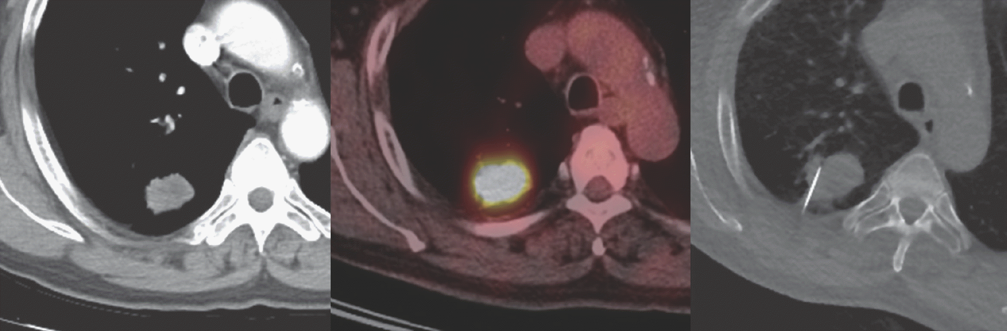 | Fig. 1A 62-year-old male with a mass located at the right upper lobe posterior segment. PET/CT scan shows homogeneous uptake (SUVmax of 14.3), with no definite additional benefit before percutaneous transthoracic needle aspiration biopsy. Pathologic results revealed squamous cell carcinoma. |
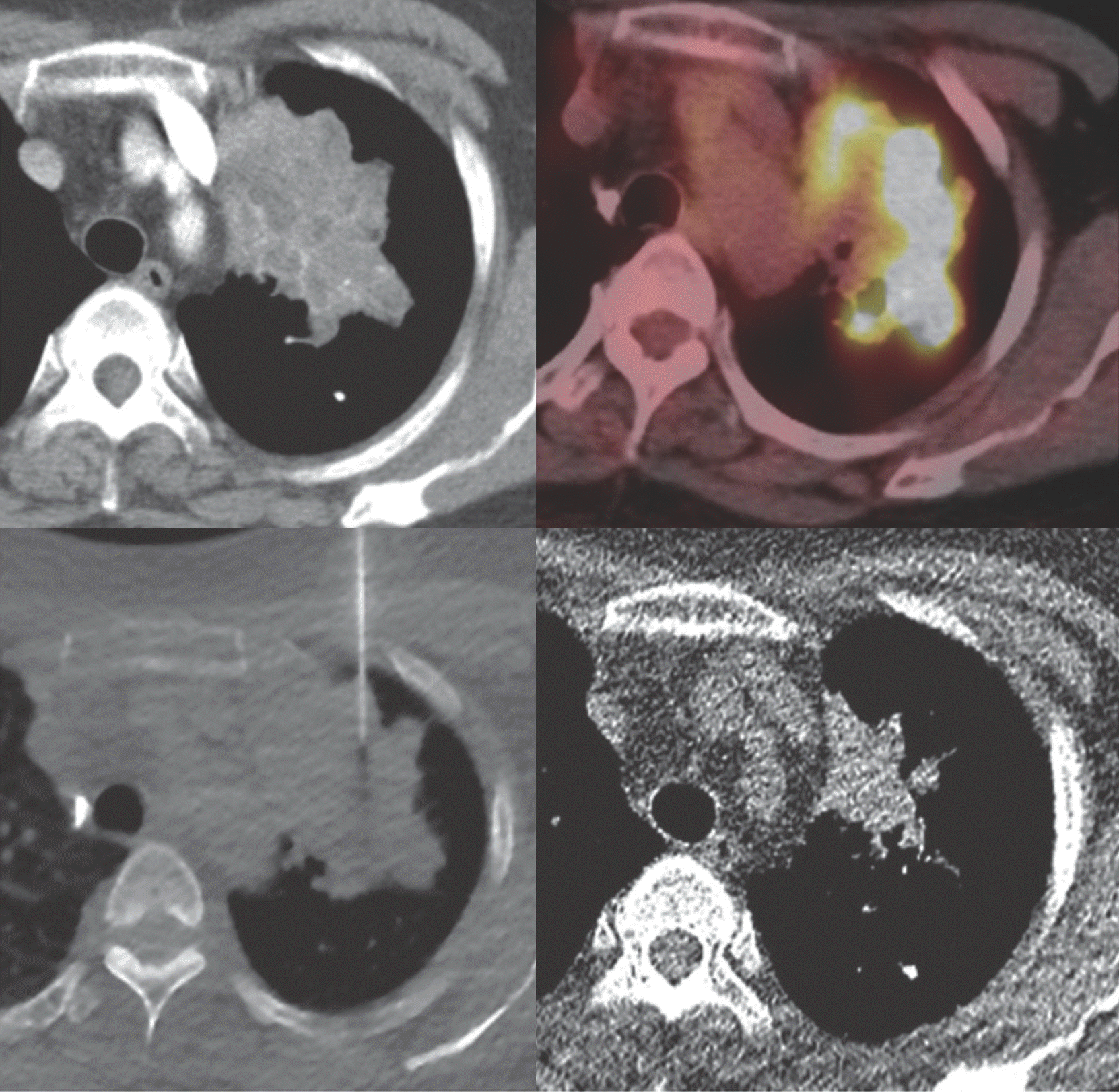 | Fig. 2A 61-year-old female with a heterogeneous, lobulated mass with suspicious necrosis, located at the left upper lobe. PET/CT scan demonstrates high metabolic uptake at the peripheral area of the lesion (SUVmax of 16.7). Thus, the biopsy needle tip is placed at the lesion with highest metabolic uptake. Pathologic results revealed pulmonary tuberculosis. Low-dose chest CT scan (right lower quadrant) after 6 months of anti-tuberculous treatment shows interval decrease of the lesion. |
Table 1.
Clinical Characteristics, Procedure Characteristics, and Pathologic Results of Patients who Underwent Percutaneous Transthoracic Needle Aspiration Biopsy between Those with and without PET Scans
Table 2.
Comparison of Radiological Characteristics among Patients with Initial Negative Percutaneous Transthoracic Needle Aspiration Biopsy Results
Table 3.
Comparison of Characteristics between False-Negative and True-Negative Lesions
Table 4.
Results of Univariate and Multivariate Logistic Regression Analysis for Predictors of False-Negative Lesions at Percutaneous Transthoracic Needle Aspiration Biopsy
 XML Download