

 PDF
PDF ePub
ePub Citation
 Print
Print


INTRODUCTION
Patients with malignancy are at increased risk for vascular thromboembolism; thromboembolic events are a significant cause of morbidity and mortality (1). In patients undergoing chemotherapy, the risk for thromboembolism is approximately six times higher than that in the non-cancer population (1), and most of these thrombotic events occur in the venous system. Although the incidence of arterial thromboembolism is lower, it has, nevertheless, been reported to be 3.8% among cancer patients (2).
Capecitabine is an oral prodrug of 5-fluorouracil (5-FU), which is commonly used to treat gastric, colon, and breast cancers. The most common complications associated with capecitabine administration are hand-foot syndrome, neutropenia, fatigue and gastrointestinal symptoms, such as, diarrhea, vomiting, nausea, and abdominal pain (3). Cardiovascular complications are uncommon; however, cardiac toxicities, such as arrhythmias, cardiomyopathy, angina, and myocardial infarction, have been reported (4). However, acute aortic thrombus is an extremely rare toxicity that requires early diagnosis and prompt treatment because it can be a potential source of cerebral, visceral, or lower extremity emboli. We report a case of acute aortic thrombosis in a patient who underwent capecitabine treatment for colon cancer.
CASE REPORT
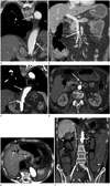
A 63-year-old man was diagnosed with adenocarcinoma in the ascending colon and associated bowel obstruction. Right hemicolectomy was performed and pathologic staging revealed stage IIA (pT3N0M0) disease. His medical history included hypertension, pulmonary tuberculosis, and chronic obstructive pulmonary disease. Two months after surgery, he underwent adjuvant chemotherapy with capecitabine monotherapy (1250 mg/m2 twice daily on days 1–14, every 3 weeks). At the time of colon cancer diagnosis, initial computed tomography (CT) revealed atherosclerotic change in the aorta and superior mesenteric artery (SMA) (Fig. 1A, B). After the second cycle of chemotherapy, he developed anorexia and asthenia. Physical examination revealed only ulcers in the oral cavity, and laboratory findings revealed mild leukocytosis (white cell count, 10520/mm3) and a slightly increased prothrombin time (PT, 16.6 sec). At a routine surveillance CT examination, long segmental, irregular, low-attenuation was observed in the right wall of the descending thoracic aorta and suprarenal abdominal aorta, suggestive of in situ arterial thrombosis (Fig. 1C), with acute thrombosis evident in the SMA (Fig. 1D). There was no evidence of perfusion abnormality in the abdominal solid organs or bowel. The patient was treated conservatively with acetylsalicylic acid and clopidogrel, and continued to take capecitabine.
After 2 months of further treatment, the patient visited the emergency department of the authors' institution complaining of severe abdominal pain and distension. At the time of admission, his systolic blood pressure was 80 mm Hg. Complete blood count revealed a white cell count of 1760/µL, a hemoglobin level of 10.9 g/dL, and a platelet count of 46 × 103/µL. Coagulation tests revealed a prolonged PT (32.4 sec), an elevated activated partial thromboplastin time (57.7 sec), normal fibrinogen levels, an increased D-dimer concentration (3.02 ug/mL), and decreased antithrombin III activity (21%). An abdominal CT scan revealed diffuse small bowel wall necrosis with pneumatosis intestinalis (Fig. 1E, F). At emergency exploratory laparotomy, the entire small bowel was necrotic and nonviable; consequently, an open-and-close operation was performed. The patient was then treated conservatively but died four days later.
DISCUSSION
We report a case of long segmental, acute aortic thrombus that developed in a patient with colon cancer after a second cycle of capecitabine chemotherapy. Arterial thrombosis is rare, and is usually associated with underlying atherosclerosis and/or aneurysmal change (5). At the time of colon cancer diagnosis, baseline CT revealed atherosclerotic changes in the aorta and SMA, presumably related to older age, hypertension, and tobacco smoking. Mesenteric artery stenosis is usually due to atherosclerosis, and results in insufficient blood flow to the small intestine, causing chronic intestinal ischemia (5).
Chemotherapy is a well-known risk factor for thromboembolism in cancer patients; however, in most cases, it involves the venous system (1). In situ arterial thrombosis is rarely associated with cytotoxic chemotherapy, and is regarded to be a potential side effect of anti-angiogenic therapies. The risk for arterial thrombosis in patients undergoing vascular endothelial growth factor (VEGF) receptor-targeted therapy has been reported to be approximately twice that of control patients (67). Bevacizumab is a recombinant humanized monoclonal antibody against VEGF, and is used in combination chemotherapy for metastatic colorectal cancer. Although the exact mechanism has not been documented, VEGF acts to maintain the integrity of the vascular endothelium (67).
Capecitabine is an anti-metabolite, oral prodrug of 5-FU, and arterial thrombosis has been reported to be an uncommon adverse event following 5-FU chemotherapy (4). Furthermore, 5-FU can increase fibrinopeptide A levels and reduce protein C activity and, thus, render blood vessels more susceptible to thrombus formation. In addition, the vascular endothelium is susceptible to free radicals generated by 5-FU, and several animal studies have reported endothelial damage caused by 5-FU (8).
Because complications of arterial thromboembolism (abdominal organ and limb ischemia) are usually fatal, early diagnosis and treatment is imperative. The use of low-dose acetylsalicylic acid is recommended for prophylaxis in high-risk patients without contraindications to its use (9). Thrombolysis, anticoagulation, and surgery could be considered treatment options for arterial thromboembolism; however, the continuance of chemotherapy in patients with chemotherapy-induced thrombosis is controversial. Clinical decision making based on considerations of risks and benefits of further chemotherapy is difficult. Previous study suggested that arterial embolism can often be managed successfully using conservative management, and that further treatment with the same chemotherapy regimen is possible with minimal interruption (10). However, considering the fatal outcome of arterial thrombosis in our case, further evidence supporting the safety of ongoing treatment is still needed.
In conclusion, capecitabine-induced aortic thrombosis is a rare but important complication. Physicians who treat patients with capecitabine need to be mindful of the possibility of arterial thrombosis, especially in patients with underlying atherosclerosis.
 XML Download