

 PDF
PDF ePub
ePub Citation
 Print
Print


INTRODUCTION
The traditional method of placing an implant is to construct a radiological guiding stents and then converting it to a surgical guiding device after Cone-beam computed tomography (CBCT) were taken.1 However, this traditional surgical guiding stent has complicated and inaccurate lab procedure, and difficult in placing the implant fixture as planned.2 The digital surgical guide was introduced to compensate this. Scanned image of intraoral cavity and CBCT image are used to plan the placement of implant considering bone, mucosa, and tooth.3 Using digital surgical guide, drilling and placing implant at a preset position is possible, which makes less error compare to the traditional method, but only when surgical guide is maintained accurate and stable.4
The accuracy of implant placement with a digital surgical guide is evaluated by the deviation in the planned implant and the placed implant.5 A previous study showed coronal deviation of 1.09 mm, apical deviation of 1.28 mm, and axis angle deviation of 3.9°. The deviation may vary in different studies.6 Most previous studies were conducted with the full edentulous ridges. There are little studies about the accuracy test in the partial edentulous ridges. Furthermore, the accuracy studies were conducted with the digital guide stent only for the only one company. Also, many studies were conducted in the laboratory study, there is no clinical study. In previous studies, the accuracy of implant was assessed by overlapping CBCT before and after surgery. Analysis of implant error is not accurate due to resolution and distortion of CBCT, and error in superimposing two CBCT images. In addition, resolution is decreased due to the metal artifact when there are many metal structures.78
The primary purpose of this study is to assess the implant placement error by using CBCT and plaster cast after placing implant in the posterior tooth with universal digital surgical guide and kit.
MATERIALS AND METHODS
This study was approved by the Institutional Review Board, Chonnam National University Dental Hospital (IRB No. CNUDH-2016-007). To calculate the number of subjects required for this study, in vitro experiment9 was performed using a partial edentulous epoxy model (M. Tech, Seoul, Korea). The number of the subjects is 26, which is calculated using G*power 3.1 program (Heinrich Heine University, Düsseldorf, Germany). 28 implants were selected in this study considering 10% failure rate. The following criteria were used to recruit 28 implants placement (Table 1).
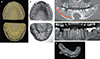
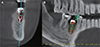
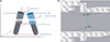
Before placing an implant, patient's preliminary impression was taken and the diagnostic model was fabricated using hard plaster. 3D model scanner (Freedom HD, Degree of Freedom, Seoul, Korea) was used to scan the diagnostic model and the information of patient's intraoral soft tissue surface was saved as Surface Tesselation Language (STL) file. Patient's hard tissue information was obtained by taking CBCT (Alphard-3030, ASAHI Rogentgen, Kyoto, Japan) and saved as Digital imaging and communications in medicine (DICOM) file. After superimposing the STL and DICOM files on the remaining natural teeth using In2guide (Cybermed, Seoul, Korea) software, the surgical guide was fabricated using 3D printer considering the diameter, length, and position of implant (Fig. 1). 4 weeks after placing the implant with digital surgical guide, CBCT was taken to evaluate the implant accuracy (Fig. 2). The term ‘planned implant’ is used to describe the preset position of implant before the surgery, and the ‘inserted implant’ is the actual placement of fixture after the surgery. The coronal deviation (in mm), apical deviation (in mm), and angle deviation (in °) of planned implant and inserted implant were measured to evaluate the accuracy (Fig. 3).
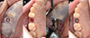
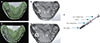
Eight weeks after the implant placement, polyvinylsiloxane (Honigum, DMG, Hamburg, Germany) was used to take impression to make hard plaster model and be scanned; this was called inserted model (Fig. 4). Lab analog inside the plaster was removed after scanning. A lab cylinder screw, a lab cylinder body, and a lab analog were connected to the metal sleeve of the surgical guide used for implant placement, and the lab analog was fixed with a hard plaster to regenerate the planned model, the model of planned position of implant before the surgery. The STL file obtained by scanning pre-operative and postoperative models was superimposed and analyzed with a 3D analysis program (Geomagic control X, 3D Systems, Morrisville, NC, USA). Depth deviation is obtained by subtracting the z-axis coordinate of the post-operative model from the pre-operative model. If the value is positive, the implant fixture is located more apically and if the value is negative, the implant is located more on coronal (Fig. 5).
All statistical analysis is performed by SPSS Ver 23.0 (SPSS Inc., Chicago, IL, USA). Angle deviation, coronal deviation, and apical deviation measured by CBCT and cast model are statistically analyzed by Independent t-test after performing normality test using Kolmogorov-Smirnov test. All results were tested for significance at the level of P < .05. Pearson correlation analysis on angle, coronal and apical deviation was also used in searching for the correlativity of the two methods.
RESULTS
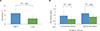
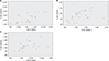
The accuracy measured with CBCT and cast model is as follows (Table 2). The means for CBCT analyses were: angle deviation: 4.74 ± 2.06°, coronal deviation: 1.37 ± 0.80 mm, and apical deviation: 1.77 ± 0.86 mm. The means for cast analyses were: angle deviation: 2.43 ± 1.13°, coronal deviation: 0.82 ± 0.44 mm, apical deviation: 1.19 ± 0.46 mm, and depth deviation: 0.03 ± 0.65 mm. The angular, coronal, and apical deviations were significantly smaller in measurement using cast model than those measured using the CBCT method (P = .01). Also, apical deviation was bigger than coronal deviation (Fig. 6). Angular, coronal and apical deviation in CBCT and cast showed positive correlation and there were significant difference among them (P = .001, .024) (Fig. 7).
DISCUSSION
While digital surgical guide makes it possible to drill and place an implant in a preset position, the procedure has to include accurate position and accurate analysis before the surgery. Effort has been made to improve the accuracy of digital surgical guide as well as maintenance of surgical guide and errors occurring during the manufacturing process.
During implant procedure, 1 angle deviation makes 0.34 mm length deviation in the 10-mm fixture apical area. 5° angle deviation makes 1.7 mm length deviation. If the space between implant and tooth root were set to 1.5 mm during implant planning, 5° angle error will impair the tooth root. Thus the angle deviation should be no more than 3° to implant installed safely without the tooth damaged.10 If the important anatomical structure such as inferior alveolar nerve is close by, acceptable surgical guide's maximum angle deviation is less than 3° and maximum vertical error is less than −1.5 mm.10 Less loosening of implant and passive fit is possible when the angle between the hex of fixture and hexagonal freedom of abutment is less than 5° and the distance is 150 µm.1112 This study shows that angular, coronal, and apical deviation are accurate enough to avoid the damage of major anatomical structure during the procedure, but less accurate to connect directly to pre-manufactured hexagonal implant prosthesis. Therefore, it is necessary to manufacture prosthesis by taking an impression after the implant placement, or to use non-hexagonl implant fixture.1
In this study, deviation measured by CBCT is similar to that of other studies but angle deviation is somewhat higher. The reason for this is that the previous studies used surgical kit and implant fixture of the subsidiary company that makes surgical guide, while universal surgical guide kit is used in this study. Also, compared the previous studies, the more rearmost molars are included in this study. Reference marker was not used when taking CBCT, so higher error occurred during overlapping preoperative and postoperative CBCT.
The angle, coronal, and apical deviation were statistically significantly smaller than those of CBCT in this study (P < .05). Not only there is no error occurred while superimposing pre-operative and postoperative CBCTs, but also can additional radiation be decreased by using cast model to analyze accuracy. However, errors can occur when taking impression or making plaster cast model. There was more error in overlapping CBCTs because of the absence of marker in CBCT analysis. Error can be decreased by using only one cast model to reproduce preoperative and postoperative cast model. Further studies in comparing these two methods are required by adding marker to improve accuracy of superimposition. In cast model analysis, depth deviation can be measured by setting axis of the implant as z-axis and obtaining difference in the z-axis. Average of depth deviation is −0.03 ± 0.65 mm, which is more on the coronal side than planned position. This result is similar to that of the previous study, especially when those who have less experience with digital guided surgery tend to have less reliability in the accuracy of the surgical guide.13
This study shows that the deviation obtained by the plaster cast is significantly smaller, which can be useful in evaluation of implant placement accuracy. In addition, the angular deviation may become larger toward the farthest tooth in implant placement.


 XML Download