Abstract
Ecthyma gangrenosum (EG) is a rare skin manifestation which starts with a maculopapular eruption and followed by a necrotic ulcer covered with black eschar. EG usually occurs in immunosuppressed patients with Pseudomonas aeruginosa sepsis. We present a pre-viously healthy 12-month-old girl with EG by P. aeruginosa and agranulocytosis due to influenza A and then rhinovirus infection, without bacteremia. It is important for allergists to culture wound and differentiate EG from other skin disorders including Tsutsuga-mushi disease and initiate appropriate empiric antipseudomonal antibiotic treatment, and to evaluate for possible immunodeficiency, even in a healthy child.
REFERENCES
1. Vaiman M, Lazarovitch T, Heller L, Lotan G. Ecthyma gangrenosum and ecthyma-like lesions: review article. Eur J Clin Microbiol Infect Dis. 2015; 34:633–9.

2. Lee MK, Yoo SY, Hwang PH. Ecthyma gangrenosum caused by Klebsiella pneumoniae in immunocompromised patient associated with severe aplastic anemia. Clin Pediatr Hematol Oncol. 2013; 20:59–61.
3. Cohen N, Capua T, Bilavsky E, Dias-Polak H, Levin D, Grisaru-Soen G. Ecthyma gangrenosum skin lesions in previously healthy children. Acta Paediatr. 2015; 104:e134–8.

4. Murray TS, Baltimore RS. Pseudomonas, burkholderia, and stenotroph-omonas. Kliegman R, Stanton B, Behrman R, Geme JS, Schor N, editors. Nelson textbook of pediatrics. 20th ed.Philadelphia (PA): Elsevier;2016. p. 1412–5.
5. Koo SH, Lee JH, Shin H, Lee JI. Ecthyma gangrenosum in a previously healthy infant. Arch Plast Surg. 2012; 39:673–5.

6. Seo JY, Kim SY, Han MY, Lee KH. A case of ecthyma gangrenosum associated with liver abscess and renal abscess. Korean J Pediatr Infect Dis. 2002; 9:104–9.

7. Juern AM, Drolet BA. Ecthyma. Kliegman R, Stanton B, Behrman R, Geme JS, Schor N, editors. Nelson textbook of pediatrics. 20th ed.Philadelphia (PA): Elsevier;2016. p. 3207–8.
8. Vaiman M, Lasarovitch T, Heller L, Lotan G. Ecthyma gangrenosum versus ecthyma-like lesions: should we separate these conditions? Acta Der-matovenerol Alp Pannonica Adriat. 2015; 24:69–72.

9. Bozkurt I, Yuksel EP, Sunbul M. Ecthyma gangrenosum in a previously healthy patient. Indian Dermatol Online J. 2015; 6:336–8.

10. Pacha O, Hebert AA. Ecthyma gangrenosum and neutropenia in a previously healthy child. Pediatr Dermatol. 2013; 30:e283–4.

11. Sharon N, Talnir R, Lavid O, Rubinstein U, Niven M, First Y, et al. Tran-sient lymphopenia and neutropenia: pediatric influenza A/H1N1 infection in a primary hospital in Israel. Isr Med Assoc J. 2011; 13:408–12.
12. Abraham TY, Dat N, John NG. Rhinovirus infection among patients with hematologic malignancy at a cancer center. Infect Dis Clin Pract. 2016; 24:29–30.
13. Celkan T, Koç BŞ. Approach to the patient with neutropenia in childhood. Turk Pediatri Ars. 2015; 50:136–44.

14. Lanzkowsky P. Disorders of white blood cell. Lanzkowsky P, editor. Manual of pediatric hematology and oncology. 5th ed.Philadelphia (PA): Elsevier;2011. p. 275–95.
15. Biscaye S, Demonchy D, Afanetti M, Dupont A, Haas H, Tran A. Ecthyma gangrenosum, a skin manifestation of Pseudomonas aeruginosa sepsis in a previously healthy child: a case report. Medicine (Baltimore). 2017; 96:e5507.
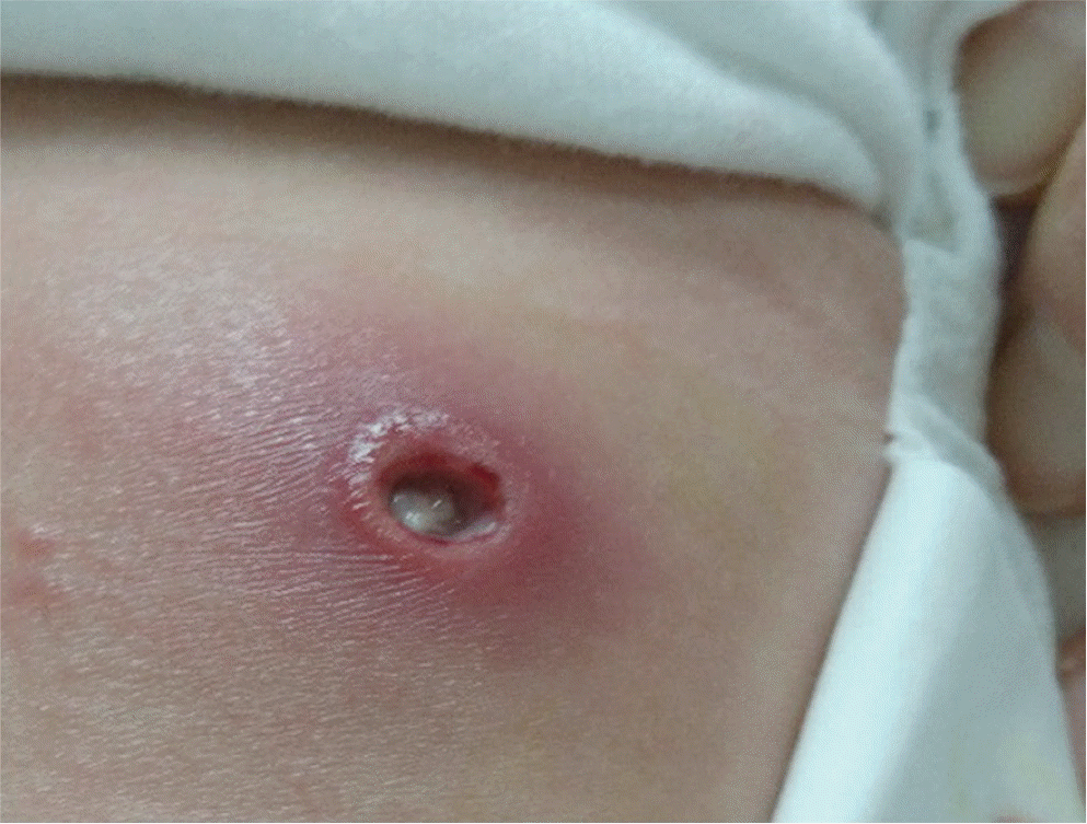
Fig. 1.
A 12-month-old girl had a round shaped skin lesion of a necrotic ulcer with surrounding erythematous rim at the Emergency Department on the 8th day.

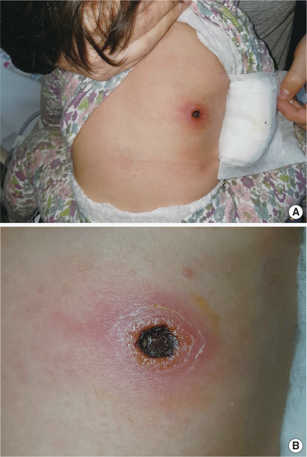
Fig. 2.
(A) A black eschar in central area surrounded by erythematous rim reap-peared on the 9th day. Eschar had occurred on the 4th day of skin lesion. (B) A black eschar in central area surrounded by erythematous rim. Picture zoomed in panel A.

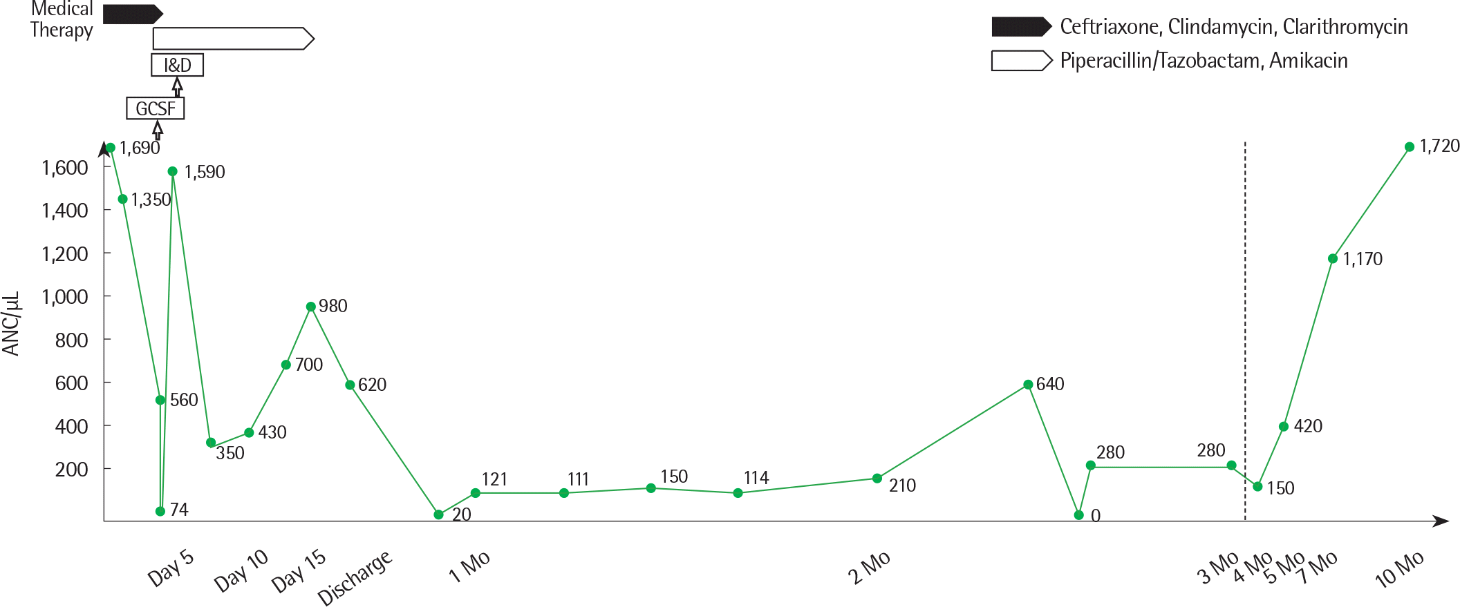
Fig. 4.
Changes in neutrophil count (ANC) by date and then month. GCSF, granulocyte colony stimulating factor; I&D, incision and drainage.

Table 1.
Summary of lab findings


 PDF
PDF ePub
ePub Citation
 Print
Print


 XML Download